
Neokamalomyces indicus Sanjay & Raghv. Singh, 2022
|
publication ID |
https://doi.org/10.11646/phytotaxa.571.2.3 |
|
DOI |
https://doi.org/10.5281/zenodo.7293275 |
|
persistent identifier |
https://treatment.plazi.org/id/03DB87C3-1B33-762E-80B1-FBF076AFA2BB |
|
treatment provided by |
Plazi (2022-11-04 09:06:14, last updated 2024-11-28 17:02:07) |
|
scientific name |
Neokamalomyces indicus Sanjay & Raghv. Singh |
| status |
sp. nov. |
Neokamalomyces indicus Sanjay & Raghv. Singh , sp. nov. Figs 2–5 View FIGURE 2 View FIGURE 3 View FIGURE 4 View FIGURE 5
MycoBank: MB 843768
Type:— INDIA. Uttarakhand: Haridwar, Har Ki Pauri , 29.9567°N 78.1710°E, July 2019, coll. Sanjay Yadav, on living leaves of Ficus benghalensis L. ( Moraceae ), AMH 10233 (holotype) GoogleMaps , MH-BHU 13 (isotype), NFCCI 4870 (ex-type living culture) GoogleMaps .
Diagnosis:— Differs from Parapallidocercospora colombiensis by its conidiomata pycnidial type, with conidiogenous cells developing from inner lining of conidiomatal wall.
Etymology:— indicus , referring to India, the country where the fungus was discovered.
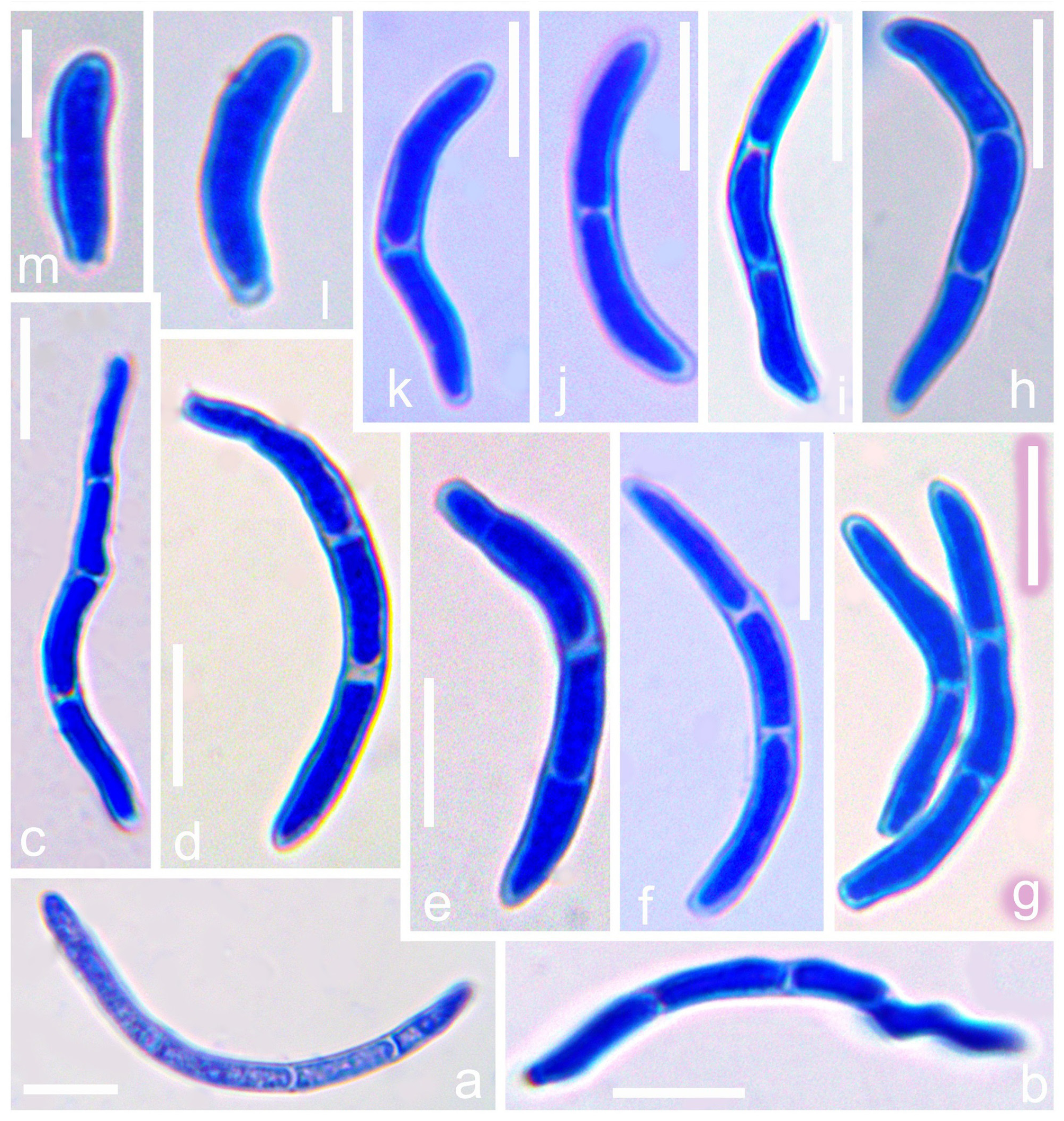
Leaf spots numerous, amphigenous, circular to subcircular, brown, with dark brown border with greyish white centre (with many blackish dots of conidiomata), 5–10 mm diam., later becoming irregular and covering entire leaf surface. Conidiomata pycnidial, brown to dark brown, subepidermal, epigenous, visible on upper sides of the leaf, several in each lesion, immersed to erumpent, subglobose to globose, 50–180 µm diam., with a central ostiolum, releasing a hyaline conidial mass; conidiomatal wall without distinctly differentiated layers of textura angularis, outer cells with brown, somewhat thickened walls, inner cells hyaline, thin-walled. Ostiole single, circular, central. Conidiophores reduced to conidiogenous cells, lining the inner cavity. Conidiogenous cells hyaline, tightly aggregated, cylindrical and tapering gradually toward the apex, ampulliform or lageniform with a relatively long neck, holoblastic, proliferating sympodially, smooth, percurrent proliferations not observed, 8–12(–16) × 3–7 μm, scars unthickened. Conidia cylindrical, weakly to strongly curved, or flexuous, gradually attenuated to a rounded apex, gradually or more abruptly attenuated into a broadly truncate base, 0–4-septate, not or indistinctly constricted around the septa, hyaline to light olivaceous, (10–)23–36(–50) × (1.5–)2–4(–5) μm, hila unthickened to slightly thickened. Sexual morph not seen.
Culture characteristics:— Colonies on PDA restricted and erumpent, surface folded, cerebriform to irregularly pustulate, mostly covered by a dense woolly floccose mat of smoke grey aerial mycelium, dark blackish green, reverse fuscous-black, with an irregular margin, reaching 8 mm diam. after 28 days at 25 °C with an even to slightly ruffled and glabrous margin. Only sterile mycelium was found without development of any kind of fruiting body or spores.
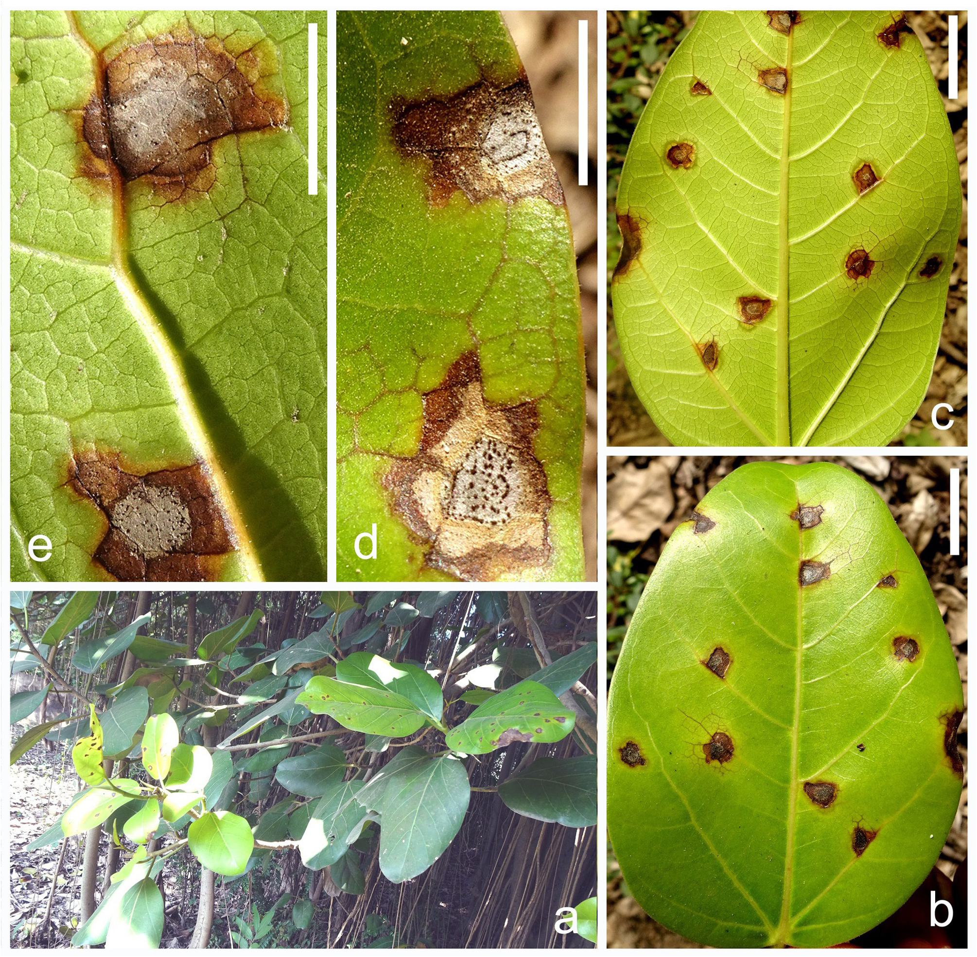
FIGURE 2. Neokamalomyces indicus on Ficus benghalensis. a. Host plant in natural habitat. b. Initial stage of symptom on upper surface of leaf. c. Initial stage of symptom on lower surface of leaf. d, e. Conidiomata on host tissue. Bars: b, c = 20 mm, d, e = 10 mm.

FIGURE 3. Neokamalomyces indicus on PDA (ex-type culture, NFCCI 4870). a, b. Colony on PDA top view. c. Colony on PDA reverse view. d–h. Germinated conidia in water droplet in cavity slide after 12–15 hours. i, j. Germinated conidia on PDA (stained with cotton blue). k. Development of mycelia on PDA. Bars: a = 10 mm, b = 5 mm, c = 10 mm, d–h = 20 μm, i–k = 10 μm.
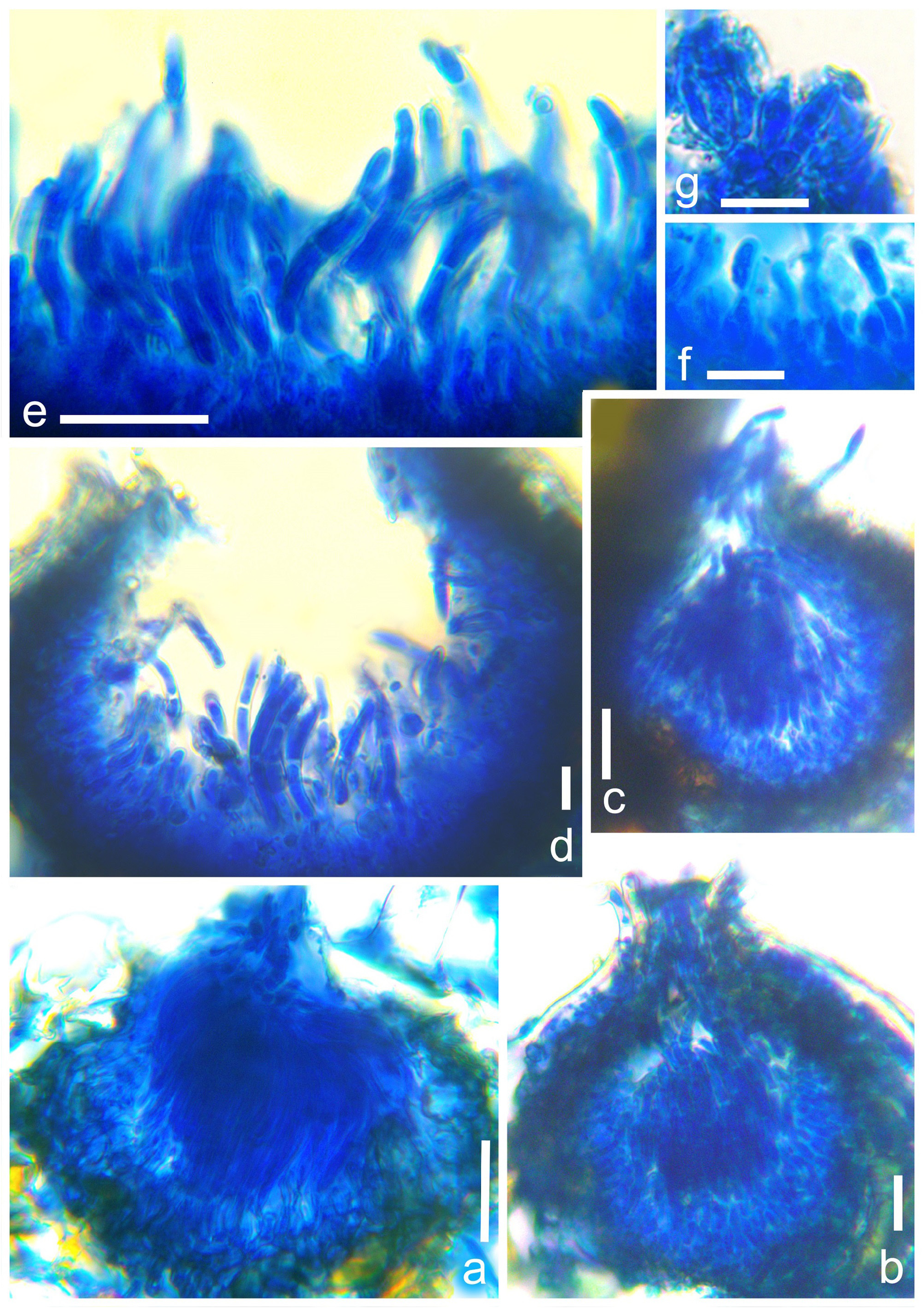
FIGURE 4. Fruiting body of Neokamalomyces indicus (AMH 10233, holotype). a–d. Vertical section through conidiomata. e, f. Conidia with conidiophores. g. Conidiogenous cells. Bars: a–e = 20 μm, f, g = 10 μm.
No known copyright restrictions apply. See Agosti, D., Egloff, W., 2009. Taxonomic information exchange and copyright: the Plazi approach. BMC Research Notes 2009, 2:53 for further explanation.
|
Kingdom |
|
|
Phylum |
|
|
Class |
|
|
Order |
|
|
Family |
|
|
Genus |
1 (by plazi, 2022-11-04 09:06:14)
2 (by ExternalLinkService, 2022-11-04 09:13:37)
3 (by jonas, 2022-11-04 13:41:01)
4 (by ExternalLinkService, 2022-11-04 13:51:09)
5 (by ExternalLinkService, 2022-11-04 15:16:17)
6 (by ExternalLinkService, 2022-11-04 19:51:37)
7 (by plazi, 2023-11-07 21:30:18)
8 (by ExternalLinkService, 2023-11-08 02:14:25)