
GYMNOTIFORMES, Berg, 1940
|
publication ID |
https://doi.org/ 10.1111/zoj.12142 |
|
persistent identifier |
https://treatment.plazi.org/id/03DB6116-5304-A226-FB98-D0B424A07F92 |
|
treatment provided by |
Marcus |
|
scientific name |
GYMNOTIFORMES |
| status |
|
GYMNOTIFORMES View in CoL View at ENA
Description
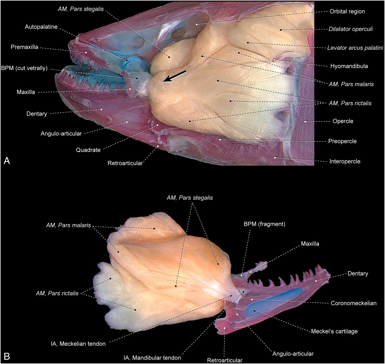
Gymnotus carapo ( Fig. 19 View Figure 19 )
The rictalis is readily distinguishable and almost fully separated from the remainder of the segmentum facialis, with the rictalis continuous with the malaris and stegalis only along its anterodorsal portion. Fibres of the rictalis are arranged into several distinguishable bundles. The entire rictalis section arises from the quadrate, preopercle, and hyomandibula and inserts primarily on the coronoid process formed by the dentary and angulo-articular in addition to the buccopalatal membrane. An additional flat anterolateral tendon emerges from the rictalis immediately internal to the third infraorbital, and grades into a strong subcutaneous layer of connective tissue.
The malaris and stegalis are largely continuous with each other at their common origin from the metapterygoid, hyomandibula, frontal, sphenotic, and parasphenoid, with this origin situated medial to the levator arcus palatini. Towards their insertion, the malaris and stegalis become more obviously differentiated from each other. The anterolateral fibres of the malaris insert onto a superficial anterodorsal aponeurosis of the rictalis, whereas most fibres of the malaris converge onto a strong mandibular tendon that inserts on the medial face of the coronoid processes of the dentary and angulo-articular. The stegalis inserts on the coronomeckelian via the meckelian tendon.
The ramus mandibularis trigeminus nerve could not be located in the examined specimen.
The segmentum mandibularis is absent.
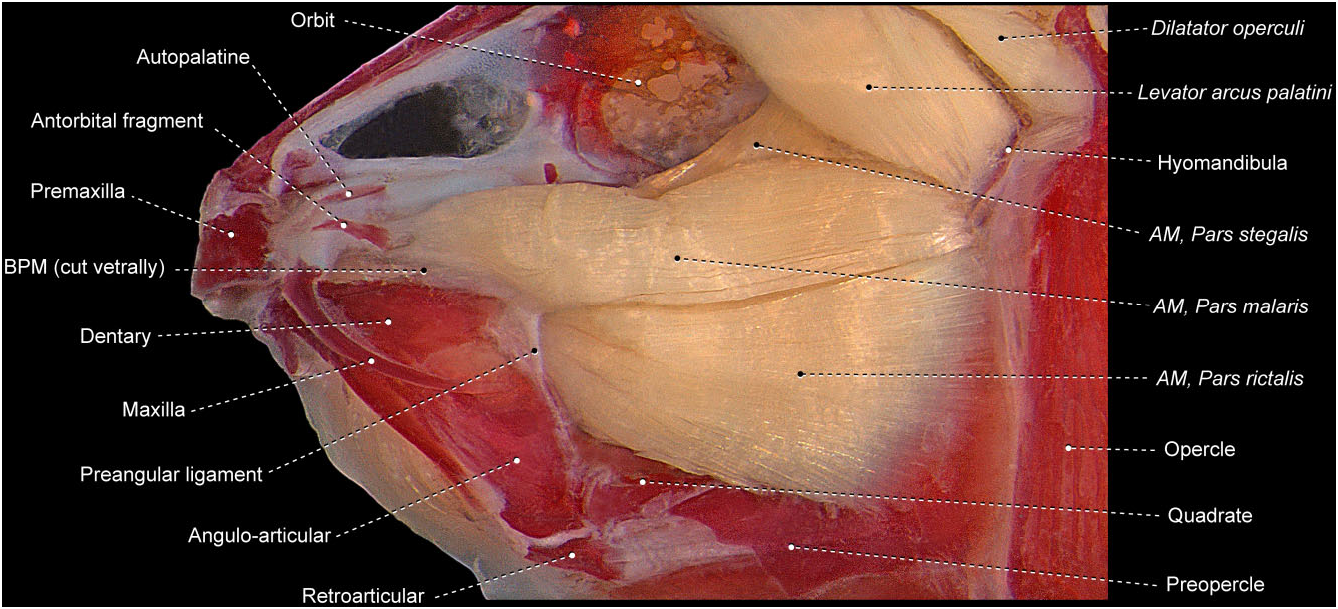
Brachyhypopomus pinnicaudatus ( Fig. 20 View Figure 20 )
Except for a limited intermingling of muscle fibres between the ventral portions of the rictalis and stegalis, all of the three primary facial sections are completely separated from each other. The rictalis originates from the quadrate and preopercle and inserts on the coronoid process of the dentary.
The malaris originates from the hyomandibula, with the posterodorsal portion of the section running between the lateral and the medial sections of the levator arcus palatini. The malaris inserts onto the buccopalatal membrane, primarily onto its anterodorsal portion that firmly attaches to the antorbital and maxilla. This membrane further has a well-differentiated preangular ligament, which anchors to the angulo-articular and also receives a few muscle fibres of the malaris.
The stegalis is the most massive of the facial sections with a broad origin from the quadrate, metapterygoid, hyomandibula, sphenotic, orbitosphenoid, and parasphenoid. This section converges anteriorly onto the intersegmental aponeurosis, which, in turn, subtly differentiates into a ventral portion that inserts on the coronomeckelian (thus, corresponding to the meckelian tendon) and a dorsal component that serves as the site of origin for the segmentum mandibularis (thereby corresponding to the mandibular tendon).
The ramus mandibularis trigeminus nerve traverses the segmentum facialis by passing internal to the rictalis and malaris and external to the stegalis.
The segmentum mandibularis is undifferentiated into subsections. It arises from the mandibular tendon and inserts on the medial surface of the dentary and angulo-articular.
Remarks
Aguilera (1986) studied the adductor mandibulae of representatives of all families in the Gymnotiformes . According to that author, the facial fibres of this muscle in the Gymnotidae ( Electrophorus and Gymnotus ) are arranged in a convoluted fashion and the segmentum facialis lacks obvious subdivisions. Examination of Gymnotus carapo confirms this characterization, although we were, nonetheless, able to confidently identify the three primary facial sections that remain partially differentiated from one another in this species ( Fig. 19 View Figure 19 ). The segmentum facialis in the Gymnotidae inserts on the lower jaw and buccopalatal membrane ( Fig. 19 View Figure 19 ; Aguilera, 1986). Gymnotids have the dorsolateral portion of the segmentum facialis (= malaris) arising not only from the suspensorium but also from the neurocranium ( Fig. 19 View Figure 19 ; Aguilera, 1986), an arrangement unique amongst the Gymnotiformes . The neurocranium further serves in most gymnotiforms as a site of origin of the dorsomedial portion of the segmentum facialis (= stegalis; Fig. 19 View Figure 19 ; Aguilera, 1986).
In the Hypopomidae , Rhamphichthyidae , and Sternopygidae , the malaris is well separated from the remaining facial sections and inserts on the maxilla and/or lacrimal ( Aguilera, 1986; Datovo & Vari, 2013). The rictalis and stegalis retain their insertions on the lower jaw with these sections better separated from each other in the Hypopomidae and Sternopygidae than in Rhamphichthyidae ( Aguilera, 1986) .
The segmentum facialis of the Adontosternarchus (Apteronotidae) is overall similar to that in the Hypopomidae and Sternopygidae ( Aguilera, 1986) , with the only notable difference being that the malaris inserts on both the maxilla and lower jaw in Adontosternarchus versus on the maxilla and/or lacrimal in those two families. Identification of the facial sections elsewhere in the Apteronotidae becomes more complicated because the muscle division(s) inserting onto the maxilla may occupy different positions (ventral, posterior, or both dorsal and ventral; cf. Aguilera, 1986; Marrero & Winemiller, 1993), consequently obfuscating a resolution of whether the differences across the family are a function of migration of sections or new attachments to the maxilla. These components of the Apteronotidae are, therefore, not included in our synonymy.
A curious feature of the adductor mandibulae of some gymnotiforms is the presence of filamentous intermuscular bones within the segmentum facialis. Such ossifications have been reported for the gymnotid Gymnotus carapo , the apteronotid Orthosternarchus tamandua and the rhamphichthyids Iracema caiana , Rhamphichthys marmoratus , and Rhamphichthys rostratus ( Aguilera, 1986; Albert, 2001; Hilton et al., 2007; Carvalho & Albert, 2011). These structures probably represent ossifications of some internal tendons of the adductor mandibulae of these fishes. To our knowledge, such ossifications have not been reported elsewhere in the Teleostei.
Synonymy
Segmentum facialis
Complejo adductor mandibulae: Aguilera (1986): Electrophorus , Gymnotus .
Pars malaris
A 1: Aguilera (1986): Adontosternarchus , Rhamphichthys , Steatogenys , Sternopygus .
Pars rictalis
A 2: Aguilera (1986): Adontosternarchus , Rhamphichthys , Steatogenys , Sternopygus .
Pars ricto-stegalis A 2–3: Aguilera (1986): Rhamphichthys .
Pars stegalis
A 3: Aguilera (1986): Adontosternarchus , Rhamphichthys , Steatogenys , Sternopygus .
No known copyright restrictions apply. See Agosti, D., Egloff, W., 2009. Taxonomic information exchange and copyright: the Plazi approach. BMC Research Notes 2009, 2:53 for further explanation.