
Pediobius prominentis Cao & Zhu
|
publication ID |
https://doi.org/ 10.5281/zenodo.375759 |
|
publication LSID |
lsid:zoobank.org:pub:9A463357-EEE2-4A70-BCB2-573052DB48CC |
|
DOI |
https://doi.org/10.5281/zenodo.5694596 |
|
persistent identifier |
https://treatment.plazi.org/id/03DA87F7-3B71-FF89-FF05-FCE8FD34FE76 |
|
treatment provided by |
Plazi |
|
scientific name |
Pediobius prominentis Cao & Zhu |
| status |
sp. nov. |
11. Pediobius prominentis Cao & Zhu , sp. n.
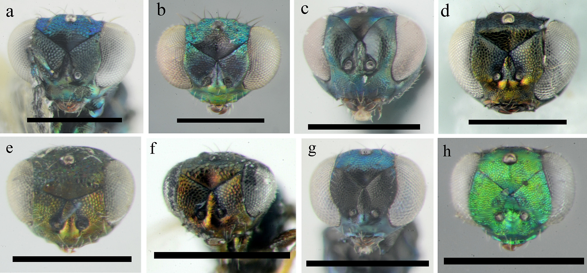
( Figs 10 View FIGURE 10 , 33 View FIGURE 33 d)
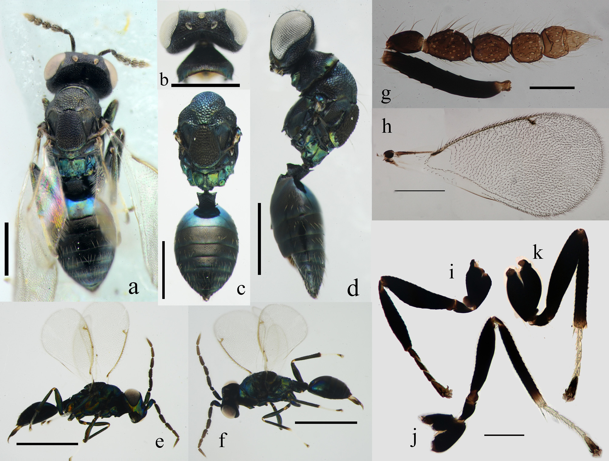
Diagnosis. Antenna with scape reaching median ocellus, funicle not clearly separated from clava and flagellum light brown ( Fig. 10 View FIGURE 10 g). Vertex, mesoscutum and scutellum strongly reticulate. Scutellum raised but not convex, distinctly higher than mesoscutum and metanotum, with a smooth posterior margin ( Figs 10 View FIGURE 10 a, 10c–d). Submedian area sculptured along submedian carina and posterior margin ( Fig. 10 View FIGURE 10 c).
Female. Body length 2.2–2.4 mm. Body large and slender.
Body black, with following parts with bluish reflections: collar of pronotum, the smooth margin of scutellum, metanotum, propodeum and Gt1. Head in anterior view with coppery reflections. Scape dark green, pedicel brown and flagellum light brown ( Fig. 10 View FIGURE 10 g). Legs, except tarsi, dark green, but tibiae of mid leg pale apically; tarsomeres in fore leg infuscate, dark brown; the first three tarsomeres pale white, and the apical tarsomere dark brown in mid and hind legs; metatibial spur white, and brown apically ( Figs 10 View FIGURE 10 i–k).
Antenna with 3-segmented funicle and 2-segmented clava, and funicle not clearly separated from clava ( Fig. 10 View FIGURE 10 g). Scape reaching median ocellus. Pedicel longer than broad, narrower and shorter than F1. F1 and F2 longer than broad, F3 subquadrate; F1 broader than F2 and F3, F2 as broad as F3; C1 transverse, C2 slightly narrower than C1, and C2 tapering to apex and bearing a distinct terminal spine. Relative measurements of scape: pedicel: F1: F2: F3: C1: C2=2.2: 0.6: 0.8: 0.6: 0.6: 0.4: 0.4. Face reticulate, and more finely so below toruli, and more coarsely so above frontofacial sutures. Toruli inserted high above lower margin of eyes. Scrobal grooves meeting below frontofacial sutures and connected with frontofacial sutures with a very short longitudinal suture. Interscrobal space weakly raised. Frontofacial sutures diverging at an obtuse angle of about 110° ( Fig. 33 View FIGURE 33 d).
Head in dorsal view transverse, more than 3× as broad as long. Vertex strongly reticulate. Ocelli arranged in about a right-angle triangle. POL 2× as long as OOL (1.2: 0.6). Occiput sharply margined throughout.
Pronotal collar rugose medially, and with anterior carina. Mesoscutum strongly reticulate, with the posterior pair of mesoscutal setae situated on notaular depressions. Notauli poorly indicated; notaular depressions shallow. Axilla finely reticulate. Scutellum distinctly raised but not convex, higher than mesoscutum and metanotum, longer than broad (3.0: 2.2) ( Figs 10 View FIGURE 10 a, 10c); strongly reticulate, and with a smooth posterior margin; reticulation slightly elongate anteriorly, more finely so along median line ( Figs 10 View FIGURE 10 a, 10c). Dorsellum large, distinctly reticulate; weakly protruding in a strongly obtuse angle apically. Propodeum with submedian carinae slightly diverging posteriorly; submedian area longer than broad, distinctly reticulate along submedian carina and posterior margin; plicae subparallel; callus with 2 setae ( Figs 10 View FIGURE 10 a, 10c). Lateral panel of pronotum and prepectus reticulate; acropleuron polished; mesepisternum partially with weak reticulation, but transepimeral sulcus with distinct meshes; upper mesepismeron strongly reticulate, lower mesepismeron only with weak meshes on basal third; transepimeral sulcus and pleural suture distinct, transepisternal sulcus indistinct; transepimeral sulcus slightly curved ( Fig. 10 View FIGURE 10 d).
Petiole subquadrate, strongly reticulate. Gaster pointed apically, as long as mesosoma; Gt1 occupying about 1/ 6 length of gaster, smooth, with posterior margin straight; Gt6 about 3× as broad as long, Gt7 about 6× as broad as long, and Gt2–7 reticulate with smooth posterior margin ( Fig. 10 View FIGURE 10 c).
Fore wing hyaline and setose, with speculum closed and very small. MV arched. PMV and STV short, and PMV longer than STV. Relative measurements of SMV: MV: PMV: STV= 2.4: 6.2: 0.8: 0.6. Metatibial spur much longer than the width of tibia and nearly as long as the basal tarsomere ( Fig. 10 View FIGURE 10 k).
Male. Body length about 2 mm. Male is similar to female and mainly differs from female as follows. Pronotal collar and mesoscutum metallic blue green. Antenna filiform, with five flagellar segments subequal in length and more than 3× as long as broad, not forming an outstanding clava; scape slightly compressed ( Figs 10 View FIGURE 10 e–f). Petiole about 2× as long as broad. Gaster distinctly shorter than mesosoma; Gt1 occupying about 1/3 length of gaster.
Etymology. This species is named for its distinctly prominent scutellum, from Latin prominentem = prominent.
Material examined. Holotype ♀, CHINA, Guangxi, Guilin, XingAn , Mao-Er Mountains , V.2013, coll. Kan Wang ( IZCAS) . Paratypes: 2♀ 1♂, same data as the holotype .
Biology. Unknown.
Distribution. CHINA: Guangxi.
Remarks. This new species runs to P. eubius in the key of Bouček (1965a) and Kamijo (1986a), and P. eubius complex in the key of Graham (1959). We include P. prominentis in the epigonus -group along with P. metallicus and P. eubius because of their similarities in sculpture of mesosoma. Pediobius prominentis is characterized by the distinctly raised scutellum and deep and strong reticulation on mesoscutum and scutellum, which separates it from other species of the epigonus -group. More differences between P. prominentis and P. eubius complex are given in the key. Additionally, P. prominentis is very similar to P. sasae in the sculpture of mesoscutum and scutellum, which is strongly and deeply reticulate (see Hansson 2006). Pediobius prominentis can be easily separated from the latter species by characters as follows. Pediobius prominentis has body not so slender; occiput distinctly excavated; scutellum distinctly raised but not convex; female gaster weakly slender. Whereas in P. sasae , body distinctly elongate, strikingly slender; occiput strongly concave in the upper half; scutellum distinctly raised but not convex; female gaster strikingly elongate and slender.
| IZCAS |
Institute of Zoology, Chinese Academy of Sciences |
No known copyright restrictions apply. See Agosti, D., Egloff, W., 2009. Taxonomic information exchange and copyright: the Plazi approach. BMC Research Notes 2009, 2:53 for further explanation.