
Microdon (Chymophila) bruchi Shannon, 1927
|
publication ID |
https://doi.org/ 10.1080/00222933.2020.1746847 |
|
persistent identifier |
https://treatment.plazi.org/id/03D9057A-136D-342C-FE8F-FE1FC0D91CF4 |
|
treatment provided by |
Carolina |
|
scientific name |
Microdon (Chymophila) bruchi Shannon, 1927 |
| status |
|
Microdon (Chymophila) bruchi Shannon, 1927 View in CoL
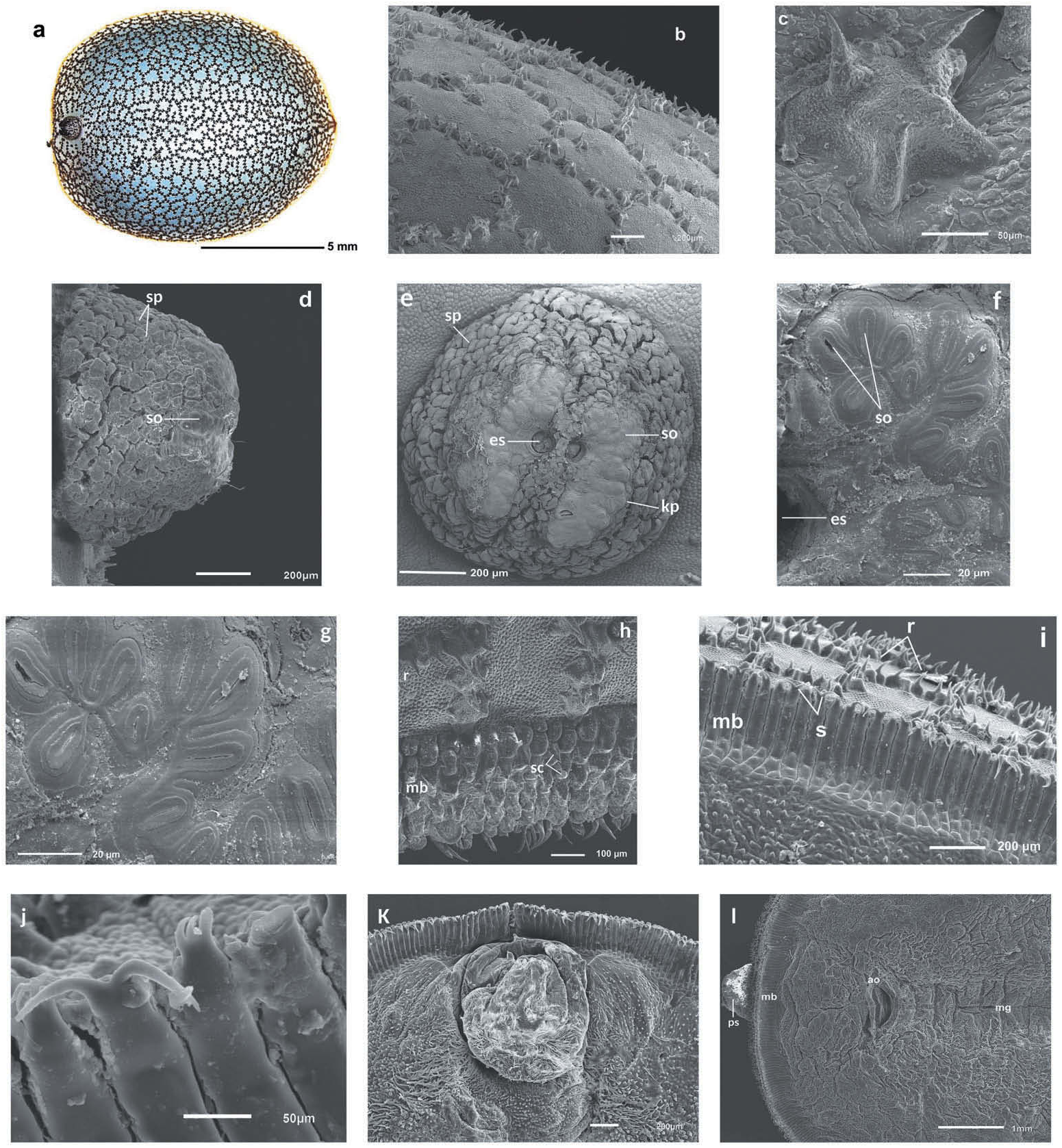
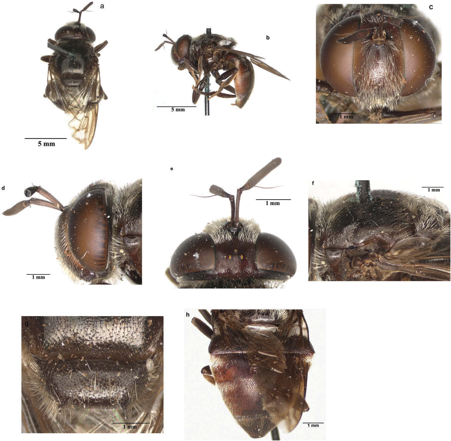
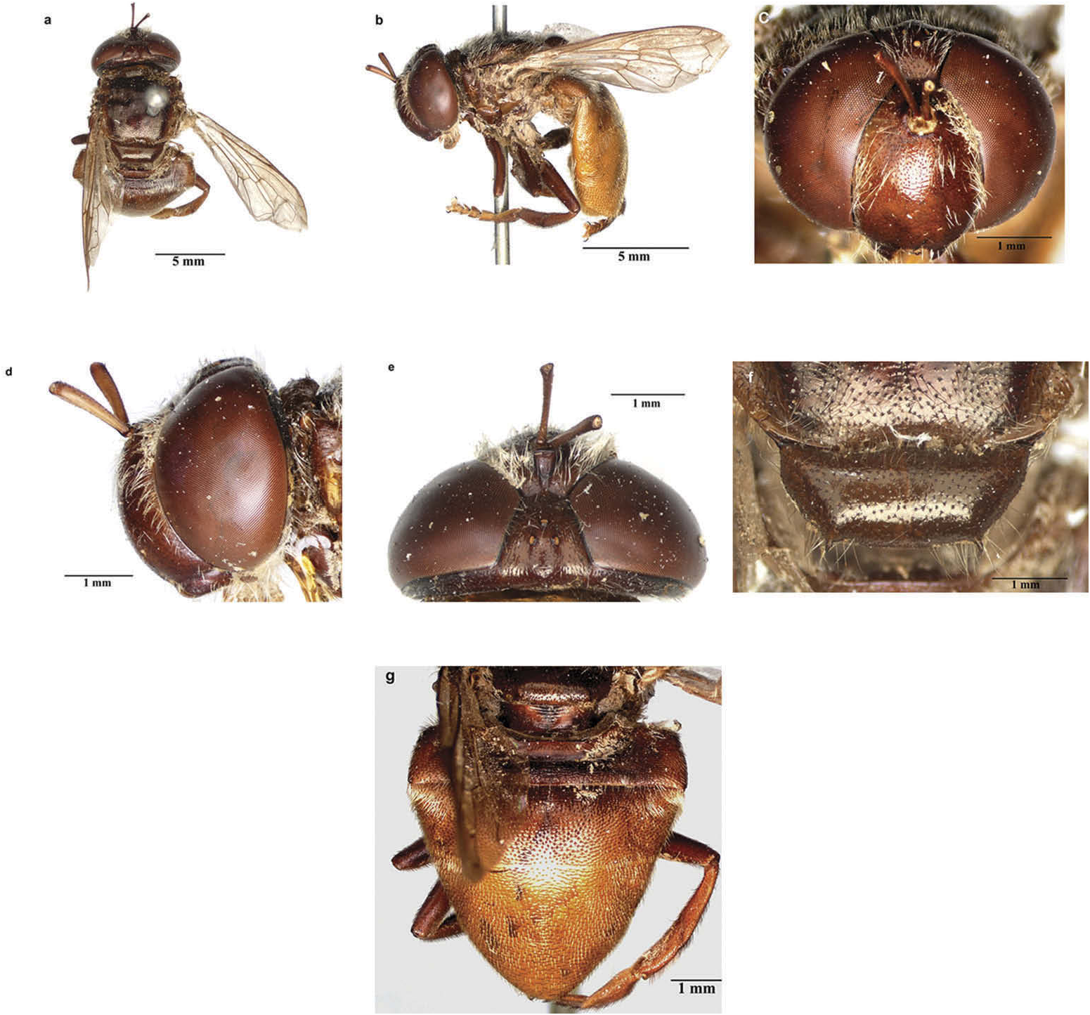
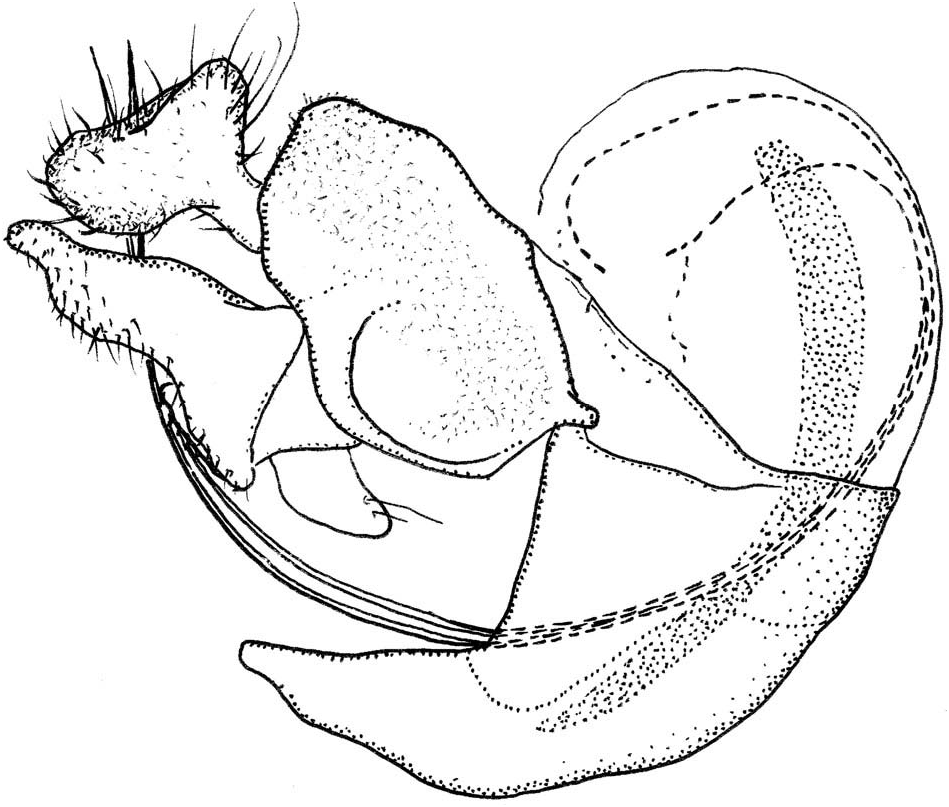
( Figures 3 View Figure 3 (a – l), 4(a – h), 5(a – g), 6)
Microdon bruchi Shannon, 1927: 38 View in CoL . (Lectotype: ♀, USNM; here designated. Type locality: Argentina, Córdoba, Alta Gracia ) .
Microdon argentinae Hull, 1937: 18 View in CoL . (Holotype: ♂, MCZ; by monotypy. Type locality: Argentina, Córdoba). New synonym.
Microdon (Chymophila) View in CoL sp. of Zubarán (2018).
Material examined
Lectotype of Microdon bruchi Shannon, 1927 . ARGENTINA: 1♀; Alta Gracia // Cord. 20.1.27 // R.C. Shannon' 'Laying eggs in // trail of ants' ' Microdon // bruchi // Shannon' 'Cotype // No. 40822 // U.S. N.M' 'USNMENT 01295601 ’ [barcode]; USNM coll. USNM type database: http:// n 2t.net/ark:/65665/395cc169c-24d2-42a0-a81c-cca5df2904f5.
Holotype of Microdon argentinae Hull, 1937 . ARGENTINA: 1♂; Cordova. // Argent. Davis. Davis; Microdon // argentina // FMH Hull; ‘ M.C.Z. Type // 22218’; MCZ coll.; MCZ type database: https://mczbase.mcz.harvard.edu/MediaSearch.cfm?action=search&media_id= 115547,115548,115549,115550,115551.
Other studied material . ARGENTINA: 1♀; Catamarca Province, Trampasacha , 8 km W Chumbicha; 28.816°S, 66.300°W; alt GoogleMaps . 650 m.; 24 October 2003; F .D GoogleMaps . Parker and M .E. Irwin leg.; CSCA coll.; hand netted in damp wash. 1♀; Mendoza Province, San Martín, Estación Experimental INTA Junín ; 32.985°S, 68.360°W; 19 September 2016; G GoogleMaps . Debandi leg GoogleMaps .; ZFMK coll.; ZFMK-DIP-00015973, ZFMK DNA voucher D291. 1♀; same locality as previous; 20 September 2016; ZFMK coll GoogleMaps .; ZFMK-DIP-00015974, ZFMK DNA voucher D293. 1♂; same locality as previous; 17 September 2016; ZFMK coll GoogleMaps .; ZFMK- DIP-00015975, ZFMK DNA voucher D296. 1♂; same locality as previous; 15 September 2016; ZFMK coll GoogleMaps .; ZFMK-DIP-00015976, ZFMK DNA voucher D298. 1♂; same locality as previous; 5 November 2016; on Camponotus mus colony; RMNH coll GoogleMaps .; ZFMK-DIP- 00046334, ZFMK DNA voucher D369. 2♀; same locality as previous; 15 October 2016; ZFMK coll GoogleMaps .; ZFMK-DIP-00046223, ZFMK DNA voucher D371; RMNH coll.; ZFMK- DIP-00046225, ZFMK DNA voucher D373.
Diagnosis
Body length: male 11 – 12 mm, female 12 – 14 mm. Microdon bruchi is assigned to the subgenus Chymophila based on the characteristic shape of wing vein M1, which has an outward angle and is anteriorly recurrent ( Figures 4 View Figure 4 (a), 5(a) and 5(b)). The morphology of the male genitalia is also distinctive for the subgenus, with both phallic processes very long and slender ( Figure 6 View Figure 6 ). Microdon bruchi belongs to the group of Neotropical non- (or only faintly) metallic species without conspicuous stripes of pale pile on the abdomen. Among these species, M. bruchi can be recognised by the combination of the following characters: metatibia with pile shorter than width of tibia; scutellum with calcars shorter than 1/3 of scutellar length; tergite 3 largely brown to black pilose with anterolateral patches of whitish pile; anterior fascia of whitish pile on mesoscutum absent or covering at most 1/3 of mesoscutum; scutellum at least partly pale pilose.
Redescription (based on lectotype)
( Figure 4 View Figure 4 (a – h)). Adult female. Body size: 12 mm. Head. Face occupying 0.44 of head width in frontal view; blackish brown; white pilose. Gena blackish brown; white pilose. Lateral oral margin weakly produced; blackish brown; white pilose. Frons and vertex blackish brown; white pilose, except vertex black pilose at level of ocellar triangle. Occiput blackish brown; white pilose. Eye bare. Antennal fossa about as wide as high. Antenna brown. Ratio of scape: basoflagellomere approximately 1:1.3. Basoflagellomere parallel sided with somewhat acute apex. Arista slender, about 2/3 length of basoflagellomere Thorax. Mesoscutum shining blackish brown; semi-erect black pilose, except for narrow uninterrupted fasciae of semierect white pile along anterior and posterior margins (width of anterior fascia is approx. 1/6 length of scutum, width of posterior fascia approx. 1/8). Postpronotum blackish brown; white pilose. Postalar callus blackish brown; white pilose. Scutellum blackish brown; white pilose; with two apical calcars of about 1/10 length of scutellum, with mutual distance approximately 1.4 length of scutellum. Pleura blackish brown. Anterior and posterior part of anepisternum separated by deep sulcus; white pilose anteriorly and posteriorly, with wide bare area in between. Anepimeron entirely white pilose. Katepisternum white pilose dorsally, bare ventrally. Other pleurae bare (except for microtrichiae). Calypter grey with black fringe, halter brown with blackish knob. Wing hyaline, brownish in antero-apical cells; veins around cell br and vena spuria yellow. Wing microtrichose, except cell br only microtrichose along vena spuria and at apical 1/8; cell bm widely bare along anterior and posterior margins, leaving wedge-shaped field of microtrichia with narrowest part at base of cell; cell cup bare on anterobasal 2/5; alula bare at basomedial 1/2. Legs blackish, pale brown pilose. Abdomen broadly oval, wider than thorax, widest at base of tergite 3. Tergites blackish brown with weak metallic sheen. Tergites 1 brown pilose. Tergites 2 brown pilose on lateral 1/4, white pilose medially. Tergites 3 – 5 brown pilose except white pilose at anterolateral corners and lateral margins. Sternites 1 – 5 brown; brown pilose.
Variation in additionally studied females
The additionally studied females (see Material examined) differ from the type of M. bruchi most notably in the colouration of the pile. The occiput is black pilose dorsally in the recently collected specimens, and all pile on the mesoscutum and pleura is black. The scutellum is white pilose, except black pilose on anterior 1/3 and along posterolateral margins. The legs are entirely black pilose. The tergites are black pilose on parts where holotype is brown pilose.
Male (based on the recently collected material from Argentina; see Material examined)
As for the female, except for the following differences: Occiput black pilose dorsally. Ratio of scape: basoflagellomere approximately 1:1.2. Width of anterior fascia of yellowish white pile on mesoscutum varies from approx. 1/8 to 1/3 of length of scutum; also, with
yellowish white pile along transverse suture; white pile along posterior margin of mesoscutum absent to covering approx. 1/8 of length of scutum. Apical tarsomeres reddish brown to black. Tergite 3 with lateral margin reddish brown. Tergite 4 reddish brown, clearly getting paler laterally and posteriorly, but to variable degree among examined specimens; yellowish pilose laterally and on posterior half. Sternites 3 and 4 reddish brown. Sternites yellow pilose. Genitalia as in Figure 6 View Figure 6 .
Colour variation and synonymy
Colouration of body pile is quite variable among the studied specimens. Most notable is the variation in width of the fascia of pale pile along the anterior margin of the scutum. In males, its width ranges from approximately 1/8 to 1/3 of the length of the scutum. In females, the anterior fascia of pale pile is entirely absent in the studied specimens from Catamarca and Mendoza provinces, whereas in the lectotype specimen of M. bruchi its width is about 1/4 the length of the scutum. In the female specimen in the photos of Zubarán (2018, figs 16 and 17), an intermediate width is found of about 1/6 the length of the mesoscutum. As the studied specimens are highly similar in many other characters, we choose to consider them conspecific. Moreover, all specimens have the same host ant species, and the COI variability among the Mendoza specimens is less than 1%. Variation in pile colouration has previously been recorded in a number of other Microdon species ( Akre et al. 1973; Thompson 1981, 2007; Wolton 2017). The male holotype of Microdon argentinae shares the diagnostic characters of M. bruchi with the holotype of the latter taxon: abdomen non-metallic without conspicuous stripes of pale pile, metatibia with very short pile, scutellum with small (shorter than 1/3 of scutellar length) calcars, tergite 3 largely black or brown pilose with anterolateral patches of pale pile, and mesoscutum with less than anterior half pale pilose. Consequently, M. argentinae is here considered to be a junior synonym of M. bruchi ( Figure 5 View Figure 5 (a – g)).
The original descriptions of M. bruchi by Shannon (1927) and of M. argentinae by Hull (1937) are not entirely accurate with regard to the colour of the pile on the mesoscutum. Shannon (1927: 39) wrote ‘ [Thorax …] clothed with short yellow hairs ’, but he did not mention the black pile clearly visible in the holotype. Hull (1937: 18) wrote ‘ Pile of thorax short, pale, appressed, with a few scattered darker hairs ’, whereas in the holotype much of the posterior half of the mesoscutum is covered with black pile. Shannon (1927: 39) observed a male Microdon at the site where he had collected the female. He noted that ‘ It was smaller in size and the abdomen was of a distinct brown colour ’. This concurs with the reddish-brown colour we observed in the recently collected Argentinean males, as well as in the male holotype of Microdon argentinae .
Lectotype designation and additional remarks
In the original description of M. bruchi, Shannon (1927) studied two females on which the description was based, but he did not mention the holding institutions of these syntypes. Thompson et al. (1976) stated that one syntype is in the USNM collection and the other is likely to be (question mark added) in the MACN collection. In our survey, only the female specimen deposited in the USNM was studied, and it is here designated as the lectotype to fix and ensure the universal and consistent interpretation of the name.
After the preparation of the identification key and the survey of the published literature and type material for Neotropical species, we could identify as M. bruchi the female specimen named as Microdon (Chymophila) sp. by Zubarán (2018, figs 16 and 17), photographed laying eggs in a decayed tree with a nest of C. mus . This female specimen represents the first record of M. bruchi in the Entre Ríos Province, Argentina.
No known copyright restrictions apply. See Agosti, D., Egloff, W., 2009. Taxonomic information exchange and copyright: the Plazi approach. BMC Research Notes 2009, 2:53 for further explanation.
|
Kingdom |
|
|
Phylum |
|
|
Class |
|
|
Order |
|
|
Family |
|
|
Genus |
Microdon (Chymophila) bruchi Shannon, 1927
| López-García, Guillermo P., Reemer, Menno, Debandi, Guillermo & Mengual, Ximo 2020 |
Microdon argentinae
| Hull FM 1937: 18 |
Microdon bruchi Shannon, 1927: 38
| Shannon RC 1927: 38 |