
Pareiorhina pelicicei, Azevedo-Santos, Valter M. & Roxo, Fábio F., 2015
|
publication ID |
https://doi.org/ 10.11646/zootaxa.3937.2.8 |
|
publication LSID |
lsid:zoobank.org:pub:942BB7F1-E8DA-494B-BE18-6CC1CC030B43 |
|
DOI |
https://doi.org/10.5281/zenodo.5696250 |
|
persistent identifier |
https://treatment.plazi.org/id/03D687BA-FF81-FF95-FF54-ADA2DD2AF98D |
|
treatment provided by |
Plazi |
|
scientific name |
Pareiorhina pelicicei |
| status |
sp. nov. |
Pareiorhina pelicicei View in CoL , new species
Table 1 View TABLE 1 , Figs. 1–2 View FIGURE 1 View FIGURE 2
Holotype. MZUSP 115495, male, 47.4 mm SL, Brazil, Minas Gerais State, municipality of Capitólio, córrego Tamborete, rio Grande basin, upper Paraná basin, 20°38'38"S 46°10'13"W, 24 May 2014, Azevedo-Santos VM, Paschoal LRP.
Paratypes. Same locality as the holotype. DZSJRP 20156, 1 male, 38.7 mm SL, 2 females, 31.3–37.0 mm SL, 16 June 2014, Azevedo-Santos VM. LBP 18990, 3 males, 39.0– 41.4 mm SL, 9 females, 27.8–42.0 mm SL, 2 females c& s 37.5 –39.9 mm SL, 16 June 2014, Azevedo-Santos VM. LBP 18899, 12 juveniles unsexes and not measured, collected with holotype. ZUEC 8604, 2 females, 37.4–40.8 mm SL, 16 June 2014, Azevedo-Santos VM.
Diagnosis. The new species, Pareiorhina pelicicei , is distinguished from P. carrancas and P. hyptiorhachis by absence of a postdorsal ridge (vs. presence of a postdorsal ridge). Additionally, it can be distinguished from all congeners, except P. hyptiorhachis , by having lower number of vertebrae = 29 (vs. 30 in P. brachyrhyncha and P. cepta and 31 in P. carrancas and P. rudolphi ).The new species differs from P. carrancas , P. hyptiorhachis and P. rudolphi by having the teeth with a minute lateral cusp (vs. unicuspid teeth); from P. brachyrhyncha and P. hyptiorhachis by having the anterior profile of the head elliptical in dorsal view (vs. rounded; see Fig. 3 View FIGURE 3 in Silva et al. 2013). Pareiorhina pelicicei differs from P. brachyrhyncha by having the tip of the snout naked (vs. the tip of the snout completely covered with small odontodes), a lower predorsal length (41.0– 44.8 mm SL vs. 45.6–48.9 mm SL), a higher number of premaxillary teeth (35–48 vs. 28–36), and a higher number of dentary teeth (33–49 vs. 26–36). Finally, the new species is distinguished from P. cepta by lacking small plates distributed on the thorax and abdomen (see Fig. 2 View FIGURE 2 a in Roxo et al. 2012a).
Description. Morphometric and meristic data are summarized in Table 1 View TABLE 1 . Small-size loricariids (mean 36.9 mm SL); bigger specimen examined with 47.4 mm SL. In lateral view, dorsal profile of body convex from snout tip to dorsal-fin origin, slightly concave and decreasing to end of caudal peduncle. Ventral surface of body straight from head to posterior end of caudal fin. Snout tip elliptical in dorsal view. Dorsal surface of caudal peduncle without keels and ventral surface flat. Greatest body depth at dorsal-fin origin. Trunk and caudal peduncle trapezoid in cross-section. Body progressively narrowing posteriorly from cleithrum.
Head flat to slightly convex in interorbital area. Eye relatively small and round, situated dorsolaterally just posterior of midpoint of head. Iris operculum present and poor developed. No ridge between eyes and nares. Nostril opening small. Supraoccipital process not elevated. Rostral margin of snout with minute, posteriorly directed odontodes; numerous small odontodes on dorsal portion of head. Tip of snout naked (i.e., without odontodes). Mouth wide; oral disk rounded; lips large with several papillae equally distributed on base of dentary and premaxilla and mildly decreasing in size distally; upper lip folded over itself. Teeth long and bicuspidate; cusps asymmetrical and minute laterally. Tooth series of premaxilla and dentary curved mesially. Buccal papillae short and digitiform located readily anterior to buccal valve. Maxillary barbel short, slender and free distally.
Dorsal region of body covered by dermal plates, except dorsal-fin base. Head and trunk plates covered with minute, uniformly sized and distributed odontodes. Ventrally largely unplated from snout to anal-fin origin. Abdomen and thorax completely naked. Lateral side of body entirely covered by plates; mid-dorsal plates slightly developed, reaching middle of anal-fin base; median plates not interrupted in median portion of body, with 1 or 2 plates not perforated before end of series; mid-ventral plates exceed end of anal-fin base.
All plates sculpted with lines of odontodes more developed posteriorly. Dorsal-fin rays ii,7; dorsal-fin originating at vertical slightly posterior through origin of pelvic-fin; its distal margin slightly convex. Spinelet present, reduced, and locking mechanism non-functional. Pectoral-fin rays i,6; distal margin slightly convex; unbranched pectoral-fin ray reaching beyond origin of unbranched pelvic-fin ray and covered with pointed odontodes (well developed in ventral portion). Pelvic-fin rays i,5; distal margin of fin slightly convex; tip of adpressed pelvic fin slightly reaching beyond anal-fin origin and covered with pointed odontodes (well developed in ventral portion and turned mesially). Adipose-fin not present. Anal-fin rays i,5; distal margin slightly convex. Caudal fin rays i,7-7,i, truncated.
Total number of vertebrae 29. Anterior portion of compound supraneural-first proximal radial of dorsal fin, contacting neural spine at 7th vertebrae. Eight pairs of ribs associated with vertebrae 8–15. Ribs slender and poorly ossified.
Supraorbital sensory canal with four pores s1, s3, s6+s6 and s8; s1 located on terminal portion of nasal plates; pore s3 located on anterior portion of nasal; pore s6+s6 located between frontal, on horizontal line through anterior most limits of eyes; pore s8 on division between frontal, sphenotic and parieto-supraoccipital, just above eyes. Five pores associated with infraorbital sensory canal; pore io2 present between first and second infraorbitals; pore io3 located in medial region between second and third infraorbitals; pore io4 located in medial region in anterior portion of eyes; pore io5 located in medial region between fourth and fifth infraorbitals, and pore io6 located between sixth and sphenotic in posterior portion of eyes. Preopercular canal composed of four pores; pore pm2 present on vertical through anterior margins of orbits; pore pm3 present between cheek plate and preopercle; pore pm4 present between preopercle and compound pterotic; pore po1 + pm5 present near io6 at edge of ventral orbital. Two postotic pores; pore po2 present above branchial opening; and po3 present on region overlying opening of swim-bladder capsule.
Coloration in life. In dorsal view, adult individuals have light brown ground color, from tip of snout to caudalfin spine origin. Lateral region with a dark brown stripe; in ventral portion with an area, mostly rectangular, with gold color. All fins with small dark spots. Individuals less than 15 mm SL, in general, present caudal-fin completely black.
Coloration in alcohol. Similar to living specimens, but with background color dark brown (see Fig. 1 View FIGURE 1 ). Sexual dimorphism. Adult males have a fleshy flap in the surface of first ray of pelvic fin (absent in females) and a conspicuous papilla in urogenital opening (absent in females).
Distribution. Known only from córrego Tamborete, a small tributary of rio Grande (upper rio Paraná basin), municipality of Capitólio, Minas Gerais State, Brazil ( Fig. 3 View FIGURE 3 ).
Ecological notes. The type locality of Pareiorhina pelicicei is a stream of clear water ( Fig. 4 View FIGURE 4 ), with bedrock and gravel, and the presence of partially submerged riparian vegetation. In general, individuals were always found in lotic environments associated with rocks, never in lentic (i.e., pools that formed on stretches of the creek). In some areas the gallery forest were partially preserved, and penetration of light was scarce. Another three species of fishes were sampled syntopically: Astyanax scabripinnis group and Trichomycterus spp. We also observed amphibian larvae, spiders, leeches, aquatic insects (Coleoptera; Hemiptera) and larval stages of insects (Diptera; Trichoptera; Megaloptera).
Etymology. The specific epithet pelicicei is in honor of Dr. Fernando Mayer Pelicice, from Universidade Federal do Tocantins, by his relevant scientific contributions on fish ecology (e.g., Pelicice et al. 2005) and impacts of dams on Neotropical ichthyofauna (e.g., Pelicice et al. 2014).

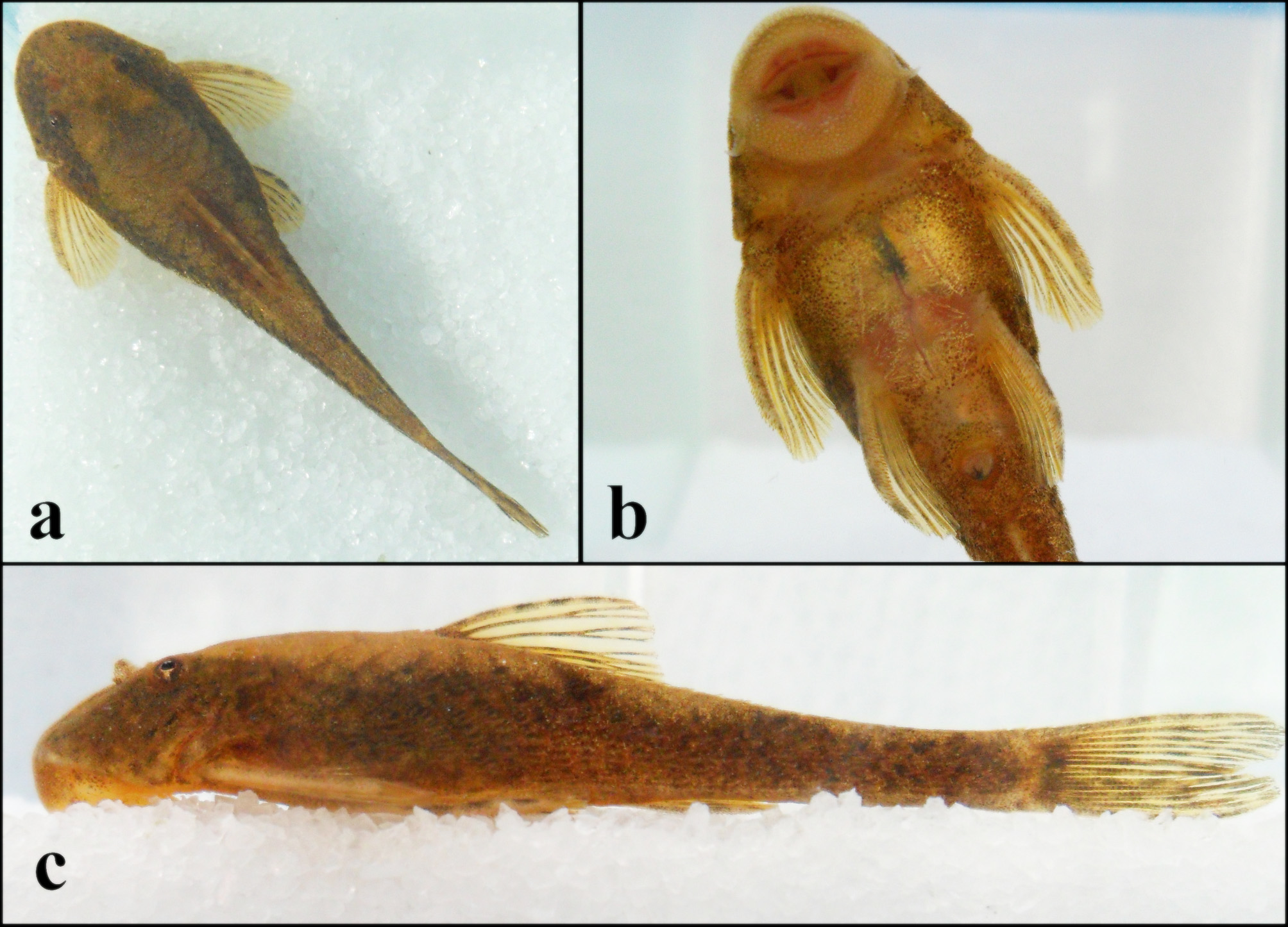
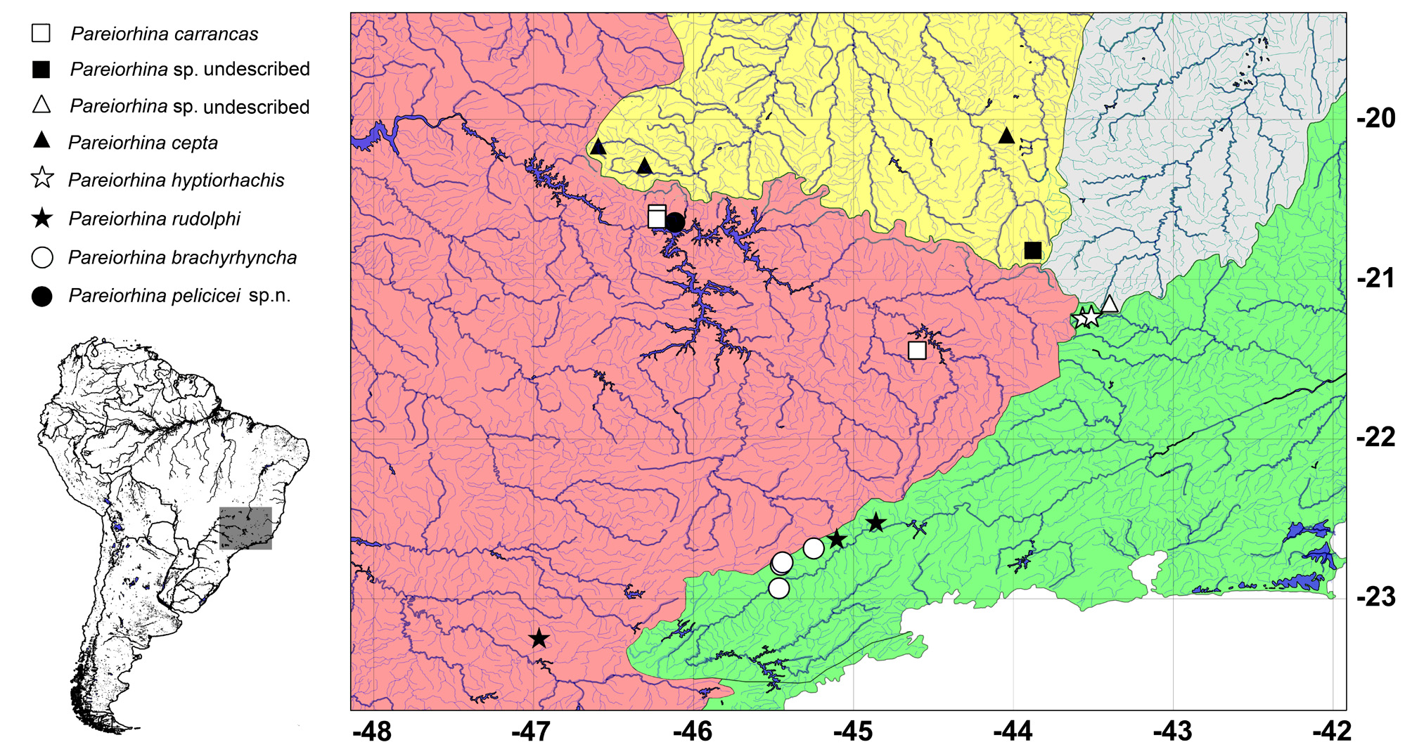

TABLE 1. Morphometric and meristic data for P. pelicicei. Range includes holotype and 19 paratypes. SD = standard deviation.
| Holotype | Range | Mean | SD | |
|---|---|---|---|---|
| Standard length (SL) Percentage of standard length Predorsal length | 47.4 41.9 | 27.8–47.4 41.0–44.8 | 36.9 42.6 | 5.0 1.0 |
| Preanal length Head length Cleithral width | 59.2 32.4 29.7 | 57.5–62.5 29.6–35.1 27.8–31.0 | 60.0 32.0 29.2 | 1.1 1.2 0.8 |
| Dorsal-fin spine length Base of dorsal fin length Thorax length | 21.3 15.1 17.9 | 21.2–23.4 13.4–16.8 15.7–18.7 | 22.3 14.9 17.1 | 0.6 0.8 0.7 |
| Pectoral-fin spine length Abdomen length Pelvic-fin spine length | 18.5 24.4 22.3 | 18.5–24.3 21.4–26.2 17.6–23.7 | 21.4 23.8 20.4 | 1.6 1.1 1.4 |
| Anal-fin spine length Lower caudal spine Caudal peduncle depth | 15.4 23.4 8.0 | 13.5–17.1 22.9–28.7 7.6–10.0 | 15.4 25.5 8.4 | 1.1 1.5 0.6 |
| Postanal length Anal width | 33.5 12.8 | 33.4–42.1 12.7–15.0 | 37.6 13.8 | 2.9 0.6 |
| Percentage of head length Head width Head depth | 90.9 46.7 | 82.2–97.0 43.8–55.8 | 90.8 48.4 | 3.9 2.7 |
| Snout length Interorbital width Orbital diameter | 56.4 36.3 10.5 | 52.5–60.8 34.7–44.3 10.5–14.9 | 56.0 39.3 12.4 | 1.9 2.4 1.3 |
| Meristics | Holotype | Range | Mode | SD |
| Dorsal plates Mid-dorsal plates Median plates | 24 17 24 | 24–25 16–19 24–26 | 24 17 24 | – – – |
| Mid-ventral plates Ventral plates Predorsal plates | 17 20 4 | 17–19 10–21 4 | 18 20 4 | – – – |
No known copyright restrictions apply. See Agosti, D., Egloff, W., 2009. Taxonomic information exchange and copyright: the Plazi approach. BMC Research Notes 2009, 2:53 for further explanation.