
Pseudopannota
|
publication ID |
https://doi.org/10.11646/zootaxa.4169.1.1 |
|
publication LSID |
lsid:zoobank.org:pub:F15C0038-DF14-4E4B-98F5-FE1BD7A5759F |
|
DOI |
https://doi.org/10.5281/zenodo.5616281 |
|
persistent identifier |
https://treatment.plazi.org/id/03D48C3E-E637-FFC9-76EC-B550FDD8F99F |
|
treatment provided by |
Plazi |
|
scientific name |
Pseudopannota |
| status |
s. l. |
Apomorphies of Pseudopannota View in CoL View at ENA s. l.
(1) Modification of labrum. Labrum has the following peculiar structure ( Figs 37–39 View FIGURES 34 – 37 View FIGURES 38 – 49 , 74–76 View FIGURES 74 – 79 ). Distal margin lost sclerotized seta-bearing band, and outer side lost constant pair of submedian setae and paired latero-distal setae, which are initially characteristic for Baetidae [see apomorphies “Tetramerotarsata (6)” and “Turbanoculata (4)” in Kluge 2004]. Instead of this, distal margin is formed by a soft area with membranous cuticle covered by numerous irregularly arranged fine colorless setae ( Figs 74–76 View FIGURES 74 – 79 ); inner side near anterior margin bears a pair of transverse rows of stout pointed spine-like setae, accompanied by a pair of transverse sclerotized ridges; median end of right ridge asymmetrically continues as longitudinal ridge running in proximal direction ( Figs 37 View FIGURES 34 – 37 , 74, 76 View FIGURES 74 – 79 ). I was unable to find correlation between this asymmetric element of labrum structure with asymmetric elements of adjacent mouthparts—mandibles and hypopharynx.
(2) Reduction of mandibular incisors. On both left and right mandibles, incisors are more or less reduced, so that the most projected apical denticle of mandible belongs to kinetodontium ( Figs 6, 7 View FIGURES 4 – 7 , 40, 41 View FIGURES 38 – 49 , 77–79 View FIGURES 74 – 79 , 92–94 View FIGURES 91 – 99 ).
This was described as “mandibles with incisors fused to apex” ( Waltz & McCafferty 1987) or “mandibles with fused canines” (Elouard et al. 1990), where the terms “incisors” or “canines” were referred to incisor and kinetodontium taken together. Actually, in Pseudopannota fusion of incisor and kinetodontium is not greater than in Labiobaetis and many other Baetidae , but allusion of their fusion appears because incisor is greatly reduced. Kinetodontium retains its usual structure with three large denticles, the most distal of which becomes the main biting denticle of mandible. This fact becomes clear if compare the mandible usual for Labiobaetis and other Baetidae ( Figs 4, 5 View FIGURES 4 – 7 ) with mandible of a primitive representative of Pseudopannota , where two denticles of incisor are retained ( Fig. 6 View FIGURES 4 – 7 ) and with mandible of a derived representative of Pseudopannota , where incisor is reduced to a single small denticle ( Fig. 7 View FIGURES 4 – 7 ).
(3) Protuberance on right mandible. Right prostheca is attached in a cavity, just proximad of which a sclerotized protuberance is located ( Figs 6, 7 View FIGURES 4 – 7 , 42 View FIGURES 38 – 49 , 78, 79 View FIGURES 74 – 79 , 93 View FIGURES 91 – 99 ).
(4) Shape of maxilla. Maxilla is different from maxilla of « Baetis -type» (which is characteristic for Labiobaetis and most other Baetungulata) and is similar to maxilla of « Cloeon -type» ( Crass 1947: 64): its biting margin is long, with all three canines and all three dentisetae long, pointed and thin-walled ( Figs 9, 12, 15 View FIGURES 8 – 15 , 36 View FIGURES 34 – 37 , 70 View FIGURES 68 – 73 ). Change between « Baetis -type» and « Cloeon -type» in both directions repeatedly took place in evolution of Baetidae .
(5) Modification of apical setae of maxilla. Apex of maxilla just laterad of canines bears long setae, which form a regular transverse row, with bases of setae pressed one to another; this row locates either on ventral-apical side only ( Figs 36 View FIGURES 34 – 37 ), or surrounds bases of canines from ventral, apical and dorsal sides ( Fig. 70 View FIGURES 68 – 73 ).
(6) Pseudo-three-segmented maxillary palp. Maxillary palp looks as thee-segmented, because its first primary segment is divided into two subsegments movably connected one with another; muscle, running from base of maxillary palp to base of the primary second segment, passes through both subsegments of the primary first segment ( Fig. 36 View FIGURES 34 – 37 ). Among Pseudopannota , only in P. camerunense maxillary palp is vestigial, very thin, lost intrinsic muscle and lost division of the first segment (see below).
Initial for Ephemeroptera is three-segmented maxillary palp with muscle present in the first segment only (instead of initial for insects 5-segmented maxillary palp with muscles in four segments). Such 3-segmented palp is retained in a part of Baetidae . As its 2nd segment always lacks intrinsic muscle, articulation between 2nd and 3rd segments does not provide active movements; in several non-related mayfly taxa this articulation undergoes reduction, so that palp becomes 2-segmented. Particularly, such 2-segmented maxillary palp is characteristic for the taxon Baetungulata, to which Labiobaetini belong ( Kluge & Novikova 2011). Maxillary palp of Pseudopannota is not true 3-segmented, but pseudo-3-segmented, because its first two segments originated from the initial first segment and both contain its muscle.
The 2-segmented maxillary palp of Labiobaetis has cuticle of both segments entirely membranous, with numerous campaniform sensilla (looking as small rings) located on the first segment ( Fig. 8 View FIGURES 8 – 15 ).
The pseudo-3-segmented maxillary palp of examined species of Pseudopannota has cuticle partly sclerotized: it is sclerotized at least on outer side of 2nd segment and on outer side of the distal subsegment of 1st segment. The proximal subsegment of 1st segment has cuticle either entirely membranous, or membranous at least on inner side; campaniform sensilla are concentrated on membranous cuticle of its inner side ( Fig. 14 View FIGURES 8 – 15 ; Gattolliat 2002: Fig. 1 View FIGURES 1 – 3 d). In P. f u s ca sp. n. proximal subsegment has sclerotized cuticle on outer side ( Fig. 9 View FIGURES 8 – 15 ); on shed exuviae it keeps its form, and only its base is inserted into stipes because of inversion of membranous cuticle of palpiger ( Fig. 10 View FIGURES 8 – 15 ). The same in P. pannota ( Fig. 14 View FIGURES 8 – 15 ). In the related species Pseudopannota sp. U, proximal subsegment has cuticle entirely membranous, so that on shed exuviae it is completely inverted and inserted into stipes ( Fig. 15 View FIGURES 8 – 15 ).
In P. camerunense both labial and maxillary palps are weakened and narrowed, so that maxillary palp lost its secondary articulation between subsegments of the first segment. Two parts of the first segment, corresponding to two subsegments, retain difference: distal part retains sclerotization of outer side, and proximal part retains entirely membranous cuticle and campaniform sensilla on its inner side ( Figs 11–13 View FIGURES 8 – 15 , 70–71 View FIGURES 68 – 73 ). Besides this, in penultimolarva the proximal part is thicker than the distal part ( Figs 11 View FIGURES 8 – 15 , 71 View FIGURES 68 – 73 ). Because of this, on shed exuviae of penultimolarva, the proximal part of first segment is completely inverted and inserted into stipes ( Fig. 12 View FIGURES 8 – 15 ), while on shed exuviae of ultimolarva maxillary palp keeps its form, and only its base is inserted into stipes because of inversion of membranous cuticle of palpiger ( Fig. 13 View FIGURES 8 – 15 ). These differences between proximal and distal parts of the first segment testify about their origin from the proximal and distal subsegments, which are expressed in other species of Pseudopannota .
Near its apex, second segment of maxillary palp has incision on inner side, which separates a small apical portion ( Figs 9–15 View FIGURES 8 – 15 , 70, 71 View FIGURES 68 – 73 ). Similar incision is present in certain species of Labiobaetis ( Fig. 8 View FIGURES 8 – 15 ), but not in other Baetungulata. Possibly, the apical portion separated by this incision, is a remnant of the primary third segment.
(7) Modification of labium. Glossae and paraglossae of labium are more or less elongated, with apical setae elongated, situated either irregularly ( Figs 34 View FIGURES 34 – 37 , 96 View FIGURES 91 – 99 ), or forming ring-like row ( Figs 68, 69 View FIGURES 68 – 73 ).
(8) Two-segmented labial palp. The 2nd and 3rd segments of labial palp are completely fused together, and intrinsic muscle of 2nd segment is completely lost ( Figs 35 View FIGURES 34 – 37 , 68 View FIGURES 68 – 73 , 96 View FIGURES 91 – 99 ). The same in some other taxa of Baetidae , but not in Labiobaetis , whose intrinsic muscle of 2nd segment is always retained.
(9) Pannote condition. Fore protoptera are widened in such a manner that their bases are brought together ( Figs 18–20 View FIGURES 16 – 20 ); in certain species fore protoptera are fused together at a long distance ( Fig. 17 View FIGURES 16 – 20 ). Unlike Pseudopannota , in other baetid taxa, including Labiobaetis , fore protoptera are separated by wider distance ( Fig. 16 View FIGURES 16 – 20 ).
(10) Modification of larval coxa. Larval coxa has more or less prominent lobe anteriad of trochanter base; shape of this lobe is species-specific; it is either similar on all legs, or different on legs of different pairs ( Figs 50– 52 View FIGURES 50 – 53 ).
(11) Differentiation of larval legs. Larval fore leg differs from hind leg by presence of additional spine-like setae on anterior side of femur; middle leg has intermediate structure ( Figs 50–52 View FIGURES 50 – 53 , 82–83 View FIGURES 80 – 84 , 110–111 View FIGURES 110 – 112 ).
Variable characters of Pseudopannota . Larval shape varies from the «swimming type » ( Figs 59, 60 View FIGURES 59 – 64 ) to «crawling type », i.e. shortened and robust with strengthened legs ( Fig. 31 View FIGURES 26 – 33 ). Larval claws either with usual number (6–8) of subequal denticles, or with reduced number (3–4), or with 2 unequal denticles ( Table 1 View TABLE 1 ). Larval abdominal terga either smooth with scales in semicircular sockets (e.g., in P. m a c u l o s a, P. camerunense ), or without scales and with prominent relief (e.g., in P. pannota sp. n.), or with both scales and prominent relief (e.g., in P. f us ca sp. n.) ( Table 1 View TABLE 1 ). Tergalii with or without scales and/or relief similar to that of abdominal terga, with or without additional rib along costal margin. Number of tergalii pairs varies from 7 to 6: tergalii of first pair either as large as others, or diminished, or lost ( Table 1 View TABLE 1 ). Subimaginal tarsomeres 1–4 other than on male fore legs, either with pointed microlepides, or with blunt microlepides ( Table 2 View TABLE 2 ). Hind wings are lost in all known species; vestiges of larval hind protoptera either present (in P. camerunense ), or absent.
Distribution and composition. The genus Pseudopannota as defined here, is restricted in its distribution by Afrotropical Region and includes the following species: P. maculosum ( Crass 1947 [ Pseudocloeon ]) (see below); P. berthrandi ( Demoulin 1967 [ Pseudocloeon ]); P. vinckei ( Demoulin 1973 [ Pseudocloeon ]); P. camerunense ( Ulmer 1920 [ Pseudocloeon ]) comb. n. (see below); P. muganinani Elouard & Gillies (in Elouard, Gillies & Wuillot) 1990 ; P. modesta Elouard & Gillies (in Elouard, Gillies & Wuillot) 1990 ; P. c a m i l l a e Gattolliat 2002. Two more species are described in this paper, P. pannota sp. n. and P. f u s c a sp. n., and one species is reported here without formal name as Pseudopannota sp. U.
Besides this, two species, known only as male imagoes, were attributed to the genus Pseudopannota : bergerardi Elouard & Hideux 1991 [ Pseudopannota ] and sartorii Elouard & Hideux 1991 [ Pseudopannota ]. Imaginal characters do not allow to determine their real systematic position. Possibly, bergerardi [ Pseudopannota ] belongs to Labiobaetis , and sartorii [ Pseudopannota ] does not belong to Labiobaetini.
Discussion. Waltz & McCafferty (1987), besides the genus Pseudopannota , established a separate genus Ophelmatostoma based on its autapomorphies in structure of larval glossae and paraglossae. This genus included a single species O. kimminsi Waltz & McCafferty 1987 , recently synonymized with P. camerunense ( Ulmer 1920) (see below). The genus Pseudopannota , as formerly outlined (i.e., without P. camerunense ), was characterized by two characters: (1) 3-segmented maxillary palp and (2) broad second segment of labial palp ( Gattolliat 2002: 27).
At first glance, the pseudo-3-segmented maxillary palp of Pseudopannota without P. camerunense could be regarded as an autapomorphy, and 2-segmented palp of P. camerunense could be regarded as a plesiomorphy.
However, maxillary palp of P. camerunense markedly differs from the plesiomorphically 2-segmented maxillary palps of other Baetungulata: both its segments have unusual proportions, being very thin and long, with no space for intrinsic muscle in the first segment ( Fig. 70 View FIGURES 68 – 73 ). It originated from the pseudo-three-segmented maxillary palp of other Pseudopannota by means of reduction in thickness and loss of intrinsic muscle; as a result of this, the secondary articulation is smoothed out; about this testify traces of division of the first segment into two subsegments [see above, Pseudopannota (6)].
The second segment of labial palp of P. camerunense also has derived structure, being weakened, narrowed and petiolate. In most other Baetidae this segment is wide; in other species of Pseudopannota second segment of labial palp has various shape (see Table 1 View TABLE 1 ), that does not allow to formulate a common character for Pseudopannota without P. camerunense .
fore leg ♂ fore leg ♀ mid & hind leg If regard all Pseudopannota without P. camerunense to constitute a taxon (genus), this taxon appears to be a plesiomorphon without any autapomorphic characters; possibly, this taxon is paraphyletic (see below). The genusgroup name Ophelmatostoma , being applied to a single species, is unnecessary, because the species name Pseudopannota camerunense can be used instead of it.
According to Elouard, Gillies & Wuillot (1990), the subgenus Pseudopannota was characterized by the derived character—fused fore protoptera, in contrast to the subgenus Hemipannota Elouard et al. 1990 . However, holophyly of the subgenus Pseudopannota is questionable. Possibly, P. muganinani (originally placed to the subgenus Pseudopannota ) has relationship with P. camerunense (originally placed to Ophelmatostoma ): in both species glossae are unusually bent laterally, and in both species fore tibiae bear dense long setae ( Fig. 83 View FIGURES 80 – 84 ) (see Table 3 View TABLE 3 ). Possibly, P. berthrandi (formerly placed to the subgenus Pseudopannota ) and P. pannota sp. n. are closely related with P. camillae (formerly placed to the subgenus Hemipannota ) and with P. fusca sp. n.; all these four species have peculiar structure of claws, not found in other taxa: claw constantly has only two denticles; the proximal denticle is larger, always has shape of wide triangle and rests on internal sclerotized thickening of the claw ( Figs 23–25 View FIGURES 21 – 25 ) (see Table 3 View TABLE 3 ).
In the descriptions of male imagoes reared from larvae of P. berthrandi and P. muganinani, Elouard, Gillies and Wuillot (1990) wrote that “forceps base lacking internal protuberance”. Based on this, Elouard & Hideux (1991) stated that “Le genre Ophelmatostoma ... caractérise ... par les forceps des imagos mâles qui possèdent une exctroissance interne développé” and regarded that the genera Pseudopannota and Ophelmatostoma can be distinguished by genital structure. However, male imagoes of P. camerunense (type species of Ophelmatostoma ) and P. pannota sp. n. (which is closely related to the type species of Pseudopannota ) have very similar structure, with equally developed internal protuberances on unistyligers (= forceps bases); their genital structure is very similar to that of some representatives of the plesiomorphon Labiobaetis ( Figs 1–3 View FIGURES 1 – 3 ).
The most peculiar characters of Pseudopannota coincide with characters of the Neotropical genus Guajirolus Flowers 1985 : In both taxa labrum has numerous fine setae on outer side, soft distal margin and pair of rows of spine-like setae on inner side (but Guajirolus has no asymmetric sclerite on inner side of labrum). In both taxa mandibular biting denticles are formed by kinetodontium, while incisor is greatly reduced; prostheca (at least right one) is inserted into incision (but Guajirolus has no protuberance proximad of this incision). In both taxa maxilla has long biting margin with long and delicious maxillary canines and dentisetae, and distal setae are long (but in Guajirolus these distal setae don't form a regular row). In both taxa maxillary palp is pseudo-three-segmented, with primary first segment divided into two subsegments. In both taxa glossae and paraglossae are elongated, with elongated apical setae. In both taxa larval fore femur is strengthened and bears additional spine-like setae on anterior side. Monotypic Neotropical genus Chane Nieto 2003 is closely related to Guajirolus and also has these characters. Unfortunately, imaginal structure of Guajirolus and Chane remains to be unknown: only for two species, G. ektrapeloglossa Flowers 1985 and Chane baure Nieto 2003 , imagoes were presumably associated without rearing. Judging by the original description, genital structure of G. ektrapeloglossa is similar to that of Labiobaetini ( Flowers 1985: Fig. 2 View FIGURES 1 – 3 ), but such important characters, as pose of developing gonostyli in mature larva and presence/absence of sterno-styligeral muscle, remain to be unknown. Collecting mayflies in Peru, I was able to find only two female larvae of Guajirolus .


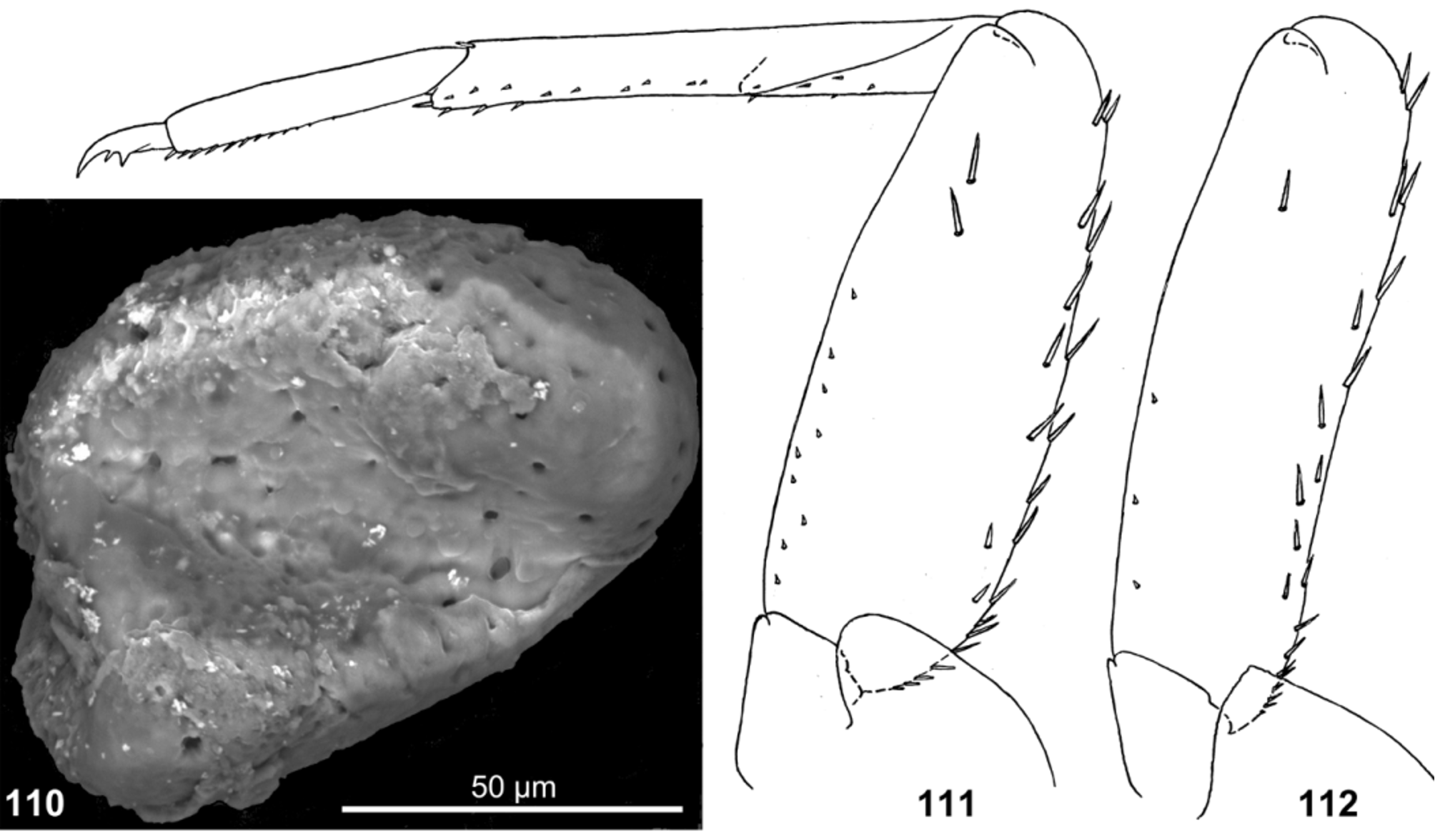


TABLE 1. Characters of known species of Pseudopannota.
| Species P. camerunense | glossa of modification tubular | glossa on setae dorsal 0 | paraglossa on setae dorsal 0 | labial of segment widest distal palp distally | protoptera fore of fusion – | tibia fore on setae long + | tarsus fore on setae long – | claw on denticles 4–7 | terga abdominal on scales + | I pair of tergalii small | ♂ tergum abdominal dark first V | ♀ tergum abdominal dark first II | subimaginal legs hind of 4 – and microlepides 1 tarsomeres middle of pointed |
|---|---|---|---|---|---|---|---|---|---|---|---|---|---|
| P. muganinani | widened | ? | ? | medially | + | + | + | 6–8 | + | + | – | – | ? |
| P. vinckei | shortened | ? | ? | distally | + | + | + | 4 | ? | + | ? | ? | ? |
| P. berthrandi | – | ? | ? | proximally | + | – | – | 2 | ? | + | V | VI | ? |
| P. pannota sp. n. | – | 1 | 1 | proximally | + | – | – | 2 | – | + | VI | II | blunt |
| P. sp. U | – | 1 | 1 | proximally | + | – | – | 2 | – | ? | V | ? | ? |
| P. f us c a sp. n. | – | 2–4 | 1 | medially | – | – | – | 2 | ± | small | V | II | pointed |
| P. camillae | – | 3 | 1 | medially | – | – | – | 2 | ? | – | ? | ? | ? |
| P. maculosum | – | 3 | 1 | medially | – | – | – | 3–4 | + | small | VIII | ? | |
| P. modesta | – | ? | ? | medially | – | – | – | 3–4 | + | – | ? | ? | ? |
TABLE 2. Texture of subimaginal legs and apical spines of imaginal and subimaginal legs.
| 1 | 2 | 3 | 4 | 5 | 1 | 2 | 3 | 4 | 5 | 1–2 | 3 | 4 | 5 | |
|---|---|---|---|---|---|---|---|---|---|---|---|---|---|---|
| P. pannota sp. n. | U | U | U | U | Y | U | U | U | U | Y | U | U | U | Y |
| apical spine: | – | – | – | – | – | + | + | – | + | + | – | |||
| P. camerunense | U | U | U | U | Y | Y | Y | Y | Y | Y | Y | Y | Y | Y |
| apical spine: | – | – | – | – | – | + | + | – | + | + | – | |||
| P. f us c a sp. n. | U | U | U | U | Y | Y | Y | Y | Y | Y | Y | Y | Y | Y |
| apical spine: | – | – | – | – | – | + | + | – | + | + | – | |||
| L. tenuicrinitus | U | U | U | U | Y | I | U | U | U | Y | I U | U | U | Y |
| apical spine: | – | – | – | – | – | + | + | – | + | + | + |
TABLE 3. Pattern of possible synapomorphies among species of Pseudopannota.
| Species | Former systematic position | glossa bent laterally | setae on fore tibia | 2-dentate claw fore protoptera fused together |
|---|---|---|---|---|
| P. camerunense | Ophelmatostoma | + | + | – – |
| P. muganinani | Pseudopannota s.str. | + | + | – + |
| P. vinckei | Pseudopannota s.str. | – | + | – + |
| P. berthrandi | Pseudopannota s.str. | – | – | + + |
| P. pannota sp. n. | – | – | – | + + |
| P. sp. U | – | – | – | + + |
| P. f us c a sp. n. | – | – | – | + – |
| P. camillae | Hemipannota | – | – | + – |
| P. maculosum | Hemipannota | – | – | – – |
| P. modesta | Hemipannota | – | – | – – |
No known copyright restrictions apply. See Agosti, D., Egloff, W., 2009. Taxonomic information exchange and copyright: the Plazi approach. BMC Research Notes 2009, 2:53 for further explanation.
|
Kingdom |
|
|
Phylum |
|
|
Class |
|
|
Order |
|
|
Family |