
Eutichurus, Simon, 1897
|
publication ID |
https://doi.org/ 10.11646/zootaxa.4382.2.6 |
|
publication LSID |
lsid:zoobank.org:pub:53C1FBC6-8A60-4C58-A8B9-47311BE186D1 |
|
DOI |
https://doi.org/10.5281/zenodo.5979804 |
|
persistent identifier |
https://treatment.plazi.org/id/03D487C3-6503-1E16-94EC-FCFFFA0D41CE |
|
treatment provided by |
Plazi |
|
scientific name |
Eutichurus |
| status |
|
Key to species of Eutichurus (updated from Bonaldo 1994)
1 Males (those of E. arnoi , E. manu and E. saylapampa , unknown)................................................ 2
- Females (those of E. abiseo , E. cuzco , E. pallatanga and E. yalen , unknown)..................................... 26
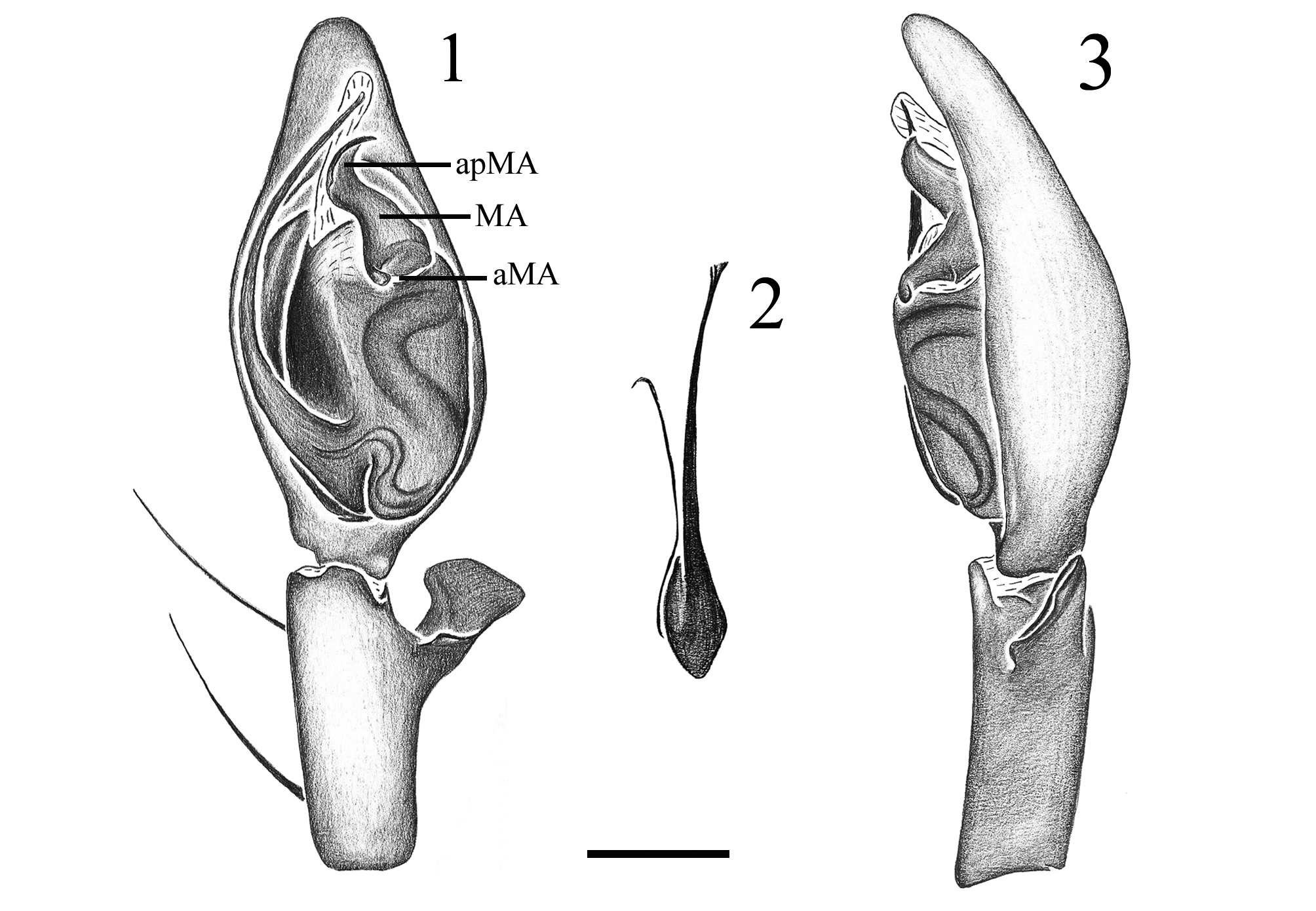
2(1) Retrolateral tibial apophysis represented by a single branch ( Figs 1 View FIGURES 1 , 6, 25; Bonaldo 1994: figs 19, 29, 38, 48, 56, 64)....... 3
- Retrolateral tibial apophysis bifid or trifid (Figs 14, 17; Bonaldo 1994: figs 34, 66, 76, 86, 91)........................ 19
3(2) Retrolateral tibial apophysis tuberculate, with a small apical projection ( Bonaldo 1994: figs 56, 57)....... E. furcifer Kraus
- Retrolateral tibial apophysis otherwise ( Figs 1 View FIGURES 1 , 25; Bonaldo 1994: figs 19, 29, 38, 48, 64)............................ 4
4(3) Retrolateral tibial apophysis short, excavated; median apophysis with a longitudinal median keel ( Bonaldo 1994: figs 43, 45, 48, 49).............................................................................................. 5
- Retrolateral tibial apophysis long, not excavated; median apophysis without such keel (Figs 6, 9; Bonaldo 1994: figs 19, 22, 26, 29, 35, 38, 62)..................................................................................... 6
5(4) Embolus with a prolateral median process ( Bonaldo 1994: figs 43, 44)........................... E. tropicus (L. Koch)
- Embolus without process ( Bonaldo 1994: figs 48, 49)...................................... E. valderramai Bonaldo
6(4) Embolar base fused to the tegulum ( Fig. 1 View FIGURES 1 ; Bonaldo 1994: Fig. 35).............................................. 7
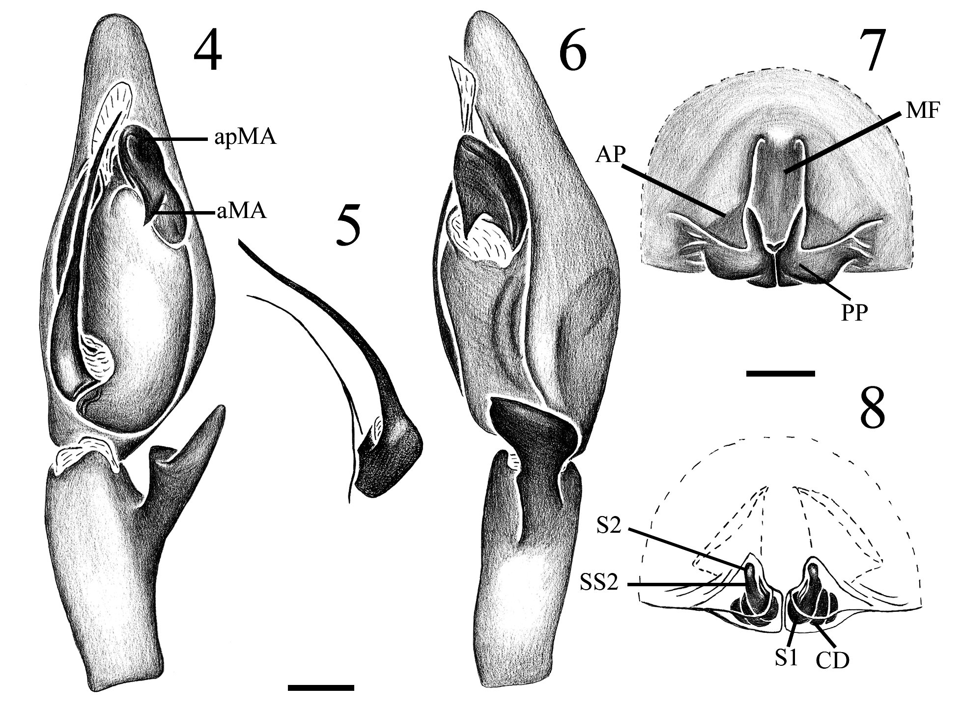
- Embolus articulated, connected to the tegulum by a membrane ( Figs 4 View FIGURES 4 , 9 View FIGURES 9 , 12 View FIGURES 12 ; Bonaldo 1994: figs 19, 22, 30, 26, 38, 62)... 8
7(6) Embolus with lamellar tip ( Bonaldo 1994: figs 35, 36).......................................... E. abiseo Bonaldo
- Embolus filiform ( Figs 1 View FIGURES 1 , 2)................................................................ E. paredesi n. sp.
8(6) Embolus with a small retrolateral sub-apical process ( Bonaldo 1994: figs 62, 63; Laborda & Simó 2015: fig. 1A)................................................................................................ E. ibiuna Bonaldo
- Embolus without processes (Figs 5, 10; Bonaldo 1994: figs 19, 22, 26) or with a prolateral process (Figs 16, 20, 28; Bonaldo 1994: figs 30, 39)..................................................................................... 9
9(8) Embolus with a prolateral process (Figs 16, 21, 28; Bonaldo 1994: figs 30, 39).................................... 10
- Embolus without process (Figs 5, 10; Bonaldo 1994: figs 19, 22, 26)........................................... 15
10(9) Median apophysis strongly sculptured, with several conspicuous sulci ( Bonaldo 1994: figs 29, 31)........ E. cuzco Bonaldo
- Median apophysis not strongly sculptured ( Figs 15 View FIGURES 15 , 20 View FIGURES 20 , 27 View FIGURES 27 , 58 View FIGURES 58 ; Bonaldo 1994: figs 38, 40).......................... 11
11(10) Embolar prolateral process inserted medially, pars pendula absent (Fig. 28; Bonaldo 1994: fig. 39).................... 12
- Embolar prolateral process inserted sub-apically, pars pendula present (Figs 21, 59)................................ 13
12(11) Median prong of median apophysis short ( Bonaldo 1994: figs 38, 40; Ramírez 2014, figs 147A, C).... E. lizeri Mello-Leitão
- Median prong of median apophysis long ( Figs 27 View FIGURES 27 , 29)........................................ E. marquesae Bonaldo
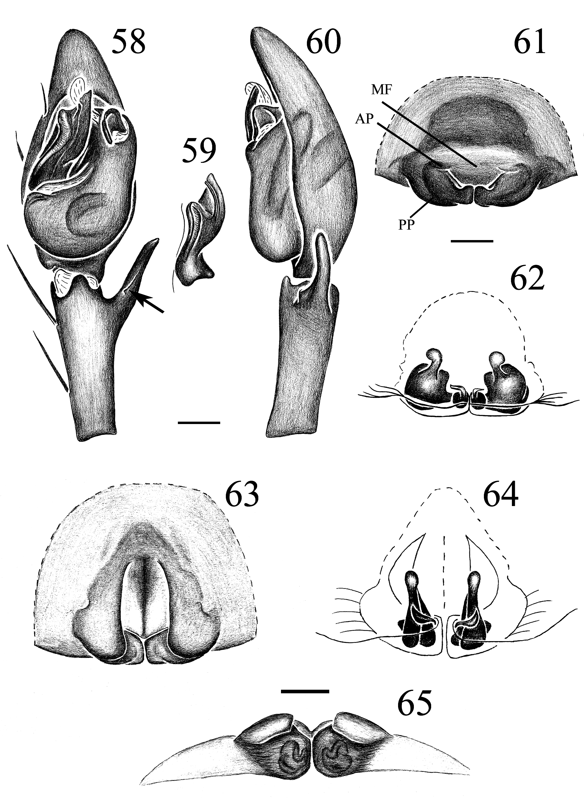
13(11) Embolus wide and short relative to tegulum ( Figs 58 View FIGURES 58 , 59)......................................... E. nancyae n. sp.
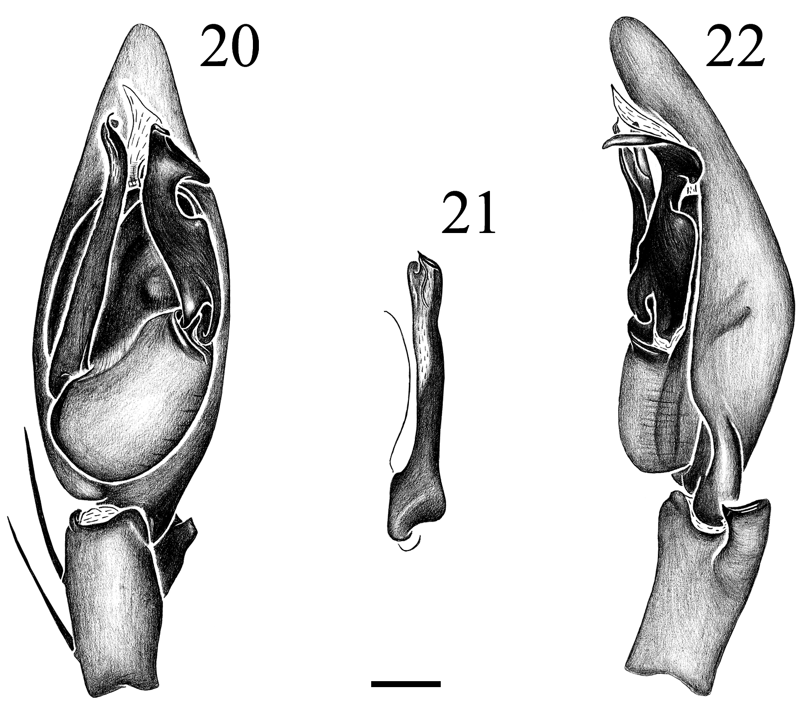
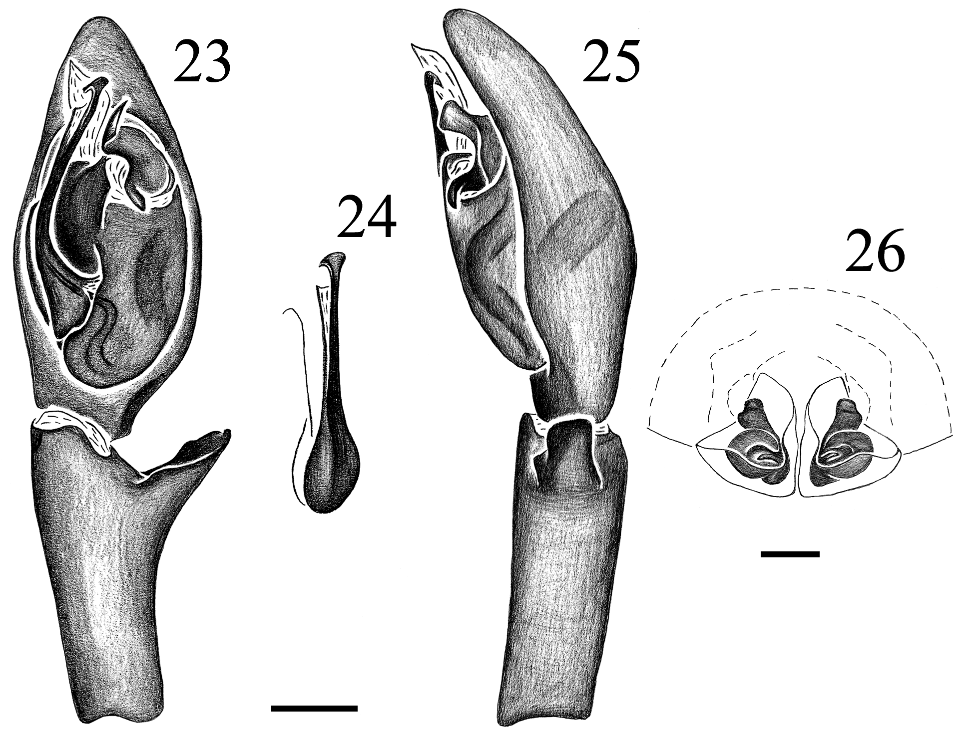
- Embolus narrow and long relative to tegulum ( Figs 20 View FIGURES 20 , 21, 23, 24)............................................. 14
14(13) Median prong of median apophysis with a pair of apical projections ( Figs 20 View FIGURES 20 , 22)................... E. tequendama n. sp.
- Median prong of median apophysis without apical projections ( Figs 23 View FIGURES 23 , 25)......................... E. madre Bonaldo
15(9) Embolus flattened; prolateral margin of median apophysis with transversal ridges ( Bonaldo 1994: figs 26, 28).................................................................................................... E. yalen Bonaldo
- Embolus conic, median apophysis without transversal ridges (Figs 5, 10; Bonaldo 1994: figs 19, 22)................... 16
16(15) Retrolateral tibial apophysis expanded distally (Figs 6, 11)................................................... 17
- Retrolateral tibial apophysis tapering toward the apex ( Bonaldo 1994: figs 20, 23)................................. 18
17(16) Conductor hyaline ( Fig. 4 View FIGURES 4 ).................................................................. E. murgai n. sp.
- Conductor partially sclerotized ( Fig. 9 View FIGURES 9 )...................................................... E. zarate Bonaldo
18(16) Retrolateral tibial apophysis gradually tapering from base; embolus inserted basally ( Bonaldo 1994: figs 19, 20).................................................................................................... E. ferox Simon
- Retrolateral tibial apophysis abruptly tapering from distal third; embolus inserted medially ( Bonaldo 1994: figs 22, 23)............................................................................................. E. silvae Bonaldo
19(2) Retrolateral tibial apophysis bifid (Figs 14, 17; Bonaldo 1994: figs 34, 67, 76).................................... 20
- Retrolateral tibial apophysis trifid ( Bonaldo 1994: figs 86, 91)................................................. 25
20(19) Retrolateral tibial apophysis split medially; median prong of median apophysis present (Fig. 17; Bonaldo 1994: figs 32, 3 4).................................................................................................... 21
- Retrolateral tibial apophysis split basally; median prong of median apophysis absent (Fig. 14; Bonaldo 1994: figs 67, 72, 76).................................................................................................... 22
21(20) Embolus without prolateral process, tapering distally ( Bonaldo 1994: figs 32, 33).................. E. pallatanga Bonaldo
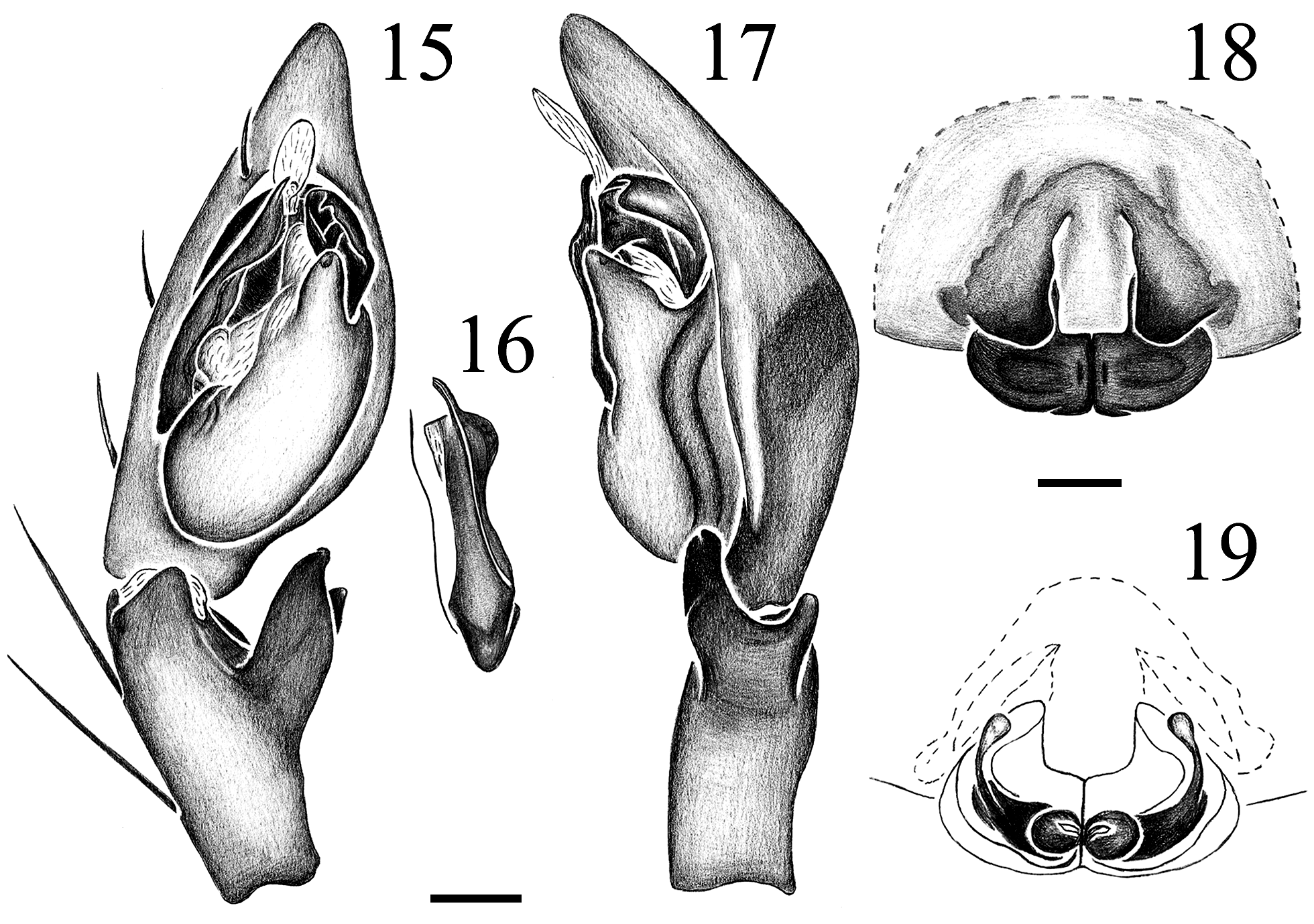
- Embolus with prolateral process, distally wide ( Figs 15 View FIGURES 15 , 16)........................................ E. yungas n. sp.
22(20) Embolus conic, without processes ( Bonaldo 1994: figs 75, 76)..................................... E. luridus Simon
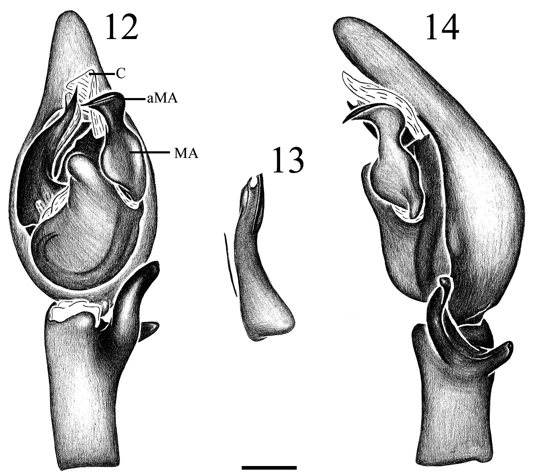
- Embolus flattened, with prolateral process (Fig. 13; Bonaldo 1994: figs 65, 70)................................... 23
23(22) Median apophysis long, with the apex prolaterally oriented; dorsal prong of retrolateral tibial apophysis shorter than the ventral prong ( Figs 12 View FIGURES 12 , 14)....................................................................... E. cumbia n. sp.
- Median apophysis short, with the apex oriented proximally; ventral and dorsal prongs of retrolateral tibial apophysis similarly sized ( Bonaldo 1994: figs 67, 72)........................................................................ 24
24(23) Apex of embolar process acute; apex of embolus apically oriented ( Bonaldo 1994: figs 65–67)........... E. ravidus Simon
- Apex of embolar process rounded; apex of embolus prolaterally oriented ( Bonaldo 1994: figs 70–72).. E. itamaraju Bonaldo
25(19) Apex of median apophysis wide; embolar apex bifid ( Bonaldo 1994: figs 89, 91)........... E. putus O. Pickard-Cambridge
- Apex of median apophysis narrow; embolar apex trifid ( Bonaldo 1994: figs 84, 86)............... E. sigillatus Chickering
26(1) Epigynal posterior plates fused, with a longitudinal median suture (Fig. 18; Bonaldo 1994: figs 41, 82, 87, 92)........... 27
- Epigynal posterior plates not fused medially (Figs 7, 52; Bonaldo 1994: figs 21, 46, 59, 68).......................... 31
27(26) Epigynal anterior projections relatively large; median field longer than wide (Fig. 18; Bonaldo 1994: figs 41, 42)....... 28
- Epigynal anterior projections relatively small; median field wider than long ( Bonaldo 1994: figs 82, 87, 92)............. 29
28(27) anterior projections covering the anterior half of the posterior plates in ventral view; anterior margin of posterior plates con- verging medially in angle ( Bonaldo 1994: fig. 41; Ramírez 2014: fig. 175G)...................... E. lizeri Mello-Leitão
- Anterior projections covering less than the anterior half of the posterior plates in ventral view; anterior margin of posterior plates converging medially in a straight line (Fig. 18)............................................. E. yungas n. sp.
29(27) Median field longer than wide, with a posterior median projection; anterior margins of posterior plates W-shaped ( Bonaldo 1994: figs 87, 88)................................................................... E. sigillatus Chickering
- Median field as long as wide, without projection; anterior margins of posterior plates otherwise ( Bonaldo 1994: figs 82, 92)..................................................................................................... 30
30(29) Anterior projections projected over the median field; anterior margins of posterior plates slightly oblique ( Bonaldo 1994: figs 82, 83)................................................................................. E. manu Bonaldo
- Anterior projections not projected over the median field; anterior margins of posterior plates straight ( Bonaldo 1994: figs 9 2, 93)......................................................................... E. putus O. Pickard-Cambridge
31(26) Epigynal anterior projections reduced, inconspicuous (Fig. 61)..................................... E. nancyae n. sp.
- Epigynal anterior projections conspicuous (Figs 7, 52, 63; Bonaldo 1994: figs 21, 46, 54).......................... 32
32(31) Anterior projections fused to posterior plates (Figs 63, 65; Laborda & Simó 2015: fig. 1D)............. E. ibiuna Bonaldo
- Anterior projections not fused to posterior plates (Figs 7, 52; Bonaldo 1994: figs 21, 46, 50)........................ 33
33(32) Each inner margin of anterior projections excavated; posterior plates laterally constricted ( Bonaldo 1994: figs 54, 55)............................................................................................. E. zarate Bonaldo
- Anterior projections and posterior plates otherwise (Figs 7, 52; Bonaldo 1994: figs 21, 46, 68, 79).................... 34
34(33) Anterior projections larger or slightly smaller than posterior plates (Fig. 7; Bonaldo 1994: figs 21, 24, 46, 50, 52, 53).... 35
- Anterior projections distinctly smaller than posterior plates ( Fig. 52 View FIGURES 52 ; Bonaldo 1994: figs 59, 68, 73, 77, 79, 80).......... 41
35(34) Anterior projections triangular, with acute apices (Fig. 7; Bonaldo 1994: fig. 52).................................. 36
- Anterior projections otherwise ( Bonaldo 1994: figs 21, 24, 46, 50, 53)........................................... 37
36(35) Posterior margin of anterior projections strongly procurve ( Bonaldo 1994: fig. 52)................ E. saylapampa Bonaldo
- Posterior margin of anterior projections nearly straight (Fig. 7)...................................... E. murgai n. sp.
37(35) Median field subtriangular, bulging between anterior projections ( Bonaldo 1994: fig. 53)................ E. arnoi Bonaldo
- Median field otherwise ( Bonaldo 1994: figs 21, 24, 46, 50)................................................... 38
38(37) Median field squared, as long as wide; anterior projections truncated ( Bonaldo 1994: fig. 46)......... E. tropicus (L. Koch)
- Median field longer than wide; anterior projections rounded ( Bonaldo 1994: figs 21, 24, 50)......................... 39
39(38) Apices of anterior projections rugose, strongly sclerotized ( Bonaldo 1994: fig. 50)............... E. valderramai Bonaldo
- Apices of anterior projections smooth ( Bonaldo 1994: figs 21, 24).............................................. 40
40(39) Median field surface plain anteriorly, with a longitudinal posterior groove ( Bonaldo 1994: fig. 24)........ E. silvae Bonaldo
- Median field surface concave anteriorly, without groove ( Bonaldo 1994: fig. 21)........................ E. ferox Simon
41(34) Each posterior plate with a posterior lateral excavation ( Bonaldo 1994: fig. 60)........................ E. furcifer Kraus
- posterior plates without excavations ( Bonaldo 1994: figs 68, 77, 79, 80)......................................... 42
42(41) Anterior projections projected over the median field ( Bonaldo 1994: figs 68, 73).................................. 43
- Anterior projections not projected over the median field ( Bonaldo 1994: figs 77, 79, 80)............................ 44
43(42) Median field as long as wide ( Bonaldo 1994: fig. 68)............................................ E. ravidus Simon
- Median field longer than wide ( Bonaldo 1994: fig. 73)....................................... E. itamaraju Bonaldo
44(42) Anterior projections not projected over posterior plates ( Fig. 52 View FIGURES 52 ; Bonaldo 1994: fig. 80)............. E. marquesae Bonaldo
- Anterior projections projected over posterior plates ( Bonaldo 1994: figs 77, 79)................................... 45
45(44) Anterior projections gently tapering ( Bonaldo 1994: fig. 77)....................................... E. luridus Simon
- Anterior projections truncated ( Bonaldo 1994: fig. 79).......................................... E. madre Bonaldo


No known copyright restrictions apply. See Agosti, D., Egloff, W., 2009. Taxonomic information exchange and copyright: the Plazi approach. BMC Research Notes 2009, 2:53 for further explanation.