
Ursus thibetanus mediterraneus MAJOR, 1873
|
publication ID |
https://doi.org/ 10.2478/if-2017-0028 |
|
persistent identifier |
https://treatment.plazi.org/id/03D487B2-646A-FFA3-0839-FE5EFA6BFB1A |
|
treatment provided by |
Diego |
|
scientific name |
Ursus thibetanus mediterraneus MAJOR, 1873 |
| status |
|
Ursus thibetanus mediterraneus MAJOR, 1873
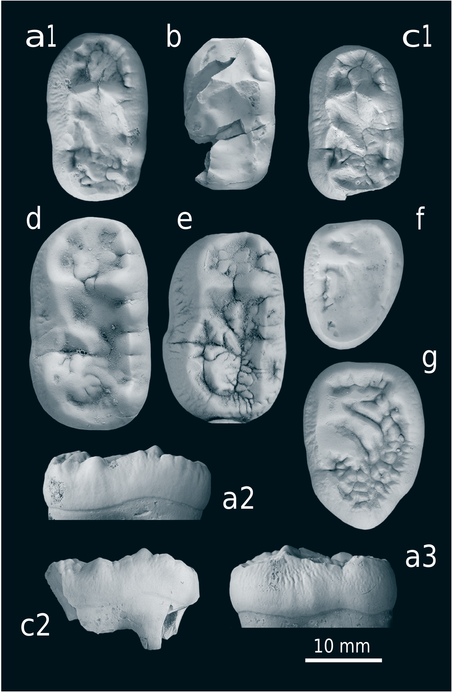
Text-figs 1 View Text-fig , 2 View Text-fig , 3a, b, f View Text-fig , 4a View Text-fig , 5b, c View Text-fig
1967 Ursus etruscus CUVIER ; Malez, pp. 57, 76. (partim)
1968a Ursus etruscus ; Malez, p. 152. (partim)
1968b Ursus etruscus ; Malez, pp. 205, 208. (partim)
1969a Ursus etruscus ; Malez, pp. 74, 78. (partim)
1969b Ursus etruscus ; Malez, p. B 21/1. (partim)
1970 Ursus etruscus ; Malez, pp. 206, 213. (partim)
1970 Ursus etruscus ; Marković- Marjanović, p. 156. (partim)
1971 Ursus etruscus ; Malez, p. 66. (partim)
1974 Ursus etruscus ; Malez, p. 79. (partim)
1974 Ursus etruscus ; Malez and Malez-Bačić, p. 6. (partim)
1975 Ursus etruscus CUV. ; Malez, pp. 183, 186, 188, 189, 197, 198, pl. IV. (partim)
Ursus (Euarctos) mediterraneus MAJOR ; Malez, pp. 186, 188, 189, 198.
Macaca florentina COCCHI ; Malez, pl. II. (partim)
1979a Ursus etruscus ; Malez, pp. 122, 143. (partim)
1979b Ursus etruscus ; Malez, p. 57. (partim)
Ursus thibetanus mediterraneus ; Malez, p. 57.
1979c Ursus etruscus ; Malez, p. 210. (partim)
Ursus mediterraneus ; Malez, p. 120.
1986 Ursus etruscus ; Malez, p. 102. (partim)
Ursus thibetanus mediterraneus ; Malez, p. 102.
1996 Ursus thibetanus ; Crégut-Bonnoure, p. 99.
1996 Ursus etruscus ; Bosinski, p. 55. (partim)
Ursus (euarctos) mediterraneus ; Bosinski, p. 55.
1998 Ursus etruscus ; Brajković, p. 8. (partim)
2000 Ursus etruscus ; Saínz de los Terreors, p. 40. (partim)
2000 Ursus [of similar stage of evolution as bears from Vallonnet and Pirro]; Spassov, p. 108. (partim)
2002 Ursus [of similar stage of evolution as bears from Vallonnet and Pirro]; Spassov, p. 234. (partim)
2003 Ursus [of similar stage of evolution as bears from Vallonnet and Pirro]; Spassov, p. 214. (partim)
2011 Ursus sp. ; Kahlke et al., p. 1376. (partim)
2012 U. t. mediterraneus ; Wagner et al., pp. 50, 51
2015 transitional stage in Ursus etruscus-U. deningeri lineage; Vislobokova and Agadjanian, p. 655. (partim)
2016a U. etruscus-U. deningeri ; Vislobokova and Agadjanian, p. 195. (partim)
U. minimus-thibetanus lineage; Vislobokova and Agadjanian, p. 195.
2016b U. etruscus ; Vislobokova and Agadjanian, p. 196. (partim)
U. mediterraneus (= U. ex gr. minimus); Vislobokova and Agadjanian, p. 196. (partim)
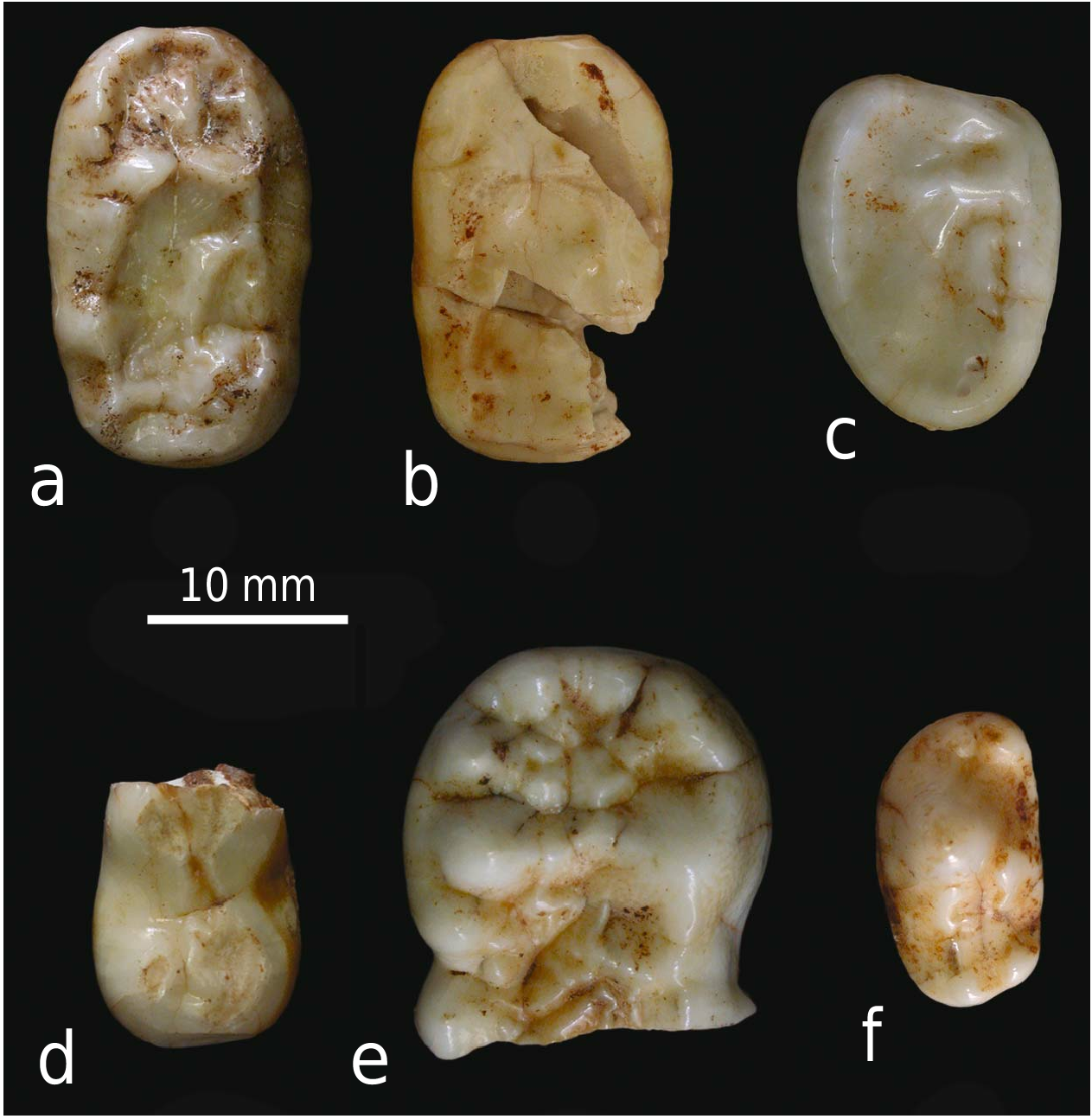
M a t e r i a l. Isolated m2 dex. (specimen A; Text-fig. 1a View Text-fig ), fragment of right hemimandible with worn m2 and m3 and distal root of m1 (specimen B; Text-figs 1b–c View Text-fig , 2a View Text-fig ), distal fragment of m1 dex. (specimen C; Text-fig. 1d View Text-fig ), mesial fragment of right hemimandible with alveoli of p1 – p3 and mesial root of p4 (specimen D; Text-fig. 2b View Text-fig ), rostral fragment of mandible with right canine, i3 dex., i2 sin. and alveoli of i1 dex., i2 dex. and i1 sin. (specimen E; Text-fig. 2d View Text-fig ) and mesial fragment of left hemimandible with canine and alveoli of p1 – p3 and mesial root of p4 (specimen F; Text-fig. 2c View Text-fig ). Specimens B – F possibly belong to the one individual. See Table 1 for tooth measurements.
D e s c r i p t i o n. The best preserved specimen is an isolated m2 dex. (specimen A), which is only moderately worn. It has an irregular oval outline, the crown is rather closed with the outer walls diverging laterally (especially the buccal one). There is no cingulum, but the outer walls are wrinkled (the buccal wall more that the lingual). The trigonid is broader than the talonid. The hypoconid area is shortened (distally compressed) compared to the part between Prd-Med and Hyd-End 1 which is relatively elongated. All the main cusps are well developed, but worn. EPrd and EMed (or their precursors) are not orientated perpendicularly to the tooth axis but are directed slightly anteriorly. Elongated swellings/crests project from the medial ends of EPrd and EMed (from the latter a smaller one) and infill most of the anterior trigonid basin. EMed is fully separated from Med, the contact between EPrd and Prd is not visible due to damage. The anterior margin of the tooth is differentiated by small wrinkles. The part of the trigonid distal to Prd-Med is simply built, the dominant structure is well developed mesolophid. The hypoconid is large, rather distally located, a well developed EHyd, fully separated from Hyd, is present. End 1 is shorter than End 2. Between End 1 and 2 is a small secondary cusp. With a maximal length 22.3 mm, this tooth is one of the largest found in Europe to date (e.g., Heller 1949, Thenius 1958, Fistani and Crégut-Bonnoure 1993, Crégut-Bonnoure 1996, Turner 2000, Baryshnikov 2007). In its general form as well as in the character of the fine enamel structures, this tooth is very similar to m2 from Grotta di Reale ( Italy), the type locality of U. t. mediterraneus ( Text-fig. 3a, c View Text-fig ), but the latter is smaller.
The right mandibular fragment (specimen B) is composed of a horizontal body broken in front of the distal root of m1 and a part of the vertical ramus (most of the ramus is missing). Processus subalveolaris is present and well developed. m2 and m3 are present. A separated distal fragment of m1 (specimen C) fits well on the preserved m1 distal root fragment. The height of the mandibular body (lingually measured) is, approximately, 38.7 mm under m1/2, 41.2 mm under m2 and 45.4 mm under m3. Malez (1975: 186) reported these specimens as a right mandible with m1 – m3, but on the photo presented ( Malez 1975: pl. IV) only m3 and a mesial fragment of m2 are present.
Although all the teeth are heavily worn, they partly bear diagnostic characters ( Text-figs 3 View Text-fig , 4 View Text-fig ). Especially important is the distal fragment of m1 with the typical thibetanus arrangement ( Text-fig. 4 View Text-fig ). There is a well developed enamel crest connecting Med and Hyd, End 1 is rather small, placed on the linguo-distal corner of the tooth and without any pre-
entoconid structures. The combination of these characters is diagnostic for U. thibetanus . The talonid of this tooth is rather wide (see Tab. 1), in fact wider than in any other studied U. thibetanus (except the type of “ U. karabach ” from Azykh Cave, Nagorno Karabakh; Text-fig. 4c View Text-fig ; see also Baryshnikov 2010) but as short (buccal length of talonid is 6.9 mm) as in other European specimens of U. thibetanus and shorter than in the studied specimens of U. etruscus (Olivola, Upper Valdarno). m2 is damaged (the talonid lateral wall and part of enamel on the occlusal surface is missing). It is shorter than specimen A and more rectangular in its shape. Almost no fine enamel structures are apparent due to the strong abrasion (and attrition). m3 is completely preserved but also heavily worn. Its shape is between triangular and egg-like, the buccal wall is wrinkled. Both teeth fit well in their general form with the situation in U. thibetanus and differ in the smaller size and simple structure (if it is possible to conclude such information from worn crowns) from U. etruscus ( Text-fig. 3 View Text-fig ).
The right mesial fragment of mandible (specimen D) shows alveoli of all three anterior premolars and of the mesial root of p4, the alveolar margins are generally damaged. The arrangement of premolar alveoli is slightly atypical. There is long diastema between p1 and p2 (10.4 mm), a short one between p2 and p3 (2.4 mm) and p3 is just in front of p4 (distance ca. 0.9 mm). Although a more regular distribution of anterior premolars is usual in Asiatic black bears, a similar situation can also be found, e.g., in one specimen from Mauer ( Germany) ( Text-fig. 5 View Text-fig ). A large foramen mentale (ca. 5.8 × 4.1 mm) is located under the alveolus of p2 ( Text-fig. 2b2 View Text-fig ) .
A rostral fragment (specimen E) bears c inf. dex., i3 dex. and i2 sin. The crowns of all teeth are heavily worn, the upper part of the canine crown is broken off. The incisor area is laterally compressed and subsequently the alveoli of both p2 sin. and dex. are apparentlylocated distally. The lingual enamel crest is present on the posterior wall in the preserved part of the canine crown. Malez (1975: 184, pl. II) assigned this specimen to Macaca florentina .
The last specimen (specimen F), which we assigned to U. thibetanus , is the anterior part of a left hemimandible with canine and alveoli of p1 – p3 and mesial alveolus of p4. The alveoli arrangement is the same as in the right fragment ( Text-fig. 5 View Text-fig ). The distance between p1 and p2 is 10.6 mm,
between p2 and p3 is 3.5 mm and between p3 and p4 only 1.2 mm. This also supports the idea that specimens B–F belongs to one individual. This specimen is also listed and figured by Malez (1975: 186, pl. IV).
D i s c u s s i o n. The bear specimens described in the previous section were originally ( Malez 1975) assigned to two species – U. thibetanus (= Ursus (Euarctos) mediterraneus in Malez 1975 and others) and U. etruscus .
Malez (1975: 186) determined as Asiatic black bear only m2 dex. (specimen A). Despite its relatively large size, its general character (especially oval outline) is rather plesiomorphic. A similar character can also be seen in several Pliocene specimens, but in more evolved species (as well as in many teeth of U. thibetanus or Pliocene bears) m2 has a more rectangular shape, with more perpendicular outer (lateral) walls. On the other hand, the fine enamel structure (e.g., well developed mesolophid and secondary structures in the anterior trigonid basin) clearly proves that this specimen is above the evolutionary level of the Pliocene taxa and corresponds to U. thibetanus .
The remaining specimens proabably belong to one individual. Although the specimens are rather fragmentary and teeth worn, a taxonomical determination is possible, especially based on the distal fragment of m1. As mentioned above, the combination of (1) End 1 located on the linguodistal corner, (2) absence of pre-entoconid structures and especially (3) well developed enamel crest connecting the distal end of Med and the basis of Hyd-complex is diagnostic for U. thibetanus . Although each of these characters can also be found separately in U. etruscus (but some of them at a very low frequency), we have not seen any tooth of this species combining all of them. It is also worth mentioning that none of these fragments/teeth bear any character, which would be known in U. etruscus but not in U. thibetanus .


No known copyright restrictions apply. See Agosti, D., Egloff, W., 2009. Taxonomic information exchange and copyright: the Plazi approach. BMC Research Notes 2009, 2:53 for further explanation.