
Aguana picinguaba, Mejdalani & Domahovski & Cavichioli, 2019
|
publication ID |
https://doi.org/ 10.11646/zootaxa.4577.1.5 |
|
publication LSID |
lsid:zoobank.org:pub:4D18F08C-F248-45A3-A3F1-1CA4FF0968D7 |
|
DOI |
https://doi.org/10.5281/zenodo.5940912 |
|
persistent identifier |
https://treatment.plazi.org/id/797CE8C1-FD8B-45A5-9E49-15406E6C5EA2 |
|
taxon LSID |
lsid:zoobank.org:act:797CE8C1-FD8B-45A5-9E49-15406E6C5EA2 |
|
treatment provided by |
Plazi |
|
scientific name |
Aguana picinguaba |
| status |
sp. nov. |
Aguana picinguaba View in CoL sp. nov.
( Figs 30–50 View FIGURES 30–40 View FIGURES 41–50 )
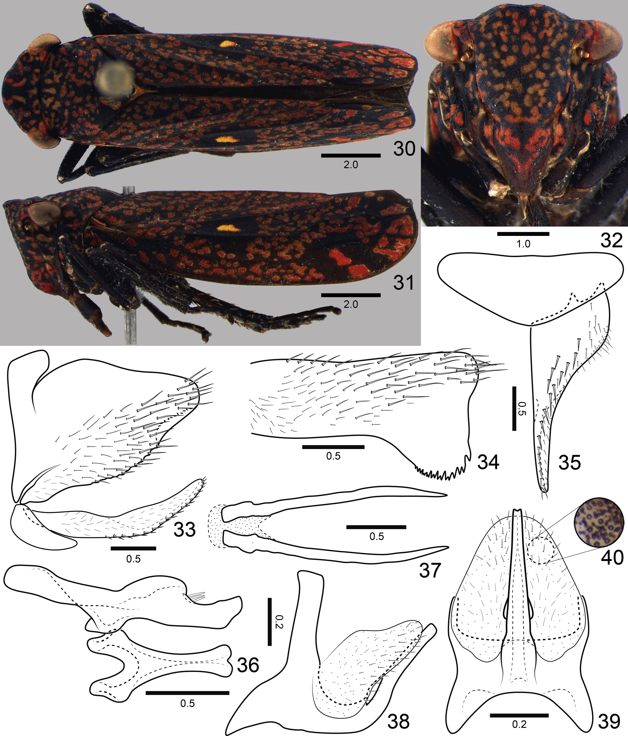
Length. Males 13.1–13.3 mm (n = 3). Females 12.1–12.5 mm (n = 3).
Head and thorax ( Figs 30–32 View FIGURES 30–40 ) much as described by Young (1977: 37) for the genus and in the diagnosis given above. Color as in Figs 30–32 View FIGURES 30–40 ; apical half of crown black at center; forewing vermiculations and spots and larger posterior spots predominantly red.
Male terminalia. Pygofer ( Figs 33, 34 View FIGURES 30–40 ), in lateral view, moderately produced posteriorly; posterior margin narrowly rounded, subangulate; apicoventral margin turned inwards, forming semicircular process with toothed apex; small macrosetae from ventrobasal portion to apex, surface of process without macrosetae. Subgenital plate ( Figs 33, 35 View FIGURES 30–40 ), in ventral view, subtriangular; broad at base and slender from basal third to apex; small macrosetae and microsetae distributed mostly along outer lateral margin; in lateral view, plate extending posteriorly as far as pygofer apex. Style ( Fig. 36 View FIGURES 30–40 ), in dorsal view, extending posteriorly slightly beyond apex of connective; with outer preapical lobe; outer margin behind lobe with setae; apex slightly expanded and subacute. Connective ( Fig. 36 View FIGURES 30–40 ), in dorsal view, Y-shaped; arms short; stalk elongate, slightly expanded apically. Aedeagus ( Figs 38, 39 View FIGURES 30–40 ) symmetrical; in lateral view, with distinct basidorsal and basiventral apodemes; shaft slender, directed dorsally, without apical processes. Large membranous area between anal tube and dorsal aedeagal surface ( Figs 39, 40 View FIGURES 30–40 ) covered by tiny tegumentary processes. Paraphyses ( Fig. 37 View FIGURES 30–40 ), in ventral view, with rami long, acute, and almost parallel to each other.
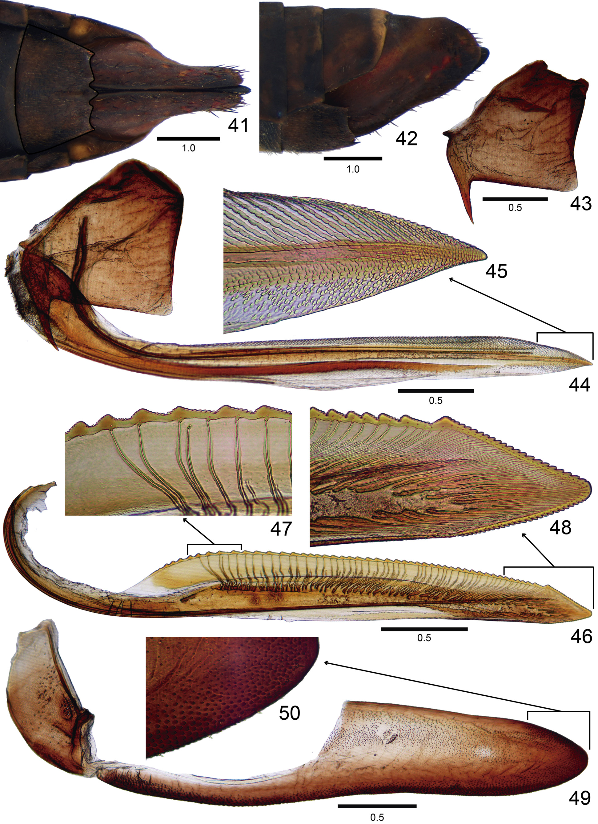
Female terminalia. Sternite VII ( Figs 41, 42 View FIGURES 41–50 ) not strongly produced posteriorly; in ventral view, with posterior margin transverse and with emargination on each side of median dentiform projection. Pygofer ( Figs 41, 42 View FIGURES 41–50 ), in lateral view, well produced posteriorly; distal margin broadly rounded; ventral margin slightly sinuous; macrosetae distributed mostly on posterior portion and extending anteriorly along ventral area. First valvifer ( Figs 43, 44 View FIGURES 41–50 ), in lateral view, subtrapezoidal, with conspicuous, basiventral spiniform process, anterior margin with small dentiform process, posterior margin almost straight. First valvula ( Figs 44, 45 View FIGURES 41–50 ), in lateral view, with dorsal margin straight and ventral margin slightly lobed; apex acute; dorsal sculptured area strigate, extending from basal portion to apex of blade; ventral sculptured area scale-like, restricted to apical portion of blade; ventral interlocking device restricted to basiventral half of blade; in ventral view, base of valvula distinctly expanded outwards. Second valvula ( Figs 46–48 View FIGURES 41–50 ), in lateral view, distinctly expanded beyond basal curvature; apex subacute; preapical prominence indistinct; dorsal margin with about 50 mostly triangular continuous teeth; denticles distributed on teeth and on apical portion of blade, except on apex (dorsal dentate apical portion slightly longer than ventral portion); ducts extending towards teeth and apex of valvula (basalmost six teeth or so do not receive ducts). Gonoplac ( Figs 49, 50 View FIGURES 41–50 ) of the usual Cicadellinae type: in lateral view, with basal half narrow and apical half distinctly expanded; apex obtuse; denticuli and setae distributed on apical portion and extending anteriorly along ventral margin.
Material examined. Holotype ♂: “ Parque Estadual da Serra \ do Mar, Picinguaba , Ubatuba, SP [ State of São Paulo] 28-30/X/2004 \ Mejdalani , Carvalho & \ Baptista ” ( MNRJ) . Paratypes: 5 ♂, same data as the holotype , 2 ♂ ( MNRJ) , 2 ♂ ( DZRJ) , and 1 ♂ ( DZUP) ; 2 ♂, 2 ♀, and 1 specimen without abdomen, “PICINGUABA \ UBATUBA- SP \ 16/XII/1992 \ M. FELIX col.” ( DZRJ, 1 ♂ DZUP, 1 ♀ MNRJ) ; 1 ♂, “ Lumiar, [ State of] Rio de Janeiro \ 22˚21’S 42˚12’W \ 16/XI/2006 V.P. Alecrim ” ( DZUP) ; 1 ♂, “ São Bento do Sul, Rio \ Natal Sta [ State of Santa ] Catarina 700m \ 01/II/2008 P.C. Grossi ” ( DZUP) .
Etymology. The species epithet refers to the type locality (Picinguaba) in the Parque Estadual da Serra do Mar, State of São Paulo, Southeastern Brazil.
Remarks. Aguana picinguaba sp. nov. can be recognized by the absence of a median spot on the apical half of the crown ( Fig. 30 View FIGURES 30–40 ), red vermiculations and spots of the forewings ( Figs 30, 31 View FIGURES 30–40 ), absence of apical aedeagal processes ( Figs 38, 39 View FIGURES 30–40 ), and posterior margin of female sternite VII with an emargination on each side of median dentiform projection ( Fig. 41 View FIGURES 41–50 ).
No known copyright restrictions apply. See Agosti, D., Egloff, W., 2009. Taxonomic information exchange and copyright: the Plazi approach. BMC Research Notes 2009, 2:53 for further explanation.
|
Kingdom |
|
|
Phylum |
|
|
Class |
|
|
Order |
|
|
Family |
|
|
Tribe |
Cicadellini |
|
Genus |