
Guajirolus ektrapeloglossa Flowers 1985
|
publication ID |
https://doi.org/ 10.11646/zootaxa.4564.2.11 |
|
publication LSID |
lsid:zoobank.org:pub:7378731F-9FB5-407A-ADB9-5430C15917B6 |
|
DOI |
https://doi.org/10.5281/zenodo.5923793 |
|
persistent identifier |
https://treatment.plazi.org/id/03CFA937-556C-1100-FF1D-E7F9FEA4FE48 |
|
treatment provided by |
Plazi |
|
scientific name |
Guajirolus ektrapeloglossa Flowers 1985 |
| status |
|
Guajirolus ektrapeloglossa Flowers 1985 View in CoL
( Figs 1–88 View FIGURES 1–3 View FIGURES 4–11 View FIGURES 12–16 View FIGURES 17–21 View FIGURES 22–28 View FIGURES 29–35 View FIGURES 36–40 View FIGURES 41–43 View FIGURES 44–50 View FIGURES 51–58 View FIGURES 59–69 View FIGURES 70–74 View FIGURES 75–80 View FIGURES 81–82 View FIGURES 83–88 )
Genus 3 nr. Pseudocloeon: Roback 1966: 135 , figs 58–63 (larva).
Guajirolus ektrapeloglossa Flowers 1985: 29 View in CoL (♂ and ♀ imago, larva).
Guajirolus nanus Lugo-Ortiz & McCafferty 1995: 58 View in CoL (larva) syn. n.
Guajirolus queremba Nieto 2003: 153 View in CoL (♀ subimago, larva) syn. n.
Guajirolus flowersi Thomas & Dominique (in Thomas, Dominique & Orth) 2005: 21 View in CoL (larva) syn. n.
Material examined. PANAMA, Provincia Chiriquí: La Esperanza (7 km NNW Gualaca), Rio Chiriquí Nuevo between dam and power station, downstream of footbridge, 19.I.2018, coll. N. Kluge: 1 L ♀; Quebrada Barrigón just upstream of the road bridge between La Esperanza and Gualaca (8°34'43''N, 82°19'24''W), 20–22.I.2018, coll. N. Kluge & L. Sheyko: 2 L-S-I ♂, 1 S-I ♂, 2 L-S ♂, 1 L/S ♂, 19 L ♂, 8 L-S-I ♀, 1 L-S/I ♀, 1 L-S ♀, 48 L ♀ GoogleMaps . PERU: Region Junun, Provincia Chanchamayo, Rio Sotorani (right tributary of Rio Perene downstream Pichanaki , 7.I.2006, coll. N. Kluge: 1 L-S-I ♀; Region Loreto, Provincia Ucayali, Pampa Hermosa, 9–21.VIII.2013, coll. N. Kluge & L. Sheyko: exuviae of 1 female larva.
Descriptions. Larva. CUTICULAR COLORATION. Head, pronotum, mesonotum and abdominal terga I–IX
at most part brown, with or without variable unpaired contrasting colorless blanks ( Figs 1 View FIGURES 1–3 , 41–42 View FIGURES 41–43 , 51–58 View FIGURES 51–58 ). Thoracic pleura and sterna with brown sclerites and colorless membranes ( Fig. 43 View FIGURES 41–43 ). Legs with colorless background and following brown markings: femur with two more or less expressed brown bands, among which distal band darker than proximal; tibia with more or less expressed brown band near base; tarsus with more or less expressed brown band at base ( Figs 44–45 View FIGURES 44–50 ); sometimes brown markings better expressed on fore legs, worse on hind legs. Abdominal terga I–IX with or without unpaired blanks, which either form integral longitudinal median line, or separated one from another; sometimes blanks on terga IV and/or VII wide, transverse and occupy most part of tergum ( Fig. 58 View FIGURES 51–58 ); sometimes blanks absent and terga I–IX entirely dark brown. Abdominal sterna I–IX brown, often lighter than terga, unicolor or with diffusive color pattern. Last abdominal segment (tergum X and paraprocts) light, often entirely colorless. Tergalii more or less colored with brown, lighter at periphery ( Fig. 67 View FIGURES 59–69 ). Caudalii with brown and colorless portions at following succession: proximally light brown—gradually turn to dark brown—contrastingly changed to colorless—contrastingly changed to dark brown—gradually become lighter toward tip ( Figs 1 View FIGURES 1–3 , 52 View FIGURES 51–58 , 59 View FIGURES 59–69 ).
HYPODERMAL COLORATION. Not expressed.
STRUCTURE. Head and thorax somewhat compressed laterally and arched dorsally ( Figs 1–3 View FIGURES 1–3 ). Frontal suture sinuate ( Fig. 2 View FIGURES 1–3 ); antennae bases brought together, frons between them keel-shaped and elevated above clypeus ( Figs 1–3 View FIGURES 1–3 ).
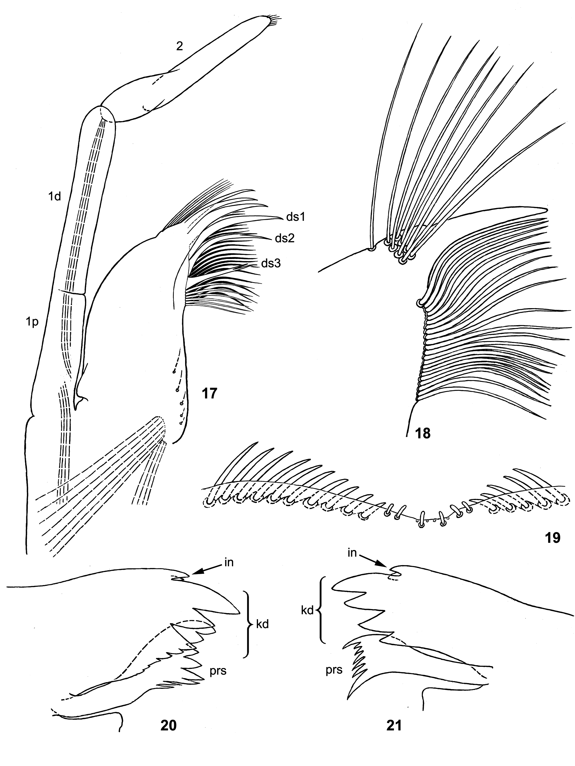
Labrum as characterized above [ Guajirolus /g1 (1)]; median emargination with 6 (i.e. 3 pairs) minute stout setae exposed on outer side ( Fig. 19 View FIGURES 17–21 ); among setae-like projections, located on inner side and directed medially, projections of distal group in right half more stout and blunt than others and form regular row ( Fig. 11 View FIGURES 4–11 ).
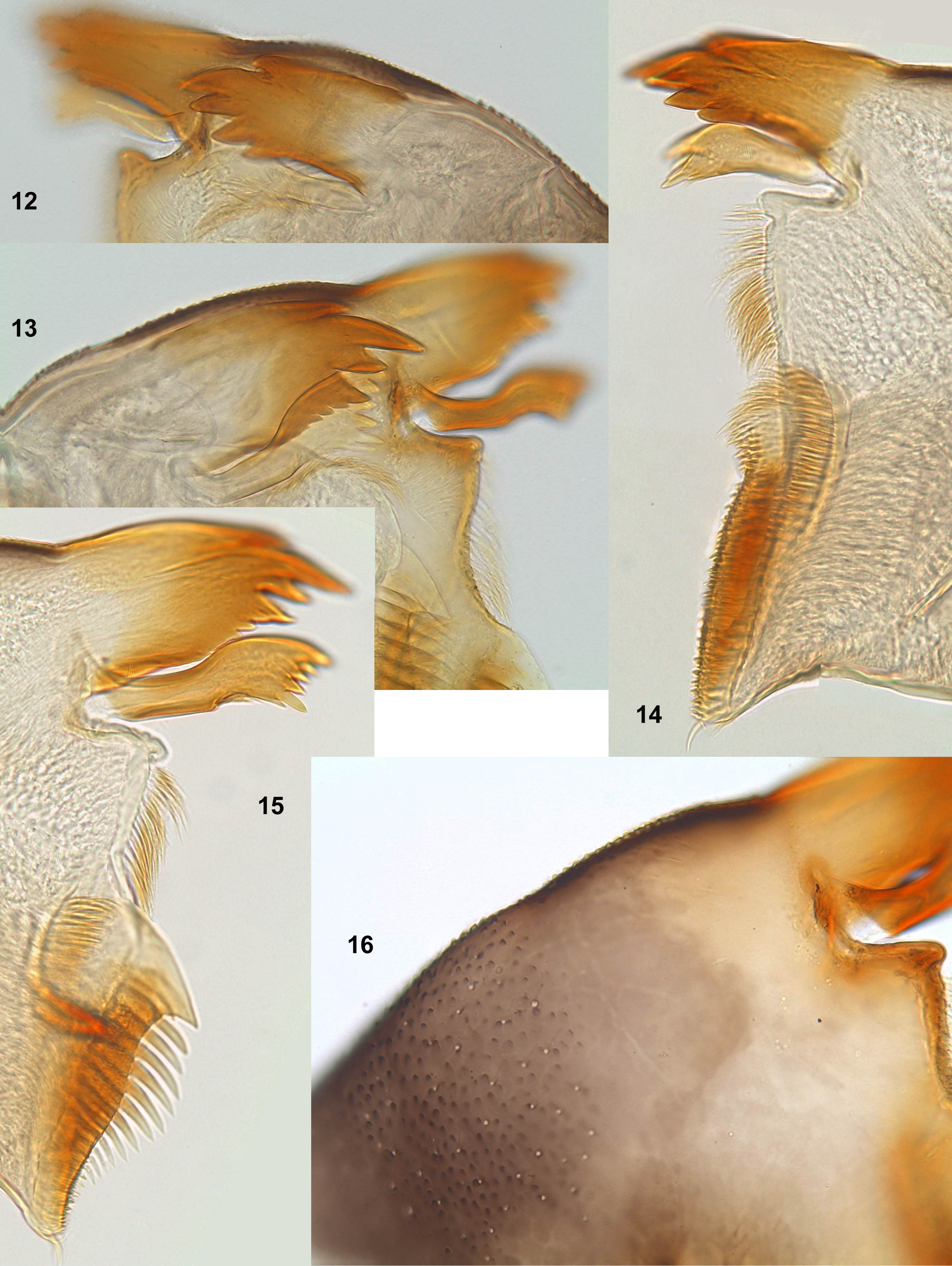
Mandibles as characterized above [ Guajirolus /g1 (2), (3)], stout and convex; lateral surface pigmented, covered with minute protuberances and sparse fine setae ( Fig. 16 View FIGURES 12–16 ); on both mandibles incisor reduced to 2 small denticles; kinetodontium (fused with incisor) on left mandible represented by 3 large and several small denticles, on right mandible by 4 large denticles; on both mandibles prostheca large and sharply widened apically ( Figs 12–13 View FIGURES 12–16 , 20–21 View FIGURES 17–21 ).
Maxilla as characterized above [ Guajirolus /g1 (5)]; galea-lacinia narrowed toward apex, proximally on lateral side with protuberance located more dorsally than base of palp ( Figs 17 View FIGURES 17–21 , 22–24 View FIGURES 22–28 ); inner-dorsal row of setae with distal end turned laterally ( Fig. 18 View FIGURES 17–21 ); apical bunch of long setae consists of 8 setae arranged in 2 rows and one seta laterad of them ( Fig. 18 View FIGURES 17–21 ).
Maxillary palp as characterized above [ Guajirolus /g1 (6)]; both subsegments of 1st palpomere long and movably articulated one with another; 2nd palpomere with proximal part swollen ( Figs 17 View FIGURES 17–21 , 22–24 View FIGURES 22–28 ).
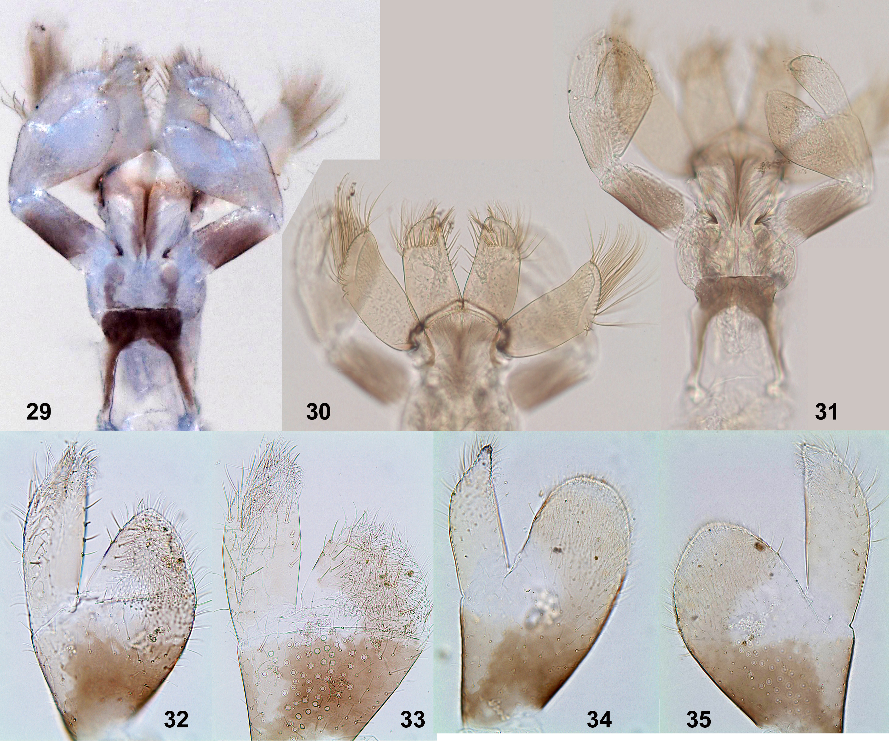
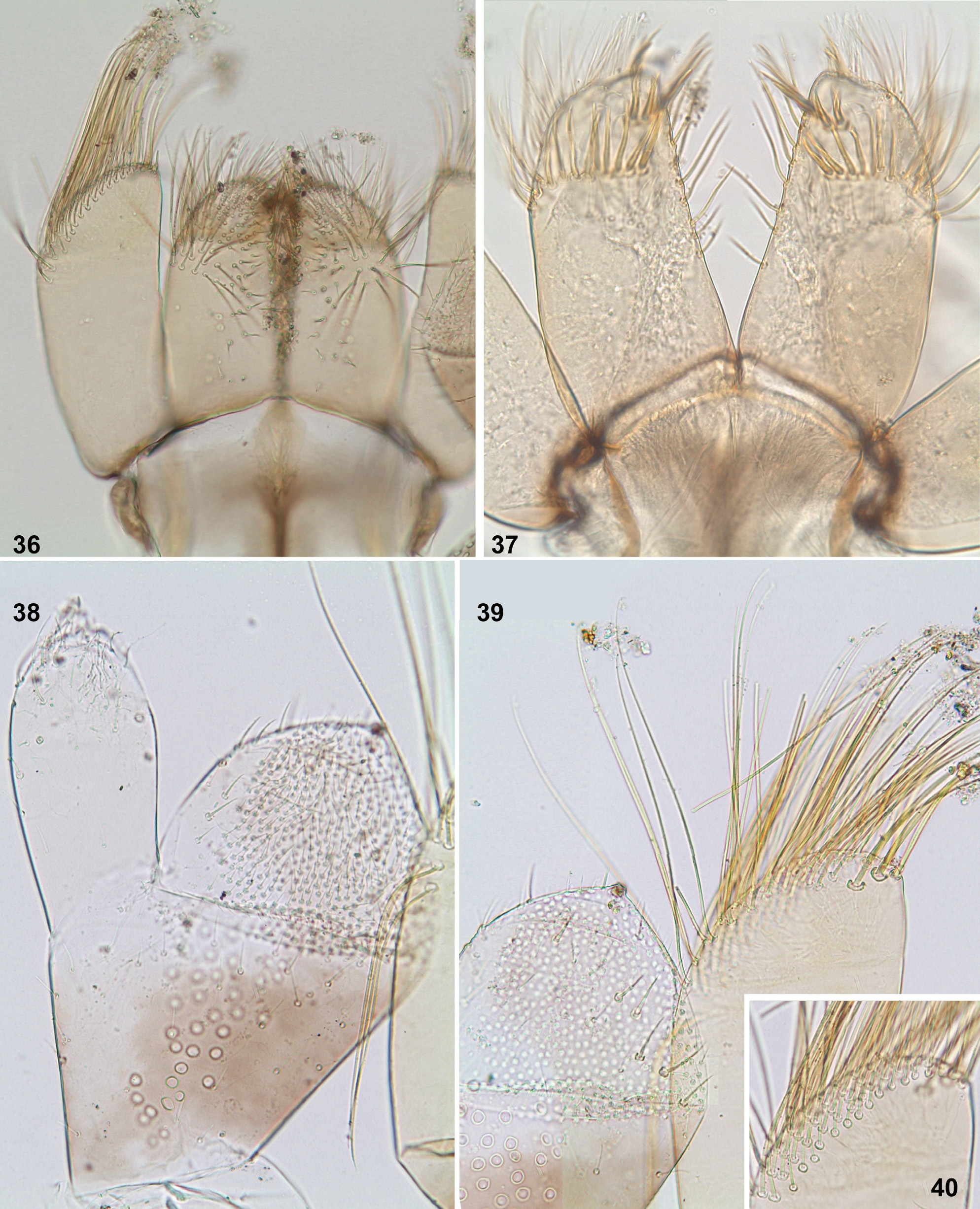
Labium with glossae and paraglossae equally wide, paraglossae slightly longer than glossae; base of glossa very slightly overlap base of paraglossa in ventral view; base of paraglossa somewhat projected laterally ( Figs 30 View FIGURES 29–35 , 37 View FIGURES 36–40 ). Ventral side of glossa with numerous irregularly situated long hair-like setae ( Fig. 36 View FIGURES 36–40 ). Dorsal side of glossa with especially stout long setae forming regular transverse row in distal part of glossa and shorter irregular transverse row more distally; middle part of dorsal side with small group of irregularly situated long hair-like setae ( Fig. 37 View FIGURES 36–40 ). Row of long setae along median side of glossa (usual for Baetidae ) either regular ( Fig. 37 View FIGURES 36–40 ), or irregular. Paraglossa without setal rows on median and lateral sides; distal margin with numerous long setae ( Fig. 40 View FIGURES 36–40 ) and with 2 somewhat thicker long setae on dorsal side close to apex ( Fig. 39 View FIGURES 36–40 ).
Labial palp as characterized above [ Guajirolus /g1 (7)]; projection of its 2nd segment very thick and soft, sacklike. 2nd and 3rd segments with irregularly situated stout pointed setae ( Fig. 39 View FIGURES 36–40 ); ventral side of projection of 2nd segment and of apex of 3rd segment with dense minute fine setae ( Fig. 38 View FIGURES 36–40 ).
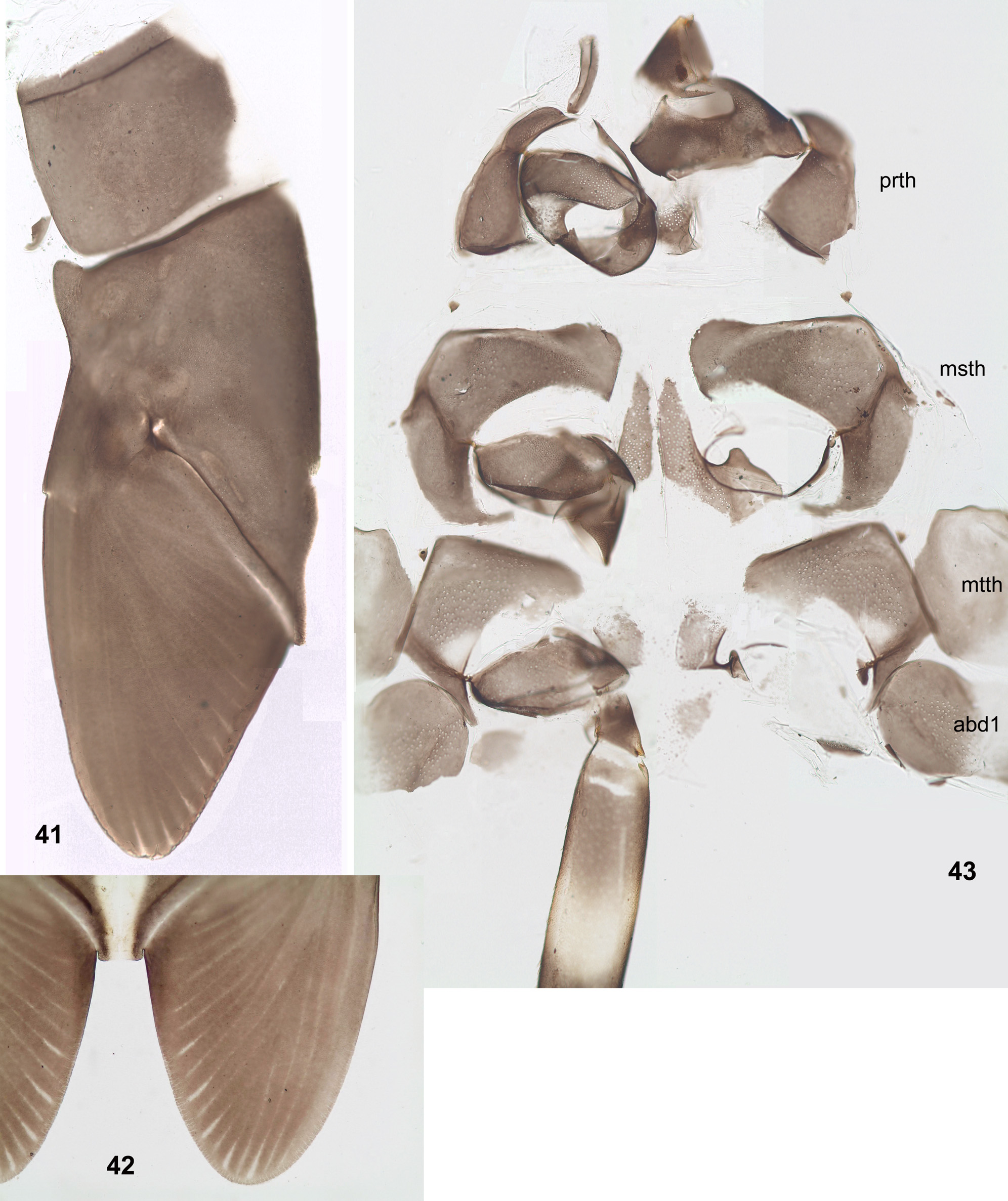
Mesonotum with anterolateral corners projected anteriorly ( Fig. 41 View FIGURES 41–43 ). Fore protoptera brought together, but not fused with posterior margin of mesonotum ( Fig. 42 View FIGURES 41–43 ) (in contrast to Pseudopannota ). Lateral margin of fore protopteron with small incision corresponding to costal brace ( Fig. 41 View FIGURES 41–43 ). Metanotum without vestiges of hind protoptera ( Fig. 43 View FIGURES 41–43 ). Thoracic sterna partly sclerotized and pigmented: prosternum with unpaired sclerite, mesosternum and metasternum each with pair of sclerites of peculiar shape ( Fig. 43 View FIGURES 41–43 ). Sclerites of thoracic pleura and sterna partly with scales in operculate sockets (similar to that of abdomen).
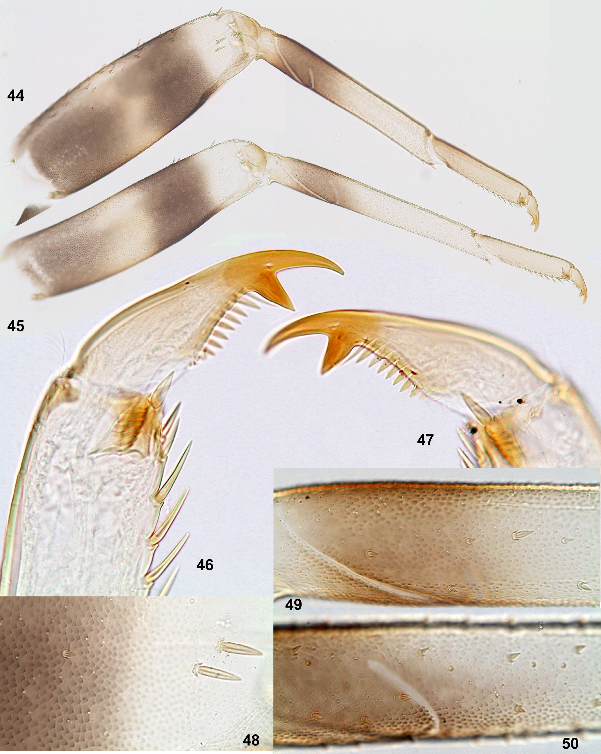
Legs with cuticle thick and denticulate ( Figs 48–50 View FIGURES 44–50 ). Femora as characterized above [ Guajirolus /g1 (8)]; fore femur, besides stout setae on outer side, with such setae also on anterior surface—in distal part and along outer side ( Figs 44, 48 View FIGURES 44–50 ). Tibiae of all legs with patella-tibial suture short, so that on inner side patella occupies only 1/3 of tibia ( Figs 44–45 View FIGURES 44–50 ). Tibia of fore leg with sparse small stout pointed setae on anterior side and few small stout setae on inner side ( Fig. 49 View FIGURES 44–50 ; Thomas & al. 2005: fig. 9). Tibiae of middle and hind legs without stout setae on anterior side, with such setae on posterior and inner sides ( Fig. 50 View FIGURES 44–50 ). Tarsus of all legs with row of stout pointed setae on inner side only. Claws as characterized above [ Guajirolus /g1 (9)] ( Figs 46–47 View FIGURES 44–50 ).
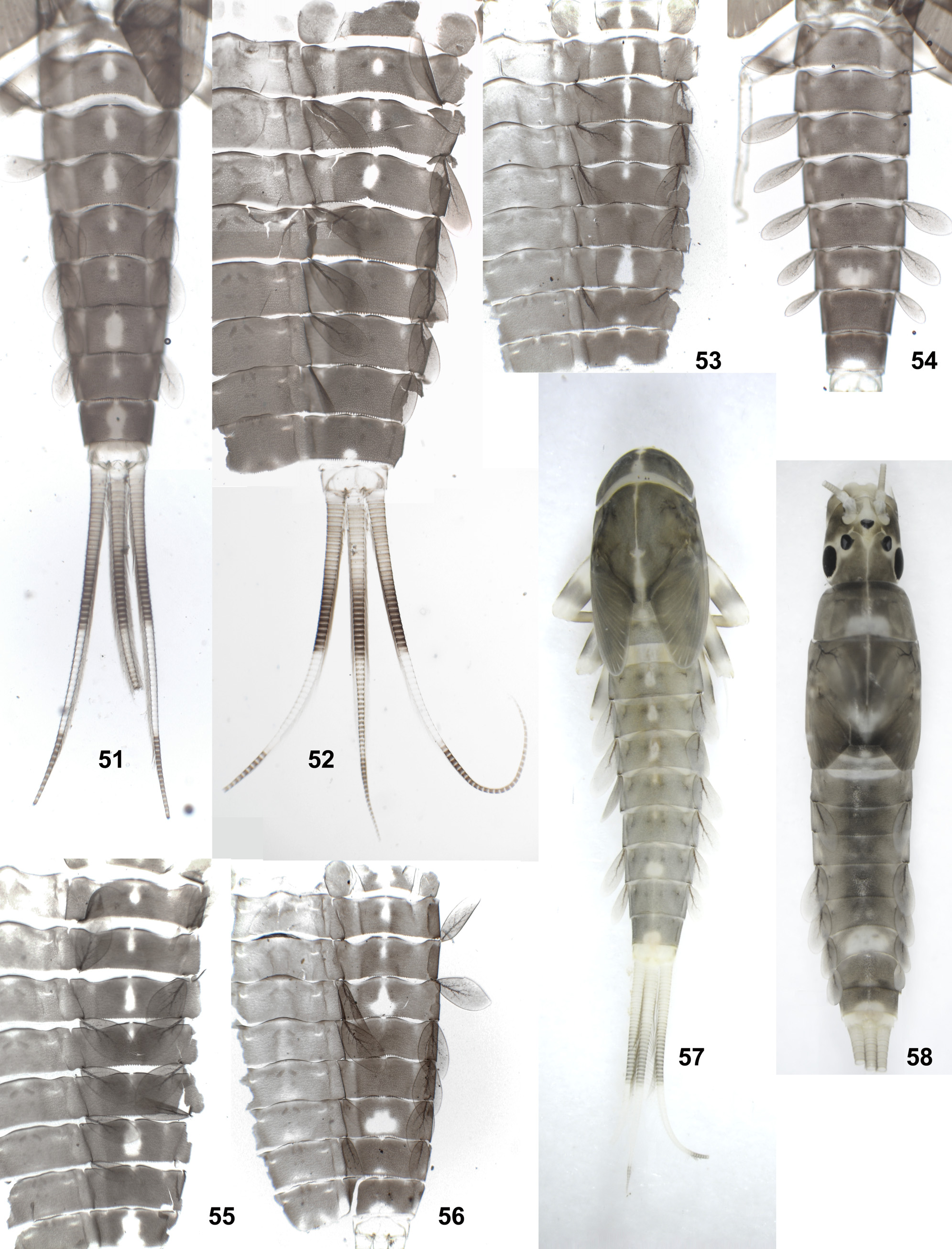
Abdominal terga and sterna with dense scales in operculate sockets ( Figs 60–61 View FIGURES 59–69 ). Posterior margin of tergum I smooth, without denticles. Posterior margins of terga II–X with row of triangular denticles; denticles of more anterior terga shorter and blunt ( Fig. 62 View FIGURES 59–69 ), denticles of more posterior terga longer and pointed ( Fig. 63 View FIGURES 59–69 ); on middle of posterior margin of tergum IX (behind submedian pair of setae) denticles smaller. Posterior margins of sterna I–VII smooth, without denticles; sternum VIII with blunt denticles on median part of posterior margin ( Fig. 64 View FIGURES 59–69 ); sternum IX with pointed denticles on posterior margin ( Figs 65 View FIGURES 59–69 , 76 View FIGURES 75–80 ). Paraprocts with 10–20 pointed denticles, increasing in size toward caudal margin ( Fig. 69 View FIGURES 59–69 ).
Tergalii I absent ( Fig. 43 View FIGURES 41–43 ). Tergalii II–V subequal, tergalii VI–VII gradually diminished ( Figs 51–58 View FIGURES 51–58 ). Each tergalius oval; costal and anal ribs fused apically, so that entire margin armed by costa; in distal half margin with pointed seta-bearing denticles ( Figs 67–68 View FIGURES 59–69 ); dorsal surface with scales in operculate sockets ( Figs 66, 68 View FIGURES 59–69 ).
Paracercus as thick as cerci and somewhat shorter ( Figs 1 View FIGURES 1–3 , 52 View FIGURES 51–58 ). Cerci and paracercus with dense primary swimming setae at most their length and with slender distal portion lacking swimming setae. In proximal and distal parts of cercus segments cylindrical, in middle part slightly oblique ( Fig. 59 View FIGURES 59–69 ); posterior margins of segments with regular rows of flat denticles, nearly equally large on dorsal, ventral and lateral sides of cercus ( Fig. 59 View FIGURES 59–69 ), on dorsal and ventral sides of paracercus.
RESPIRATORY MOVENENTS. Larva unable for rhythmical respiratory movements by tergalii; cannot live for long time in stagnant water.
Subimago. CUTICULAR COLORATION. Head and pronotum entirely colorless. Prealar bridge contrastingly dark brown ( Fig. 72 View FIGURES 70–74 ). Mesonotum partly colorless, with certain areas shed with light brownish ( Fig. 72 View FIGURES 70–74 ). Pterothoracic pleura and sterna at most part colorless, with certain sclerites contrastingly brown ( Fig. 73 View FIGURES 70–74 ). Legs entirely colorless. Abdomen, genitals and cerci entirely colorless.
HYPODERMAL COLORATION. Just after emergence from larva, most part of body lack hypodermal coloration, being pale ocher; certain areas of thorax brown; abdomen with intersegmental membranes of terga brown, terga with light brownish pigmentation forming paired diffusive maculae ( Fig. 74 View FIGURES 70–74 ). During subimaginal development, thorax and abdominal terga get brown color as in imago.
TEXTURE. On all legs of male and female last tarsomere covered by pointed microlepides, previous tarsomeres covered mainly by blunt microlepides, with pointed microlepides near apex.
Imago, male ( Figs 70–71 View FIGURES 70–74 ). Head ocher. Turbinate eyes with stalks relatively short and sharply widened toward facetted surface; facetted surfaces very wide, elliptical, greatly divergent anteriorly; stalk and facetted surface brown, border between them narrowly outlined by dark brown. Thorax brown with ocher, equally dark dorsally and ventrally. Fore wing colorless, with veins pale yellowish, only bases of costa and radius brown. Hind wing absent. All legs unicolor pale yellowish. On middle and hind legs tarsus relatively short, 0.3 of tibia length. Length of leg segments (mm): femur, tibia and tarsomeres of fore leg 0.87: 1.70: 0.06:050: 0.30: 0.20: 0.13; of middle leg 0.78: 1.05: 0.10: 0.06: 0.03: 0.13; of hind leg 0.82: 1.05: 0.10: 0.06: 0.03: 0.13. Apical spines either present on two tarsomeres, or on one tarsomere, or absent. All abdominal terga nearly unicolor brown; sterna ocher, shed with gray anteriorly and laterally. Genitals brown. Cerci unicolor pale yellowish.
Male genital structure and development ( Figs 75–80 View FIGURES 75–80 ). Sterno-styligeral muscle present, but weak; it arises from base of abdominal sternum IX and terminates near its middle, far not reaching bases of unistyligers ( Fig. 75 View FIGURES 75–80 ). Gonovectes curved S-sharply; penial bridge with small prominent slerotized median projection. Unistyligers long, narrowed at midlength and widened apically. 1st segment of gonostylus with projected right angle on inner side apically; 2nd segment widened apically; 3rd segment small, with thin stem and thickened apically. In mature larva ready to molt to subimago, gonostyli folded by the « Guajirolus - type », i.e. with 3rd segment attached caudally and retaining under larval protogonostylus, but turned by its apex medially ( Fig. 76 View FIGURES 75–80 ).
Imago, female. Coloration similar to that of male. Eyes small, widely separated ( Figs 81–82 View FIGURES 81–82 ).
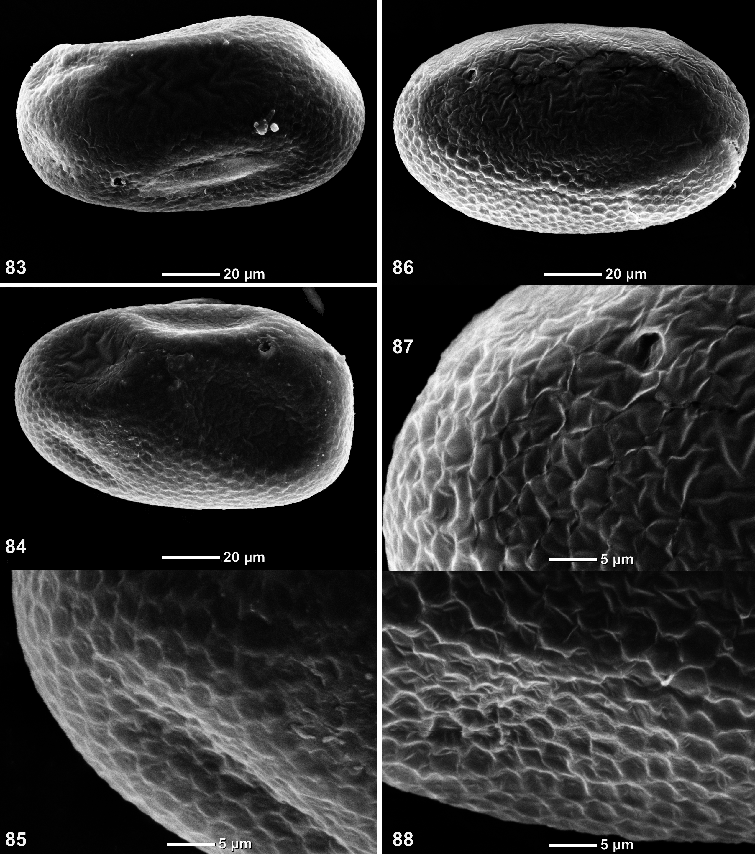
Eggs ( Figs 83–88 View FIGURES 83–88 ). Oval. Chorion partly smooth or irregularly crumpled, partly with fine net-like relief with cells small and shallow.
Dimension. Fore wing length (and body length) 4–5 mm.
Habitat and sex ratio. Most part of specimens used in this study (75 larvae and winged insects reared from them) were collected in Quebrada Barrigón, on a portion of its bottom formed by an integral flat rock. Flowers (1985) also reported that he found larvae of G. ektrapeloglossa on rocky substrates. Among all specimens collected in Barrigón, 2/3 are females and only 1/3 are males. Three other specimens examined by me (one larva from Chiriquí Nuevo and two specimens from Peru) are females. Probably, females dominate in this species.
Stage association. Flowers (1985) wrote that his association of male and female imagines with the nymphs was done «on the basis of color patterns», but did not specify, which features of coloration allowed him to do this. The brown hypodermal coloration of imaginal thorax and abdomen appears only in course of subimaginal development, being absent in larva and in just-molted subimago ( Fig. 74 View FIGURES 70–74 ), so it does not allow to associate larvae and imagines; legs and cerci have no hypodermal coloration at any stage of development. Cuticular coloration, being characteristic for larvae, is not retained in subimago and imago. Rearing imagines from larvae confirms correctness of the stage association done by Flowers.
Distribution. Central and South America (see «Discussion»).
Discussion. The examined specimens from Peru and Panama have the same structure of larva, subimago, female imago and eggs (compare Figs 83–85 and 86–88 View FIGURES 83–88 ), that allows to conclude that they belong to the same species, which is widely distributed in South and Central America. According to current literature, 6 species are reported in the genus Guajirolus : the species G. ektrapeloglossa was originally described by Flowers (1985) from Panama and Colombia as distinct from the unnamed species from Peru reported by Roback (1966); later, four other species were described— G. nanus from Costa Rica, G. queremba from Argentina, G. flowersi from French Guyana and G. rondoni from Brazil ( Lugo-Ortiz & McCafferty 1995, Nieto 2003, Thomas et al. 2005, Salles 2007). Diagnoses of all these species were based on larval characters (presumably determined imagines were described only for G. ektrapeloglossa , and subimago—only for G. queremba ). Each author, who described a new species of Guajirolus , compared its characters with the previously published descriptions and figures, but nobody compared specimens of different species. Flowers (1985) regarded G. ektrapeloglossa as different from the unnamed Roback's species because it has 6 pairs of tergalii, instead of 7 reported by Roback [see (J) below]. Lugo-Ortiz & McCafferty (1995) regarded G. nanus as different from G. ektrapeloglossa based on presence of spicules on mandibles, shape of median process on hypopharynx, presence of proximal brown band on femur and number of spines on paraprocts; however, the original description of G. ektrapeloglossa does not contain clear statements about presence or absence of these characters [see (B), (D), (F) and (H) below]. Nieto (2003) regarded G. flowersi as different from G. ektrapeloglossa and G. nanus based on combination of the same characters. Thomas et al. (2005) regarded G. flowersi as different from previously described species based on shape of distal margin of labrum [see (A)], maxillary palp interpreted as 4-segmented [see (C)] and combination of characters used by previous authors. Salles (2007) regarded G. rondoni as different from previously described species based on combination of the same characters plus narrower projection of 2nd segment of labial palp [see (E)]. Only the last character is species-specific; all other characters reported as species-specific in Guajirolus , either vary individually, or are caused by different interpretations made by different authors. The following characters were used to distinguish species of Guajirolus :
(A) Shape of distal margin of labrum. Thomas et al. (2005) stated that the new species G. flowersi differs from the earlier described species of Guajirolus by the median («sagittal») emargination of labrum, whose width is subequal to width of lateral processes, while in G. ektrapeloglossa , G. nanus and G. queremba width of this emargination is less than width of lateral processes. Salles (2007) accepted this character in the key, to separate G. flowersi and the new species G. rondoni from three other species; he also added that in G. flowersi and G. rondoni emargination of labrum has stout setae, in contrast to the three other species. Actually labrum of Guajirolus has unusual structure [see Guajirolus /g1 (1)], being soft and lacking sclerotization on distal margin ( Fig. 6 View FIGURES 4–11 ). Because of this, shape of its distal margin depends on fixation ( Figs 4–5, 7–8 View FIGURES 4–11 ). Small stout setae, reported by Salles, are present in all specimens; if the emargination is spread and looks wide, these setae are well visible, but if the emargination is compressed and looks narrow, these setae can be overlooked.
(B) Spicules on mandibles. Lugo-Ortiz & McCafferty (1995) stated that the new species G. nanus differs from the earlier described G. ektrapeloglossa by presence of minute spicules on lateral margins of mandibles. These authors did not examine specimens of G. ektrapeloglossa and regarded that «those spicules appear to be absent» based only on the figures in the original description. However, Flowers (1985) did not pay special attention to presence or absence of these spicules; on his figures of mandibles, he showed only fine setae ( Flowers 1985: figs 6–7), which were ignored and not figured by Lugo-Ortiz & McCafferty (1995: figs 2–3). Nieto (2003) did not report presence or absence of spicules neither in the description of a new species G. queremba , nor in the «Discussion», but used this character in the key to separate G. queremba from G. nanus . Thomas et al. (2005) repeated the thesis and antithesis from the paper by Nieto (2003) and reported presence of spicules for the new species G. flowersi . Salles (2007) reported presence of spicules for the new species G. rondoni . Actually these spicules are present in G. ektrapeloglossa ( Fig. 16 View FIGURES 12–16 ). Similar spicules, besides mandibles, are present on mesonotum, on certain areas of thoracic pleura and on all segments of legs ( Figs 48–50 View FIGURES 44–50 ; Thomas et al. 2005: figs 8–9).
(C) Number of segments of maxillary palp. Thomas et al. (2005) stated that the new species G. flowersi differs from the earlier described species of Guajirolus by 4-segmented maxillary palp, while in G. ektrapeloglossa , G. nanus and G. queremba maxillary palp is 3-segmented. Salles (2007) reported 3-segmented maxillary palp for
the new species G. rondoni and used this character to separate G. rondoni from G. flowersi . Actually, maxillary palp has 2 primary segments, the 1st of which is subdivided into two secondary subsegments with one muscle passing through both of them [see Guajirolus /g1 (6)]. The distal segment (2nd primary segment, lacking musculature) has a proximal swelling separated distally by an oblique concavity ( Figs 17 View FIGURES 17–21 , 22–24 View FIGURES 22–28 ); if this concavity is sharp, it can be interpreted as a joining between 3rd and 4th secondary segments; if it is shallow, it can be overlooked. The distal segment is soft, with very fine cuticle, so condition of this concavity depends on fixation. In the original description of G. ektrapeloglossa , the 2nd primary segment of maxillary palp is correctly shown with obtuse-angled bent between the proximal swelling and the remainder part ( Flowers 1985: fig. 9).
(D) Median projection of hypopharynx. Flowers (1985) did not give wordily characteristic of hypopharynx of G. ektrapeloglossa , but only gave its drawing, on which it is terminated by three projections, with the medial
projection pointed ( Flowers 1985: fig. 8). Lugo-Ortiz & McCafferty (1995) stated that the new species G. nanus differs from G. ektrapeloglossa by round medial projection of hypopharynx. Nieto (2003) used round medial projection to separate G. nanus and the new species G. queremba from G. ektrapeloglossa . Thomas et al. (2005) stated that the new species G. flowersi has medial projection reduced and flattened, in contrast to pointed in G. ektrapeloglossa and rounded in G. nanus and G. queremba . Salles (2007) reported rounded medial projection for the new species G. rondoni . Actually, the area between paired apical projections of hypopharynx is soft and has no constant shape; depending on condition of specimen, it can be widened and flattened ( Fig. 28 View FIGURES 22–28 ), or narrowed forming a median projection, which can be either rounded ( Fig. 27 View FIGURES 22–28 ), or pointed ( Figs 25–26 View FIGURES 22–28 ).
(E) Projection of 2nd segment of labial palp. Labial palp has unusually massive projection directed medially [see Guajirolus /g1 (7)]. Thomas et al. (2005) stated that in the new species G. flowersi this projection is narrower than in the earlier described G. ektrapeloglossa , G. nanus and G. queremba . On their drawing ( Thomas et al. 2005: fig. 6) projection of left palp has apex as broad as base, and projection of right palp has apex narrower than base. Salles (2007) stated that G. flowersi and the new species G. rondoni differ from others by projection of the 2nd segment of labial palp with apex narrower than base. The 2nd segment of labial palp is soft and sack-like, so its shape has individual variability ( Figs 34–35 View FIGURES 29–35 ) and depends upon flattening on slide ( Figs 32–33 View FIGURES 29–35 ). According to the figure of G. flowersi , it has no significant difference from G. ektrapeloglossa , but on the drawing of G. rondoni ( Salles 2007: fig. 8; Falcão et al. 2011: fig. 111) projection of 2nd segment is narrower than in examined specimens from Panama and Peru; this is the single known character in which G. rondoni differs from G. ektrapeloglossa .
(F) Brown bands on larval femur. Flowers (1985) reported and figured one brown band on larval femur. Lugo- Ortiz & McCafferty (1995) stated that the new species G. nanus differs from G. ektrapeloglossa by presence of two brown bands on femora. Nieto (2003) and Thomas et al. (2005) reported one brown band on larval femur of G. queremba and G. flowersi , and used this character to separate these species from G. nanus . Actually larval femur has more intensively colored subapical brown band and less intensive proximal brown coloration, which can be either interpreted as a second band, or not ( Figs 44–45 View FIGURES 44–50 ); if cuticular coloration of legs is poorly expressed, only the distal band is present.
(G) Light medial longitudinal strip on abdominal terga I–IX. Flowers (1985) figured integral light stripe on all abdominal terga I–IX ( Flowers 1985: fig. 4), but in the text wrote: «Abdomen brown; terga with median yellow bands which sometimes do not reach posterior margin». Thomas et al. (2005) stated that the new species G. flowersi differs from other species of Guajirolus by absence of this band, and described light median blanks on certain terga (without illustration). Actually, unpaired colorless blanks on dark cuticle of abdominal terga greatly vary individually, sometimes forming integral longitudinal stripe, sometimes not ( Figs 51–58 View FIGURES 51–58 ; Falcão et al. 2011: fig. 105).
(H) Denticles on paraprocts. Flowers (1985) did not give wordily description of paraproct of G. ektrapeloglossa , but gave its drawing ( Flowers 1985: fig. 17). Lugo-Ortiz & McCafferty (1995) interpreted this figure as «paraprocts with unorganized spination» and stated that the new species G. nanus differs by «paraprocts with organized spination». They did not explain, what means «organized» and «unorganized» spination. Nieto (2003) separated G. nanus and the new species G. queremba from G. ektrapeloglossa by the «organized spination». Thomas et al. (2005) reported for the new species G. flowersi 5–6 denticles on paraproct and used this character to separate this species from all others, for which they report from 11 to 22 denticles based on descriptions and/or figures. Salles (2007) stated that in the new species G. rondoni paraproct has «organized» spination with about 15 spines, and used these characters («organized» and «unorganized» spination and number of spines) in the species key. Actually paraprocts of G. ektrapeloglossa have normally about 15 denticles, varying approximately from 10 to 20, among which apical denticles are larger, and proximal gradually smaller ( Fig. 69 View FIGURES 59–69 ).
(J) Number of tergalii. Roback (1966) described larva of Guajirolus from the river Tulumayo in Peru under arbitrary name «Genus 3 nr. Pseudocloeon » and reported presence of all 7 pairs of tergalii as «darker gills, Fig. 65 View FIGURES 59–69 , ovate, single on 1–7» ( Roback 1966: 136). Flowers (1985) wrote: «The Peruvian nymphs are not included in G. ektrapeloglossa because they have gills on abdominal segment 1 ( Roback 1966)...». In the generic key to South American mayflies ( Domínguez et al. 1992) number of tergalii in Guajirolus is reported as variable: «Branquias en los segmentos abdominales 1 ó 2–7». In the monograph on South American mayflies ( Domínguez et al. 2006) invariably 6 pairs of tergalii are reported for Guajirolus : «abdominal gills present on segments II–VII»; in this book, the Roback's species is ignored, and the genus Guajirolus is not reported from Peru. Two larvae, collected by me in Peru, have 6 pairs of tergalii. Probably, the Roback's sentence about tergalii «1–7» is a mistake.
Thus, the existent descriptions of G. nanus , G. queremba and G. flowersi do not allow to distinguish them from G. ektrapeloglossa ; these names should be regarded as synonyms, if new species-specific characters will not be discovered.


No known copyright restrictions apply. See Agosti, D., Egloff, W., 2009. Taxonomic information exchange and copyright: the Plazi approach. BMC Research Notes 2009, 2:53 for further explanation.
|
Kingdom |
|
|
Phylum |
|
|
Class |
|
|
Order |
|
|
Family |
|
|
Genus |
Guajirolus ektrapeloglossa Flowers 1985
| Kluge, Nikita J. 2019 |
Guajirolus queremba
| Nieto, C. 2003: 153 |
Guajirolus nanus
| Lugo-Ortiz, C. R. & McCafferty, W. P. 1995: 58 |
Guajirolus ektrapeloglossa
| Flowers, R. W. 1985: 29 |