
Zebrus pallaoroi Kovačić, Šanda & Vukić, 2021
|
publication ID |
https://doi.org/10.1163/18759866-bja10018 |
|
DOI |
https://doi.org/10.5281/zenodo.8356649 |
|
persistent identifier |
https://treatment.plazi.org/id/03CF87C0-FFF3-FFB7-FD60-996C8F68FC27 |
|
treatment provided by |
Felipe |
|
scientific name |
Zebrus pallaoroi Kovačić, Šanda & Vukić |
| status |
sp. nov. |
Zebrus pallaoroi Kovačić, Šanda & Vukić sp. nov.
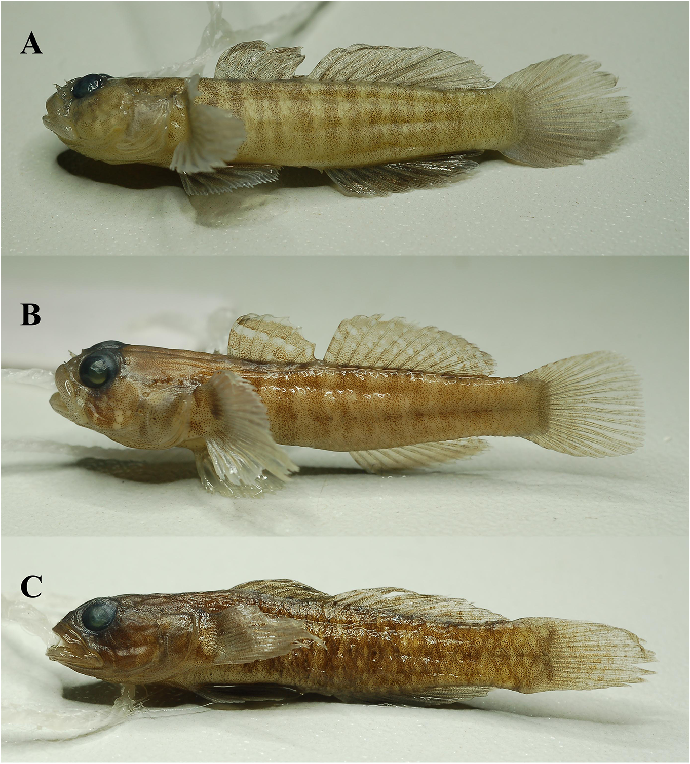
Holotype ( fig. 1A View figure 1 ). Male , 31.81 + 8.51 mm, nmp P6 V 144302 , Kostanjica , Boka Kotorska, Adriatic Sea, Montenegro, 24 August 2016, 42.485139° N, 18.670347° E, collector Šanda R. GoogleMaps
Paratypes. Male, 27.72 + 6.83 mm, nmp P6 V 144300 , and female, 26.44 + 6.39 mm, nmp P6 V 144303 , the same data as holotype GoogleMaps .
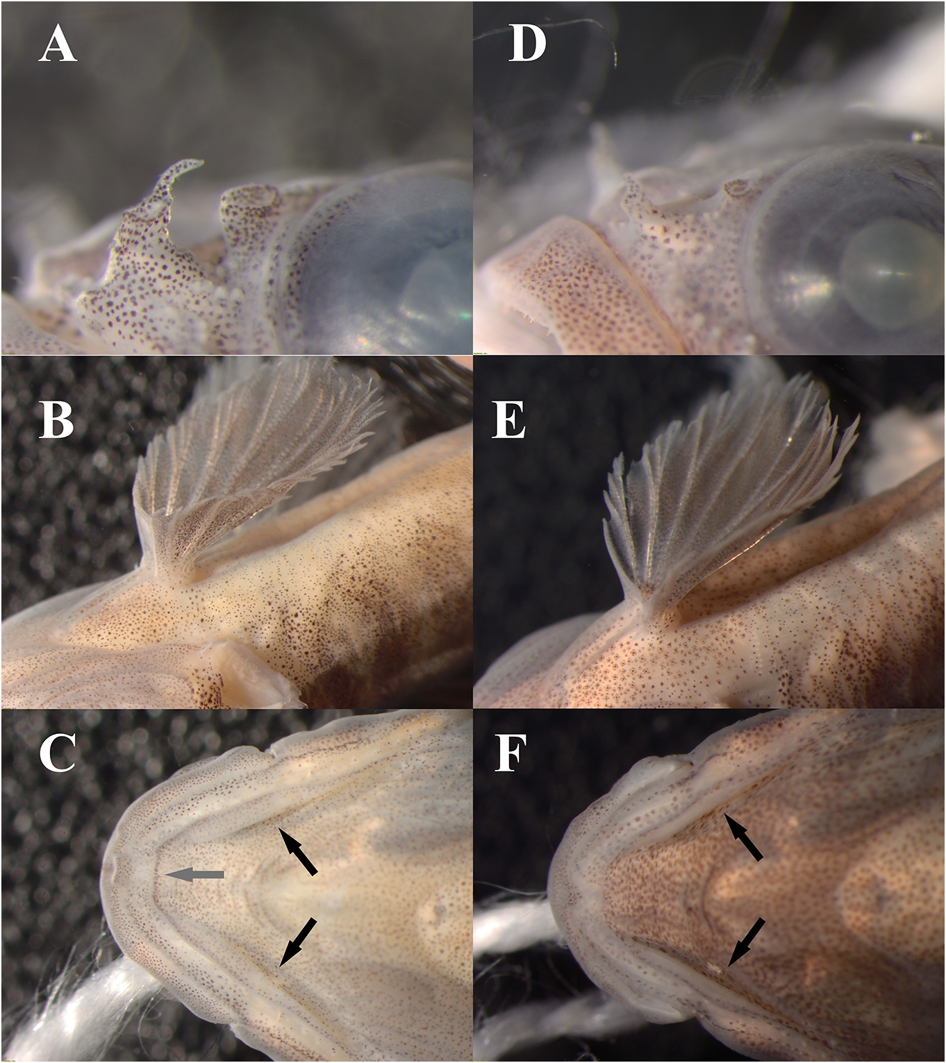
Diagnosis. Zebrus pallaoroi sp. nov. differs from the only congeneric species, Z. zebrus by each of the following characters: (1) snout longer than eye, its length 1.1‒1.2 of eye diameter; (2) posterior nostril short tube, about 0.8–0.9 of anterior nostril ( fig. 2A View figure 2 ); (3) eyes moderately small, eye diameter is 4.3‒4.7 in head length; (4) left and right ventrolateral head ridges transversally connected on anterior part by short transversal ridge ( fig. 2C View figure 2 ), (5) anterior membrane in midline depth about 2/3 of spinous ray ( fig. 2B View figure 2 ); (6) head canal pores large, pore α diameter about half of the distance between pore Ρ and Ρ1; (7) suborbital sensory papillae row 5i going downwards to or near the level of row d, distance between row 5i and row d absent or much smaller than length of row 5i; (8) body with ten to eleven vertical dark brown bands present along lateral side, first in front of the first dorsal fin, last at end of the second dorsal fin, at upper edge about equal or narrower than pale interspaces inbetween.
Description (all morphometric values and meristics in the text are presented as holotype first and paratypes, if different, in parentheses; first the male paratype, then female). General morphology ( fig. 1A View figure 1 ): Body proportions are given in table 2 View table 2 . Body elongate, its depth at pelvic-fin origin 5.3 (5.4) in sl, at anal-fin origin 5.5 (5.9) in sl, laterally compressed posteriorly, with deep caudal peduncle, caudal peduncle depth 0.6 of caudal peduncle length ( fig. 1A View figure 1 ). Head moderately large, the length 3.5 (3.2 and 3.4) in sl, width 3.9 (3.8 and 4.2) in sl, depth 6.4 (5.7 and 6.9) in sl, and depressed, its depth 1.6 (1.5 and 1.6) in width. Snout oblique, with a convex ridge along dorsal midline visible only in paratypes, longer than eye, its length 1.2 (1.1 and 1.2) of eye diameter, 3.7 (4.2 and 3.8) in head length. Anterior nostril nasal tube with a single process from the posterior rim, posterior nostril short tube, 81% (85 and 88%) of the anterior nostril ( fig. 2A View figure 2 ). Eyes dorsolateral, moderately small, eye diameter is 4.3 (4.7 and 4.6) in head length, slightly elevated above the dorsal profile. Interorbital moderately narrow, 1/2 of eye diameter. Cranial roof covered by dorsal axial musculature. Predorsal area about horizontal. Mouth oblique, jaws ending anteriorly nearly equally to lower lip. Upper lip broad, width nearly uniform, more or less as wide as the lateral preorbital area in female, wider in males. Mouth moderately large, posterior angle of jaws ending posteriorly below mideye. Cheek relatively narrow. Teeth in both jaws erect, caniniform; upper jaw with enlarged outer row, intermediate irregularly scattered small teeth and four enlarged median posterior teeth; lower jaw with an outer row of larger teeth, intermediate irregularly scattered small teeth and a short inner row of enlarged teeth laterally ending with one or two distinct canine teeth on each side. Tongue reduced. Chin with a small mental fold. Left and right ventrolateral head ridges transversally connected on the anterior part by a short transversal ridge ( fig. 2C View figure 2 ). Branchiostegal membranes fused to isthmus along the entire lateral margin of the isthmus, from immediately anterior to pectoral margin, gill openings restricted to pectoral-fin base. No spines on preopercle. Pectoral girdle without dermal flaps on anterior edge.
Fins. First dorsal fin vi, second dorsal fin I+10 (I+10–11); anal fin I+8 (I+8–9); branched caudal-fin rays 16 (15), segmented 18 (17), pectoral-fin rays 17 left fin, right fin cut (in all type material), pelvic fins I+5/5+I. Fin morphometrics in proportion to standard body length given in table 2 View table 2 . Spines of first dorsal fin not elongate or filamentous, spines ii-v about equal, the third spine longest; spines v and vi of first dorsal fin reaching the origin of the second dorsal fin when folded down. Origin of first dorsal fin behind vertical at the pectoral-fin base. Interdorsal space with a well-developed membranous connection between dorsal fins. The second dorsal fin originates above the anus in the female and above urogenital papilla in males, with the longest rays hardly reaching the base of uppermost caudal-fin rays in males, and not reaching in the female. Origin of anal fin below vertical of the first segmented ray of the second dorsal fin in males, or second in the female. Anal fin with last ray origin below penultimate ray of the second dorsal fin in the female, below third from the end in males. Posterior tip of anal fin not reaching the base of lowermost caudal-fin rays. Pectoral-fin rays all branched. Pectoral fin with three uppermost rays bifid, partially free from the membrane, and three more with shorter free tips, first ray free tip 0.6–0.7 of entire first ray length. Pectoral fins nearly extending posteriorly to below origin of the second dorsal fin. Pelvic fins disc complete, rounded, with ray 5 longest, all rays branched, anterior membrane well-developed, in midline depth about 2/3 (exactly 65‒68% in the type material) of spinous ray and without lateral lobes, but with tips of spinous ray merely visible ( fig. 2B View figure 2 ). Pelvic fins posterior edge before anus in both sexes. Caudal fin rounded, shorter than the head, 1.1 (1.2 and 1.3) in head length.
Scales. Body with ctenoid scales. Scales in lateral series 32 (male paratype 32, female paratype left 33 and right side damaged for the count) left, with one more row of small scales on caudal fin; in transverse series 11 (male paratype left 10 and 11 right side, female paratype 10); circumpeduncular scales 12. Head with cheek and opercle naked. Predorsal area and first dorsal fin base naked to the last spine, with the upper edge of the scaled area from behind upper part of pectoral axilla backward and up to the last first dorsal-fin spine. Prepectoral and breast naked. Belly naked anteriorly, with cycloid scales posteriorly and laterally. Uppermost and lowermost scales of caudal peduncle not enlarged.
Lateral line system( fig.3 View figure 3 ). Head with anterior and posterior oculoscapular canals and preopercular canal with pores σ, λ, κ, ω,α, β, Ρ,Ρ1, Ρ2,and γ, δ, Ε, respectively.Pores large, with pore α diameter about half of the distance between pore Ρ and Ρ1. Rows of head sensory papillae were counted on all type material. Preorbital rows: snout with four median preorbital series, upper row r (3) middorsally from pore σ and above posterior nostrils, s1 (3‒4) at posterior nostril, row s2 (2–3) at anterior nostril, and vertical row s3 (2) above the upper lip. Lateral series c in four parts: superior c2 (3 + 3 – 3 + 4) as two rows between the posterior nostril and anterior nasal tube, middle c1 (5‒6) as two rows or as a cluster of papillae just below the base of the anterior nasal tube, inferior rows: upper horizontal c2 (5‒6) above the upper lip and lower horizontal c1 (3) between the upper lip and row 1. Suborbital rows: no row a. Row b (8) longitudinal, short, ends anteriorly near row 4 and below pupil or pupil posterior edge and posteriorly distant from pore δ. Seven transverse suborbital rows of sensory papillae, four suborbital rows in front of row b, three above row b, two transverse suborbital rows below row b. Row 1 vertical, upper edges of rows 2‒5 in the level and close to orbit in the female paratype or, in the holotype and male paratype, rows 2–4 well separated from the lower border of orbit, row 5 divided by row b into upper ( 5s) and lower segments ( 5i), row 5i going downwards to or near the level of row d, the distance between row 5i and row d absent or much smaller than the length of row 5i, row 5i opposite row 6i, row 6 above and below row b, row 6i extending for a few papillae below the level of row d,row 7 above row b, at pore α ( 1:8‒10, 2: 6‒8, 3: 6‒7, 4: 7‒9, 5: 3 + 5 and 4 + 4, 6: 3 + 10 and 4 + 9, 7: 1). Row d (18‒22) with supralabial part and the part on cheek connected, ending backward below posterior part of the eye. Preoperculo-mandibular rows: external row e (21 + 14–26 + 16) and internal row i (8 + 7– 10 + 9) divided into anterior and posterior sections, mental row f longitudinal (4‒7). Oculoscapular rows: anterior longitudinal row x1 (7–10) ends anteriorly well behind pore β, posterior longitudinal row x2 (3‒4) above the opercular edge, transversal row z (5‒7) below the posterior end of anterior oculoscapular canal, ends close to row x1, transversal row q (2‒4) behind pore Ρ and below longitudinal row u (2), transversal row trp (3) behind row x 1, transversal row y (2‒3) below row x2. Axillary vertical rows as1 (5‒8), as2 (5‒8), and as3 (3‒5) and horizontal rows la1 (2‒3) and la2 (1‒3) present, row la2 not visible in female paratype. Opercular rows: transverse row ot (16–20), superior longitudinal row os (6‒8), and inferior longitudinal row oi (3‒5). Anterior dorsal rows: anterior transverse row n (6‒8) behind pore ω, transverse row o (3‒4) distant to the fellow in the dorsal midline, longitudinal row g (3‒4) not extending anteriorly beyond lateral ends row o, longitudinal row m (2‒3) behind below row g, longitudinal row h (7‒8) in front of the first dorsal fin anterior origin, continuous in the holotype and female paratype and divided in the male paratype. Interorbital papillae absent.
Colouration. Live colouration not documented. Colour preserved ( fig. 1A View figure 1 ). Head and body pale yellow to white brown (beige to tan), with dotted dark brown markings of larger and smaller melanophores shaping the patterns. Body with ten to eleven vertical dark brown bands present along lateral side, first in front of the first dorsal fin, last at end of the second dorsal fin. The dark vertical bands are at the upper edge about equal or narrower than paler interspaces in-between. Reticulate pattern, formed by dark markings along the scale margins also visible, better in paratypes. Caudal peduncle is more uniformly and less intensively coloured. Dorsal view also striped, no pale saddle shapes on the back. Ventral bodyside, including belly whitish, more dotted in males than in the female. Head variably dotted, with broad pale transverse stripe across anterior nape behind eyes, not including eyes, curving backward and downwards, less recognizable on opercles and well visible again on pectoral fin lobes. Posteriorly to the transverse stripe nape and predorsal area dark dotted, marbled in males, uniformly dotted in the female. The sides of the head marbled in males, uniformly dotted in the female with an anterior oblique dark band from eye to the upper lip. Snout pigmented. Eyes dark, with a grey pupil. The underside of the head and breast pigmented by fine dots in both sexes, with only anterior isthmus paler, especially in the female. Pectoral fin lobe with the broad pale oblique area and dark upper and lower corners. The dark basal pectoral mark short, restricted to the upper six rays, and the pigmented area across the entire base of pectoral rays poorly defined. The rest of the pectoral fin uniformly pigmented in males, only the basal half pigmented in the female. The first dorsal fin entirely dotted, with two hardly recognizable oblique bands of more intensive pigmentation start at the first spine at middle and at the upper part, and going backward and downward from it, the second dorsal and anal fins dotted with no recognizable pattern in both sexes, less intensively at base of rays. Caudal fin more or less uniformly dotted. Pelvic fin uniformly pigmented in males, only basal and inner part and anterior membrane pigmented in the female.
Osteology. Vertebrae (including urostyle) 27 (precaudal + caudal vertebrae: 10 + 17); dorsal pterygiophore formula 3-22110.
Etymology. Named in honour of Armin Pallaoro, a great ichthyologist from the Institute of Oceanography and Fishery Split, Croatia, who sadly passed away in January 2020. Armin unselfishly shared his knowledge and his fieldwork and laboratory skills on fishes with generations of younger Croatian colleagues at their beginnings, including one of the authors (mk).
Ecology. The types and the additional specimens were found between gravel and small boulders in very shallow infralittoral waters, just by the shore, at a maximum depth of 1 m. This species is a typical cryptobenthic fish.
Geographical distribution. Recorded in the southern part of the Adriatic Sea, the northern part of the Ionian Sea, and the northern and western part of the Aegean Sea ( fig. 4 View figure 4 ).
Remarks. At present, the genus Zebrus includes just two species: Z. pallaoroi sp. nov. and Z. zebrus . Zebrus pallaoroi sp. nov. differs in various characters from Z. zebrus (based on comparative material and Miller (1977)) as followed: snout longer than eye, its length 1.1‒-1.2 of eye diameter vs. snout shorter than eye ( Miller, 1977), its length 0.8‒0.9 of eye diameter in Z. zebrus (comparative material); posterior nostril short tube, 4/5–9/10 of anterior nostril vs. posterior nostril about 1/2 of anterior nostril reported by Miller (1977), 1/4‒2/5 of anterior nostril found in comparative material of Z. zebrus ( fig. 2A, D View figure 2 ); eyes moderately small, eye diameter is 4.3‒4.7 in head length vs. eye diameter is 3.4‒4.1 in head length ( Miller, 1977), 3.1‒4.1 in head length (comparative material) in Z. zebrus ; left and right ventrolateral head ridges transversally connected on anterior part by short transversal ridge vs. left and right ventrolateral head ridges disconnected anteriorly by midventral flat area in Z. zebrus ( fig. 2C and F View figure 2 ) (comparative material, no data in Miller, 1977, the transversal connection of ventrolateral head ridges should not be confused with the small mental fold placed more anterior on chin below lower lip that is present in Z. pallaoroi and is of variable occurrence in Z. zebrus ; both structures visible in fig. 2C View figure 2 ); anterior membrane in midline depth about 2/3 of spinous ray vs. about 1/2 of spinous ray reported in Miller (1977) and 1/4-1/2 of spinous ray found on comparative material of Z.zebrus ( fig. 2B and E View figure 2 ); head canal pores large, pore α diameter about half of the distance between pore Ρ and Ρ1 vs. head canal pores of moderate size, distance between pore Ρ and Ρ1 about three times or more longer than pore α diameter (comparative material, no data in Miller, 1977) in Z. zebrus ; suborbital sensory papillae row 5i going downwards to or near the level of row d, distance between row 5i and row d absent or much smaller than length of row 5i vs. suborbital sensory papillae row 5i ends downwards distant from row d, row 5i length equal or smaller than distance between row 5i and row d (comparative material, no data in Miller, 1977) in Z. zebrus ; body with 10‒11 vertical dark brown bands present along lateral side, first in front of the first dorsal fin, last at end of the second dorsal fin, at upper edge about equal or narrower than pale interspaces inbetween vs. 6‒9 dark vertical stripes at upper edge, broader or equal than pale interspaces in Z. zebrus ( Miller, 1977 and comparative material). In addition to these differences, the new species has a longer and more slender body which results in body depth at pelvic-fin origin 5.3 (5.4) in sl vs. body depth at pelvic-fin origin 4‒4.7 in sl in the comparative material of Z. zebrus . However, Miller (1977) reported a different range of body depth at the pelvic-fin origin in sl for Z. zebrus (4.4‒5.5) which overlaps with the new species.


| R |
Departamento de Geologia, Universidad de Chile |
No known copyright restrictions apply. See Agosti, D., Egloff, W., 2009. Taxonomic information exchange and copyright: the Plazi approach. BMC Research Notes 2009, 2:53 for further explanation.