
Dracoderes toyoshioae, Yamasaki, Hiroshi, 2015
|
publication ID |
https://doi.org/ 10.11646/zootaxa.3980.3.2 |
|
publication LSID |
lsid:zoobank.org:pub:1E024E35-8591-45AD-B23A-7A21F615A0EF |
|
DOI |
https://doi.org/10.5281/zenodo.6095456 |
|
persistent identifier |
https://treatment.plazi.org/id/3B539FCD-D18E-4431-88A6-AFE699439696 |
|
taxon LSID |
lsid:zoobank.org:act:3B539FCD-D18E-4431-88A6-AFE699439696 |
|
treatment provided by |
Plazi |
|
scientific name |
Dracoderes toyoshioae |
| status |
sp. nov. |
Dracoderes toyoshioae sp. nov.
[New Japanese name: Toyoshio tatsutogekawa] ( Figs 6 View FIGURE 6 , 7 View FIGURE 7 ; Tables 5 View TABLE 5 , 6 View TABLE 6 )
Material examined. Holotype: Exoskeleton from adult female (ZIHU-04990) used for DNA extraction, collected 23 May 2010 at station 3 ( Fig. 1 View FIGURE 1 ), mounted in Fluoromount G®. Sequences: 18S (1776 bp), GenBank accession number LC 032118 View Materials ; COI (658 bp) for holotype, LC 032119 View Materials .
Type locality. Off northern Maeshima Island, Okinawa, Japan (26° 19.550'N, 127° 29.660'E).
Diagnosis. Dracoderes with middorsal subcuticular structure (and spine?) on segment 1; paradorsal subcuticular structures (and spines/tubules?) on segments 2–9, alternately laterally displaced; paradorsal acicular spines at least on segment 5; ventrolateral acicular spines on segment 1; lateral accessory tubules on segment 2; lateral accessory subcuticular structures (and spines/tubules?) on segments 2–7; lateroventral tubules on segment 5; lateroventral subcuticular structures (and spines/tubules?) on segments 2–10.
Etymology. The specific epithet toyoshioae is from TR.V. Toyoshio-maru, a research and training vessel for Hiroshima University since 1948. Since 2008, I have collected many kinorhynch specimens from this vessel, including this species.
The Japanese name ‘Toyoshio tatsutogekawa’ alludes to TR /V Toyoshio-maru and the Japanese name of the genus.
Description. Adult with head, neck, and eleven trunk segments ( Fig. 6 View FIGURE 6 A–C). See Table 5 View TABLE 5 for measurements. Table 6 View TABLE 6 indicates the positions of cuticular structures (sensory spots, acicular spines, tubules, and glandular cell outlets).
Head consists of retractable mouth cone and introvert. Mouth cone present, but inner and outer oral styles could not examined. Introvert with spinoscalids and trichoscalids, exact numbers and arrangement of which were not examined.
Neck with four dorsal and five ventral placids ( Fig. 6 View FIGURE 6 A, B). Ventral placids closely together; dorsal placids at intervals occupied by cuticular folding ( Fig. 6 View FIGURE 6 A, B).
Trunk with eleven segments; segment 1 consists of complete cuticular ring, segments 2–11 consist of one tergal and two sternal plates ( Fig. 6 View FIGURE 6 A, B). Thickened cuticle forms pachycyclus at anterior margin of all segments ( Fig. 7 View FIGURE 7 A–E). Paradorsal acicular spine on segments 5 ( Figs 6 View FIGURE 6 A, 7C). Lateral accessory tubules on segment 2 ( Figs 6 View FIGURE 6 B, 7B). Lateroventral tubules on segment 5 ( Figs 6 View FIGURE 6 B, 7B, E). Ventrolateral acicular spine on segment 1 ( Figs 6 View FIGURE 6 B, 7B). Subcuticular structures at base of all spines and tubules. In addition, subcuticular structures middorsally on segment 2, paradorsally on segments 2–4 and 6–9, in lateral accessory position on segments 3–7, and lateroventrally on segments 2–4 and 6–10 ( Figs 6 View FIGURE 6 A, B, 7A–E). An acicular spine or tubule possibly arises from each subcuticular structure in the natural state, but spines/tubules have certainly been lost during collecting, DNA extraction, or mounting. Paradorsal acicular spine and subcuticular structures on segments 2–9, laterally displaced alternately right and left ( Figs 6 View FIGURE 6 A, 7A, C). Cuticular hairs present in central to posterior part of all segments ( Fig. 7 View FIGURE 7 B–E). Secondary pectinate fringe could not be observed.
Segment 1 with middorsal and ventrolateral subcuticular structures ( Figs 6 View FIGURE 6 A, B, 7A, B). Acicular spine from one of ventrolateral subcuticular structures ( Figs 6 View FIGURE 6 B, 7B). Possibly broken ventrolateral acicular spine close to opposite ventrolateral subcuticular structure ( Fig. 7 View FIGURE 7 B). Pairs of sensory spots in paradorsal, subdorsal, and midlateral positions ( Figs 6 View FIGURE 6 A, 7A). Three pairs of sensory spots in ventromedial position ( Fig. 6 View FIGURE 6 B). Glandular cell outlets in lateroventral (single pair) and ventromedial (two pairs) positions ( Figs 6 View FIGURE 6 B, 7B). Primary pectinate fringe with short tips along whole posterior edge of segment.
Segment 2 with unpaired paradorsal, and paired lateral accessory and lateroventral subcuticular structures ( Figs 6 View FIGURE 6 A, B, 7A, B). Lateral accessory tubules arise from corresponding subcuticular structures ( Figs 6 View FIGURE 6 B, 7B). Paired sensory spots in paradorsal, laterodorsal (two pairs), lateroventral, and ventromedial positions ( Figs 6 View FIGURE 6 A, B, 7A, B). Paired glandular cell outlets in ventromedial positions ( Figs 6 View FIGURE 6 B, 7B). Primary pectinate fringe on both tergal and sternal posterior edges, with longer tips than those on anterior segment.
Segment 3 with unpaired paradorsal, and paired lateral accessory and lateroventral subcuticular structures ( Figs 6 View FIGURE 6 A, B, 7B). Paradorsal subcuticular structure located opposite that of preceding segment ( Fig. 6 View FIGURE 6 A). Paired sensory spots in paradorsal, laterodorsal, and sublateral (two pairs) positions ( Fig. 6 View FIGURE 6 A, B). Paired glandular cell outlets in ventromedial position ( Figs 6 View FIGURE 6 B, 7B). Primary pectinate fringe similar to that of preceding segment.
Segment 4 with unpaired paradorsal subcuticular structure, on opposite side of that of preceding segment ( Fig. 6 View FIGURE 6 A). Paired subcuticular structures in lateral accessory and lateroventral positions ( Figs 6 View FIGURE 6 B, 7B, E). Unpaired sensory spot in subdorsal position close to subcuticular structure ( Fig. 6 View FIGURE 6 A). Paired sensory spots in paradorsal, laterodorsal, midlateral, sublateral, and ventromedial positions ( Figs 6 View FIGURE 6 A, B, 7B, E). Although a subdorsal acicular spine could not be examined, an unpaired subdorsal and one of the usually paired paradorsal sensory spots seem to be located perispinally. Glandular cell outlets in ventromedial position ( Figs 6 View FIGURE 6 B, 7B, E). Primary pectinate fringe at posterior edge of tergal and sternal plates with tips slightly longer than those on preceding segment ( Fig. 7 View FIGURE 7 E).
Segment 5 with unpaired paradorsal acicular spine and paired lateroventral tubules ( Figs 6 View FIGURE 6 A, B, 7B, C, E). Subcuticular structures at the base of each spine and tubule, and in lateral accessory positions ( Figs 6 View FIGURE 6 A, B, 7B, C, E). Unpaired sensory spot in subdorsal position, and paired sensory spots in paradorsal, laterodorsal, sublateral (two pairs), and ventrolateral positions ( Figs 6 View FIGURE 6 A, B, 7B, E). Unpaired subdorsal and one of the usually paired paradorsal sensory spots located perispinally. Glandular cell outlets in ventromedial position ( Figs 6 View FIGURE 6 B, 7B, E). Primary pectinate fringe similar to that on preceding segment ( Fig. 7 View FIGURE 7 E).
Segment 6 similar to segment 4, except for having unpaired sensory spot in middorsal and paired sensory spots in ventrolateral positions, and lacking paired sensory spots in laterodorsal position ( Figs 6 View FIGURE 6 A, B, 7D). Unpaired subdorsal and one of the usually paired paradorsal sensory spots located perispinally. Tips of primary pectinate fringe slightly shorter than those of segments 4 and 5.
Segment 7 with unpaired paradorsal, and paired lateral accessory and lateroventral subcuticular structures ( Figs 6 View FIGURE 6 A, B, 7C, D). Unpaired sensory spot in subdorsal position, and paired sensory spots in paradorsal, ventrolateral positions ( Figs 6 View FIGURE 6 A, B, 7D). Unpaired subdorsal and one of the usually paired paradorsal sensory spots seem to be located perispinally. Glandular cell outlets in ventromedial position ( Figs 6 View FIGURE 6 B, 7D). Primary pectinate fringe similar to that of preceding segment.
Segment 8 similar to segment 6 except for having paired laterodorsal sensory spots, and lacking middorsal and sublateral sensory spots ( Figs 6 View FIGURE 6 A, B, 7D).
Segment 9 with unpaired paradorsal and paired lateroventral subcuticular structures ( Figs 6 View FIGURE 6 A, B, 7C, D). Unpaired sensory spot in dorsal and paired sensory spots in subdorsal, laterodorsal, midlateral, ventrolateral, and ventromedial positions ( Figs 6 View FIGURE 6 A, B, 7D). Glandular cell outlets in ventromedial position ( Figs 6 View FIGURE 6 B, 7D). Primary pectinate fringe similar to that of preceding segment.
Segment 10 with lateroventral subcuticular structures ( Figs 6 View FIGURE 6 B, 7D). Sensory spots in subdorsal and ventrolateral positions ( Figs 6 View FIGURE 6 A, B, 7D). Primary pectinate fringe with shorter tips than that of preceding segment.
Segment 11 with lateral terminal spines ( Figs 6 View FIGURE 6 A–C, 7D). Lateral terminal accessory spines lacking. Gonopores not observed. One pair of subdorsal sensory spots in center of tergal plate, and two pairs of subdorsal sensory spots close to posterior ends of tergal extension ( Fig. 6 View FIGURE 6 A). One pair of ventromedial sensory spots ( Fig. 6 View FIGURE 6 B). Dorsal posterior end with triangular tergal extension.
Remarks. Dracoderes toyoshioae sp. nov. belongs to the genus Dracoderes because the species has the appropriate combination of characters, including: a neck of nine placids; segment 1 comprising a closed cuticular ring; segments 2–11 with one tergal and two sternal plates; some dorsal spines laterally displaced alternately left or right; lack of a midterminal spine; and lack of lateral terminal accessory spines. However, it differs markedly from other Dracoderes species, especially in having a dorsal spine (subcuticular structure) on segment 1, ventromedial spine on segment 1 and lateral accessory and lateroventral spines/tubules/subcuticular structures on segments 2–8. Molecular phylogenetic analyses also supported a close relationship between the new species and other Dracoderes species (see below).
Although only one specimen of Dracoderes toyoshioae was available for study, and some spines and tubules were broken or lost, this species can be easily distinguished from congeners. The most conspicuous difference is the position of subcuticular structures, and the spines and/or tubules possibly arisen from these subcuticular structures. Dracoderes toyoshioae shows middorsal and ventromedial spines on segment 1, whereas its congeners lack both. Furthermore, D. toyoshioae sp. nov. possesses two pairs of lateral (lateral accessory and lateroventral) spines, tubules and subcuticular structures on segments 2–8, whereas its congeners lack such pairs at least segments 2–4, 6 and 7 ( Higgins & Shirayama 1990; Adrianov & Malakhov 1999; Sørensen et al. 2012; Thomsen et al. 2013). Dracoderes toyoshioae also differs from its congeners in the formula of dorsal perispinal sensory spots; the other species all have perispinal sensory spots on all segments bearing a middorsal or a paradorsal spine ( Higgins & Shirayama 1990; Adrianov & Malakhov 1999; Sørensen et al. 2012; Thomsen et al. 2013), whereas D. toyoshioae bears perispinal sensory spots on segment 1 and 4–8 and the sensory spots on segments 2, 3 and 9 are not located perispinally.
Additional specimens of Dracoderes toyoshioae sp. nov. will be necessary for a more detailed examination of morphology, especially for the exact composition of spines and tubules, and sexually dimorphic characters. I searched for this species without success at stations close to the type locality and at similar depths (ca. 500–700 m). This perhaps suggests that the species occurs at very low density, is patchily distributed or occupies a microhabitat at the type locality that is not present at the other localities.
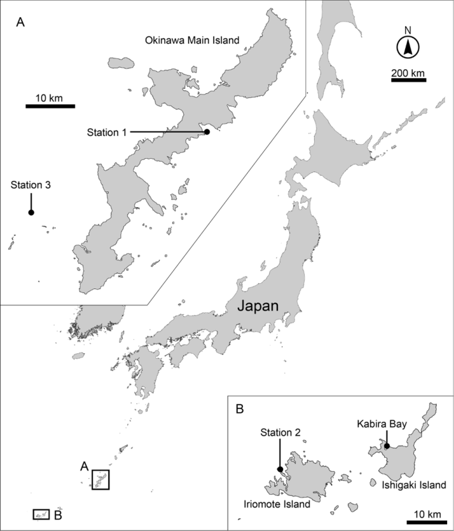
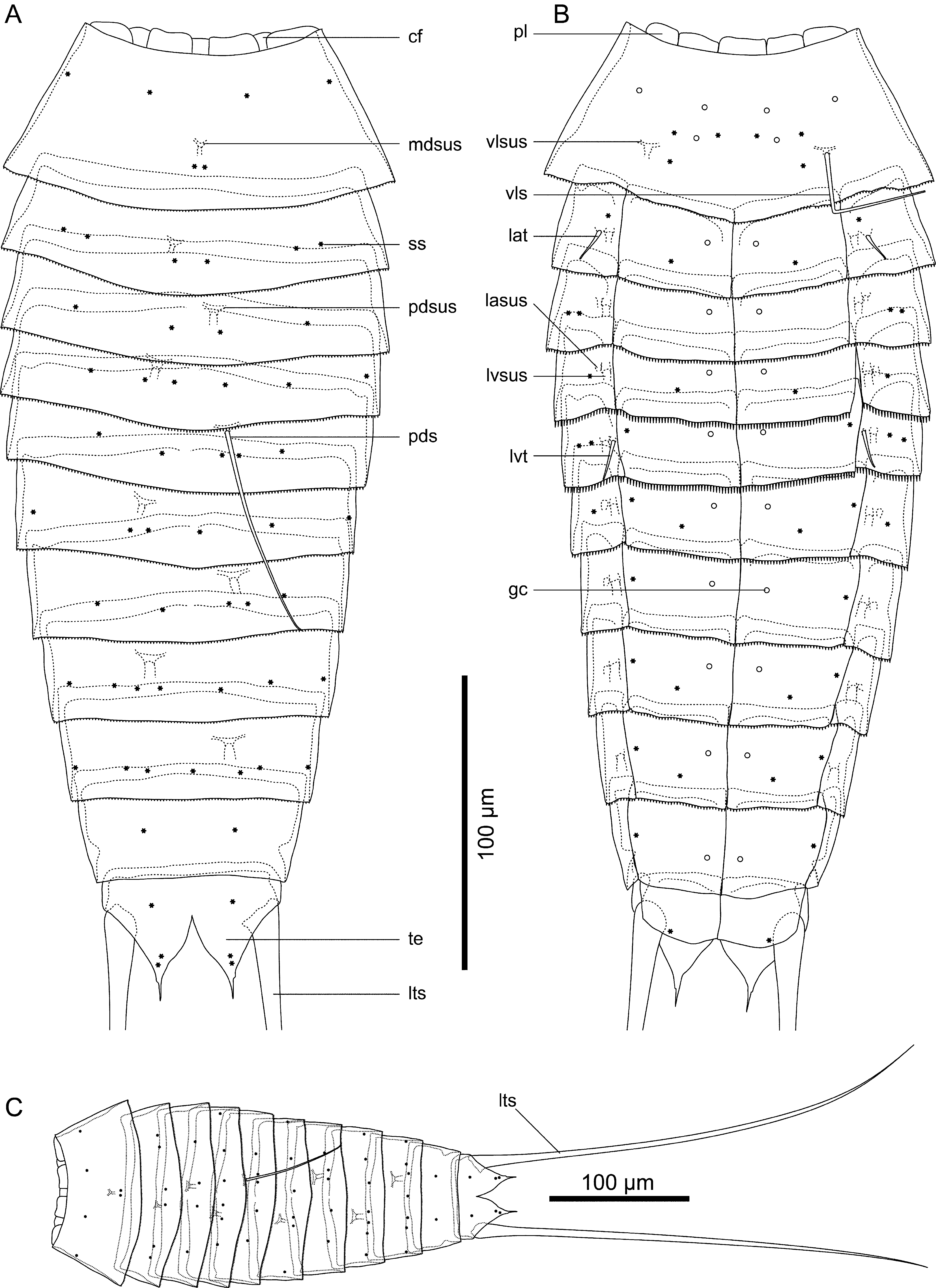
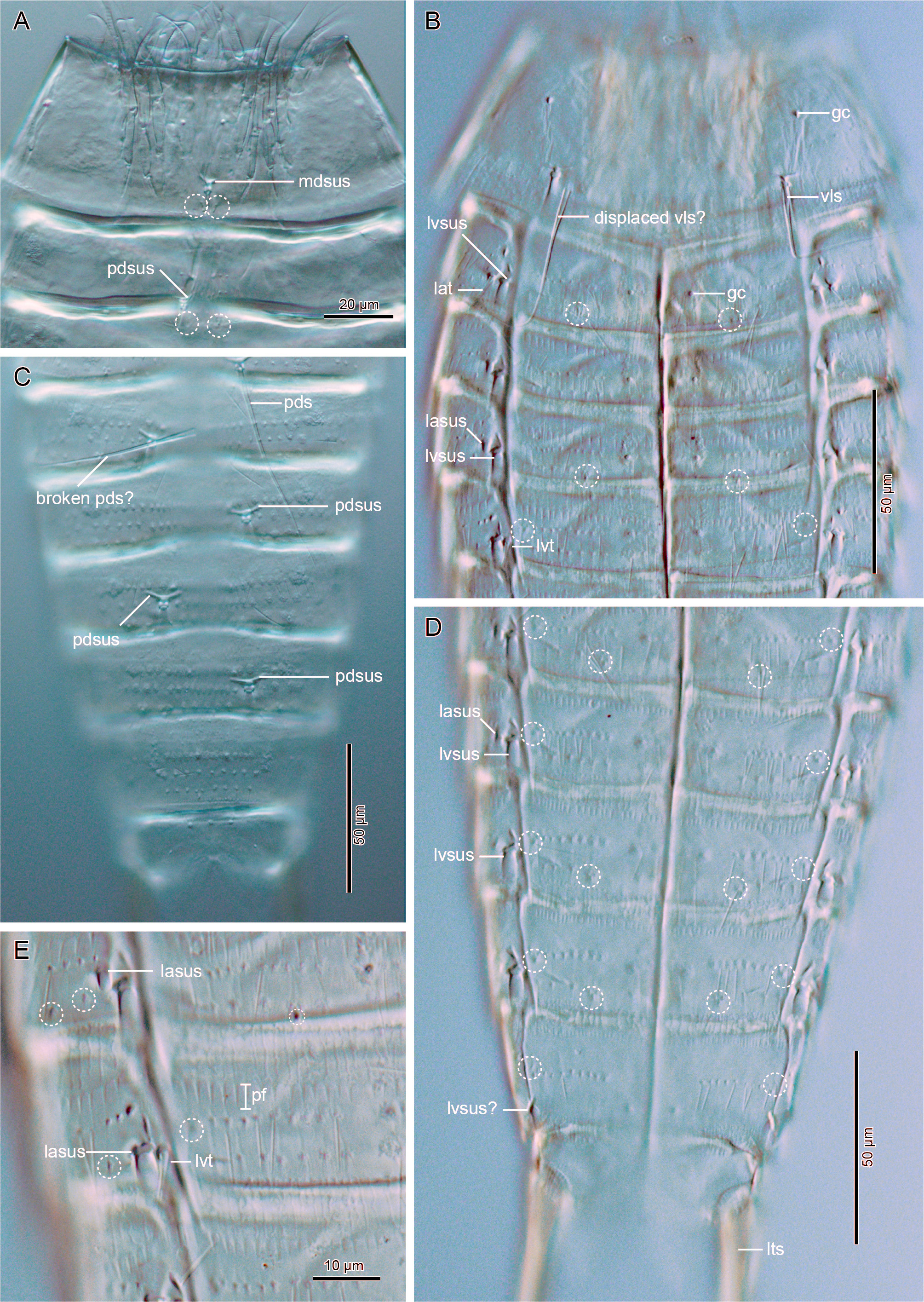
TABLE 5. Measurements for the holotype of Dracoderes toyoshioae sp. nov. (in micrometers). Abbreviations: (ac), acicular spine; LA, length of lateral accessory tubule; LTS, length of lateral terminal spine; LV, length of lateroventral tubule; MSW, maximum sternal width; S, segment length; SD, length of subdorsal acicular spine; SW, standard width; TL, trunk length; VL, length of ventrolateral acicular spine.
| Character Measurement | Character | Measurement |
|---|---|---|
| TL 331 | S7 | 36 |
| MSW-6 83 | S8 | 38 |
| MSW-6/TL 25.1% | S9 | 35 |
| SW-10 64 | S10 | 36 |
| SW-10/TL 19.3% | S11 | 44 |
| S1 57 | SD5 (ac) | 80 |
| S2 31 | VL1 (ac) | 53 |
| S3 31 | LA2 (tu) | 10 |
| S4 32 | LV5 (tu) | 14 |
| S5 33 | LTS | 341 |
| S6 34 | LTS/TL | 103% |
TABLE 6. Summary of the locations of cuticular structures, tubules, and spines in Dracoderes toyoshioae sp. nov. Abbreviations: ac, acicular spine; gc, glandular cell outlet; (l) unpaired and present on left side; LA, lateral accessory; LD, laterodorsal; lts, lateral terminal spine; LV, lateroventral; MD, middorsal; ML, midlateral; PD, paradorsal; (r), unpaired and present on right side; SD, subdorsal; SL, sublateral; ss, sensory spot; tu, tubule; VL, ventrolateral; VM, ventromedial.
| Position MD | PD | SD | LD | ML | SL | LA | LV | VL | VM |
|---|---|---|---|---|---|---|---|---|---|
| segment 1 ac? 2 3 | ss ac? (l), ss ac? (r), ss | ss | ss, ss ss | ss | ss, ss | tu ac/tu? | gc ss, ac/tu? ac/tu? | ac | ss, ss, gc, gc, ss ss, gc gc |
| 4 5 6 ss | ac? (l), ss ac (r), ss ac? (l), ss | ss (l) ss (r) ss (l) | ss ss | ss ss | ss ss, ss ss | ac/tu? ac/tu? ac/tu? | ac/tu? tu ac/tu? | ss ss | ss, gc gc ss, gc |
| 7 8 9 ss | ac? (r), ss ac? (l), ss ac? (r) | ss (r) ss (l) ss | ss ss ss | ss ss | ac/tu? ac/tu? | ac/tu? ac/tu? ac/tu? | ss ss ss | gc ss, gc ss, gc | |
| 10 11 | ss ss, ss, ss | ac/tu? lts | ss | ss |
No known copyright restrictions apply. See Agosti, D., Egloff, W., 2009. Taxonomic information exchange and copyright: the Plazi approach. BMC Research Notes 2009, 2:53 for further explanation.
|
Kingdom |
|
|
Phylum |
|
|
Class |
|
|
Order |
|
|
Family |
|
|
Genus |