
Valdasus henryi, Wolski & Chérot & Carpintero, 2020
|
publication ID |
https://doi.org/10.11646/zootaxa.4869.2.2 |
|
publication LSID |
lsid:zoobank.org:pub:DEF79389-5024-4064-9A71-837CBC28A89B |
|
DOI |
https://doi.org/10.5281/zenodo.4443139 |
|
persistent identifier |
https://treatment.plazi.org/id/D928FACA-8013-43F3-96A3-F2712815FCE6 |
|
taxon LSID |
lsid:zoobank.org:act:D928FACA-8013-43F3-96A3-F2712815FCE6 |
|
treatment provided by |
Plazi (2021-01-05 02:45:21, last updated 2024-11-27 06:03:56) |
|
scientific name |
Valdasus henryi |
| status |
sp. nov. |
Valdasus henryi n. sp.
( Figures 5 View FIGURES 1–10 , 15 View FIGURES 11–27 , 30–32 View FIGURES 28–33 , 48–51 View FIGURES 40–51 , 61–64, 67, 68 View FIGURES 61–70 )
Examined specimens: Holotype (♂): Ecuador: Napo: Tiputini Biodiversity Station , 216 m, 00º37′55″S; 76º08′39″W, 22 Oct. 1998, Erwin, T. L. et al. leg.; Insecticidal fogging of mostly bare green leaves, some with covering of lichenous or bryophytic plants, Lot 1976, Transect T-8 ( USNM); 1♀: the same data as for holotype ( USNM). GoogleMaps
Diagnosis: Recognized by the following set of features: antennal segment I shorter than vertex width, with three dark brown annulations; clypeus strongly convex basally ( Fig. 31 View FIGURES 28–33 ); mesepimeron punctate ( Fig. 31 View FIGURES 28–33 ); scutellum convex medially ( Figs 30, 31 View FIGURES 28–33 ); mesofemur and mesotibia with setae longer than leg segment diameter ( Fig. 31 View FIGURES 28–33 ); left paramere with apical process furnished with distinct process dorsobasally ( Fig. 49 View FIGURES 40–51 ); endosoma furnished with four sclerites ( Fig. 48 View FIGURES 40–51 ); sclerotized ring unpaired, encircling bursa copulatrix, its basal inner portions connected by broad sclerite (s) ( Fig. 61, 63, 64 View FIGURES 61–70 ); sclerotized areas of ventral wall of bursa copulatrix well-developed ( Fig. 62 View FIGURES 61–70 ).
Description. Measurements. Holotype (♂): Body. Total length in dorsal view (from vertex apex to membrane apex): 5.0, total width hemelytra median: 2.0. Head. Vertex width in dorsal view: 0.4, head width across eyes in dorsal view: 1.0. Antenna. Length segment: I 0.4, II 1.9, III and IV missing. Labium. Length segment: I 0.4, II 0.7, III 0.6, IV 0.2. Pronotum. Length (including collar): 0.8, posterior width (between humeral angles): 1.9. Scutellum. Length (including mesoscutum): 0.9, anterior width: 1.1. Cuneus. Length: 0.7, width: 0.4.
Paratype (1♀): Body. Total length in dorsal view (from vertex apex to membrane apex): 5.8, total width hemelytra median: 2.6. Head. Vertex width in dorsal view: 0.5, head width across eyes in dorsal view: 1.0. Antenna. Length segment: I 0.4, II 1.9 , III 3.0, IV missing. Labium. Length segment: I 0.5, II 0.8, III 0.5, IV 0.2. Pronotum. Length (including collar): 0.9, posterior width (between humeral angles): 2.2. Scutellum. Length (including mesoscutum): 1.1, anterior width: 1.2. Cuneus. Length: 0.7, width: 0.4 .
Male. COLORATION. Head. Fuscous tinged with yellow; first antennal segment yellow with three dark brown annulations situated basally, medially, and apically; second segment dark yellow on basal half and dark brown on apical half, apex with narrow, yellow annulation; third and fourth segments missing; labium dark brown ( Fig. 5 View FIGURES 1–10 , 15 View FIGURES 11–27 ). Thorax. Pronotum, mesoscutum and scutellum. Uniformly black; scutellum with small, yellow patch apically. Thoracic pleura. Black; evaporative area of metepisternum dark brown, ventral portion dark yellow. Hemelytron. Black; outer angle of exocorium and basal part of cuneus yellow; membrane fuscous, veins dark brown. Legs. Procoxa dark castaneus, narrowly yellow apically; meso- and metacoxae yellow; trochanters yellow; pro- and mesofemora dark castaneus; profemur with broad, yellow annulation subapically; mesofemur with two broad annulations: one situated basally and other subapically; pro- and mesotibiae dark castaneus with two broad annulations: on situated basally and other subapically ( Fig. 5 View FIGURES 1–10 ). Abdomen. Dark brown dorsally, laterally and ventrally. TEXTURE AND VESTITURE. Head. Covered with dense, long, erect setae; antennal segment I shiny, with several stiff setae; segment II shiny, covered with fine, sparse, semi recumbent setae, becoming longer on apical half ( Figs 15 View FIGURES 11–27 , 31 View FIGURES 28–33 ). Thorax. Pronotum. Collar covered several fine, erect setae. Mesoscutum and scutellum. Mesoscutum impunctate medially, punctate laterally. Thoracic pleura. Covered with long, erect setae; proepisternum impunctate; proepimeron deeply and densely punctate; mesepisternum rugose, mesepimeron rugopunctate; metepisternum rugose. Hemelytron. Covered with long, erect setae; margin of exocorium with well-developed spine near base. Legs. All coxae, protibia and pro- and mesofemora covered with long, erect and semi recumbent setae, not longer than diameter of these segments; mesotibiae covered with long, erect and semi recumbent setae, longer than diameter of leg segments, shorter on apical part ( Figs 5 View FIGURES 1–10 , 30, 31 View FIGURES 28–33 ). STRUCTURE. Head. Clypeus strongly convex near base; antennal segment I broadly widened medially, shorter than width of vertex; segment II weakly curved, thin, much thinner than first segment; labium reaching mesocoxae ( Figs 15 View FIGURES 11–27 , 30, 31 View FIGURES 28–33 ). Thorax. Scutellum. Medial part strongly convex. Thoracic pleura. Covered with long, erect setae; proepisternum impunctate; proepimeron deeply and densely punctate; mesepisternum rugose, mesepimeron rugopunctate; metepisternum rugose, peritreme oval, moderately raised evaporative areas. Hemelytron. Margin of exocorium with well-developed spine near base ( Figs 5 View FIGURES 1–10 , 30, 31 View FIGURES 28–33 ). Genitalia. Endosoma furnished with four sclerites, sclerotized, distal part of ductus seminis inside endosoma weakly developed, secondary gonopore rather narrow, with distinct microsculpture ( Fig. 48 View FIGURES 40–51 ); left paramere C-shaped, apical process furnished with distinct process dorsobasally ( Figs 49, 50 View FIGURES 40–51 ); right paramere sickle-shaped, apical process relatively long ( Figs 51 View FIGURES 40–51 ).
Female. Similar in coloration, structure, texture, and vestiture. Body slightly bigger and more oval. Genitalia. Bursa copulatrix ovoid; sclerotized ring unpaired, encircling bursa copulatrix, its basal inner portions connected by broad sclerite (s); lateral oviduct relatively long; sclerotized areas of ventral wall of bursa copulatrix well-developed (sv) ( Figs 61–64 View FIGURES 61–70 ).
Etymology. It gives us a great pleasure to dedicate this new species to Dr. Thomas J. Henry for his many outstanding contributions to the study of the Heteroptera .
Biology. Collected using insecticidal fogging of mostly bare green leaves, some with covering of lichenous or bryophytic plants.
Distribution. Ecuador (Napo).
Discussion. Valdasus henryi n. sp. can be separated from the other species of the genus by the following combination of characters: Size relatively reduced (total length about 5 mm), pronotum and embolium evenly coloured, dark, similar in color to clavus and corium, endosoma with four sclerites and the shape of the female genitalia

FIGURES 1–10. Habitus. 1–7.—Dorsal view; 8–10.—Lateral view. 1, 8.—Valdasus erebeus Distant (lectotype); 2, 9.—Valdasus favrei n. sp., 2: male paratype FC n° 5858, 9: male paratype, Ecuador USNM. 3.—Valdasus ferrerai n. sp. Male paratype FC n° 5965. 4.—Valdasus flavinotum (holotype); 5.—Valdasus henryi (holotype); 6, 10.—Valdasus schoenherri (female, Brazil); 7.—Valdasus stygius (lectotype). Scale = 2 mm.

FIGURES 11–27. Details of morphology. 11–17.—Head in anterior view. Scales = 0.5 mm. 18, 19.—Head in dorsal view. Scales = 0.5 mm. 20, 21.—First antennal segment. Scales = 0.1 mm. 22, 23.—Scutellum and mesoscutum. Scales 0.5 mm. 24, 25.—Hemelytron. Scales 0.5 mm. 26, 27.—Pronotum. Scales 0.5 mm. 11, 12, 18, 20, 22, 24, 26.—Valdasus favrei n. sp. Male holotype. 13, 19, 21, 23, 25, 27.—Valdasus ferrerai n. sp. Male holotype. 14.—Valdasus flavinotum (holotype); 15.—Valdasus henryi n. sp. (holotype); 16.—Valdasus schoenherri Stål, 1860 (♀, Brazil, USNM); 17.—Valdasus stygius (Distant) (lectotype).

FIGURES 28–33.Scanning electron micrographs. 28, 29, 31, 33.—Lateral views; 30.—Dorsal habitus; 32.—Metatarsus. 28, 29. Valdasus flavinotum. 30–32. Valdasus henryi. 33. Valdasus schoenherri.

FIGURES 40–51. Male genitalic structures. 40, 45, 48.—Endosoma (dorsal view). 41. Endosoma (ventral view) 42, 46, 49.— Left paramere (right lateral view). 43, 50. Left paramere apical process (dorsal view); 44, 47, 51.—Right paramere (left lateral view). 40–44.—Valdasus favrei. 45–47.—Valdasus flavinotum. 48–51.—Valdasus henryi. mw = medial weakly sclerotized part of ductus seminis distal part; ap = apical process; bp = basal process; lp = lateral outgrowth of left paramere apical process; pb = paramere body; sp1-sp4 = spicules of endosoma. Scale = 0.1.
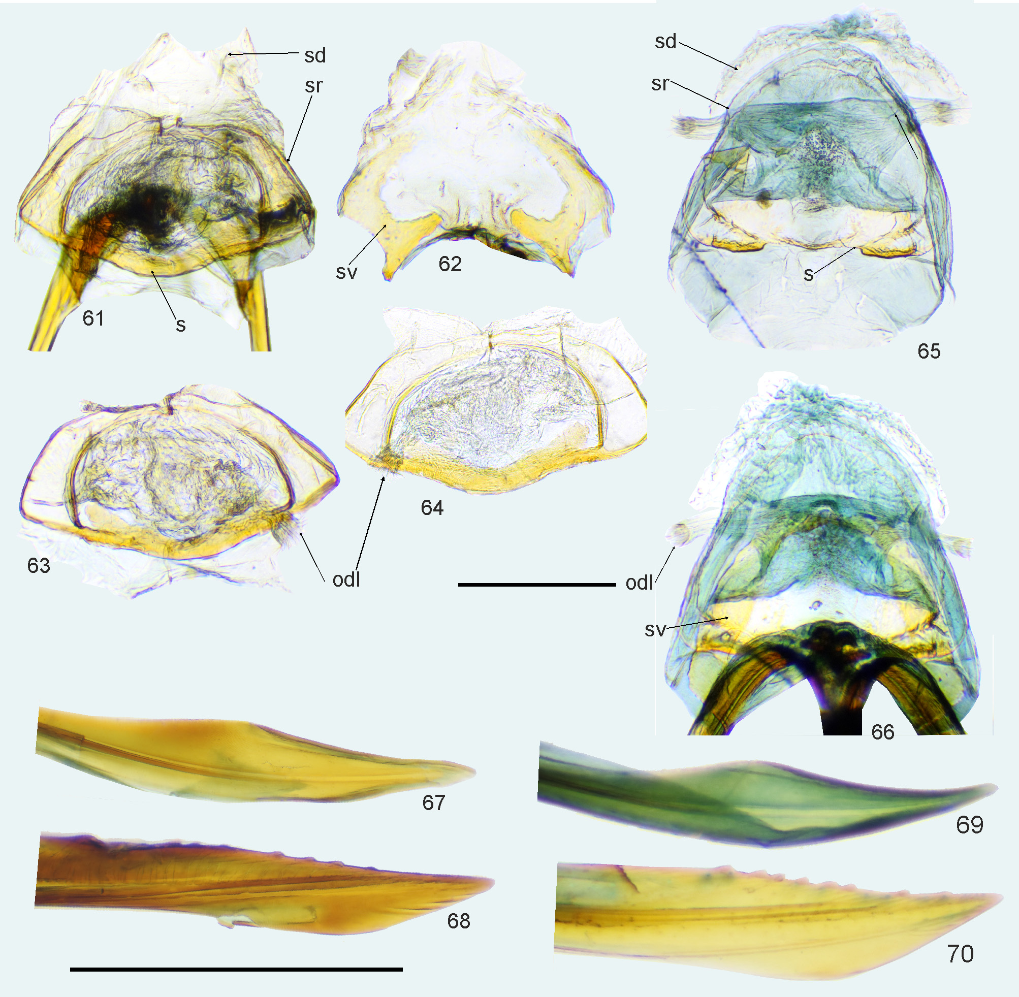
FIGURES 61–70. Female genitalic structures. 61, 63, 65. Bursa copulatrix (dorsal view), 61, 65: with ventral wall, 63: with ventral wall removed; 62, 64, 66. Bursa copulatrix (ventral view), 62: ventral wall of bursa copulatrix; 64: with ventral wall removed; 67, 68, 69, 70.—Ovipositor: 67, 69.—Gonapophysis I, 68, 70.—Gonapophysis II; odl = lateral oviduct; s = sclerite connecting posterior portions of sclerotized ring; sd = seminal depository; sg = spermathecal gland; sr = sclerotized ring; sv = sclerotization on bursa copulatrix ventral wall. 61–64, 67, 68. Valdasus henryi. 65, 66, 69, 70. Valdasus schoenherri. Scale 0.5 mm Short scale bar corresponds to bursa copulatrix; long scale bar corresponds to elements of ovipositor.
| T |
Tavera, Department of Geology and Geophysics |
| USNM |
Smithsonian Institution, National Museum of Natural History |
No known copyright restrictions apply. See Agosti, D., Egloff, W., 2009. Taxonomic information exchange and copyright: the Plazi approach. BMC Research Notes 2009, 2:53 for further explanation.
1 (by plazi, 2021-01-05 02:45:21)
2 (by ExternalLinkService, 2021-01-05 02:51:36)
3 (by ExternalLinkService, 2021-01-05 05:10:41)
4 (by tatiana, 2021-01-15 16:46:55)
5 (by ExternalLinkService, 2021-01-15 16:53:47)
6 (by ExternalLinkService, 2021-01-15 17:04:10)
7 (by ExternalLinkService, 2021-07-02 01:22:53)
8 (by ExternalLinkService, 2021-09-19 00:54:01)
9 (by plazi, 2023-11-01 10:10:16)