
Corydoras bethanae, Bentley & Grant & Tencatt, 2021
|
publication ID |
https://doi.org/10.11646/zootaxa.4948.2.2 |
|
publication LSID |
lsid:zoobank.org:pub:E36ADD5D-B461-4082-8B53-F21AD3D3750C |
|
DOI |
https://doi.org/10.5281/zenodo.4700568 |
|
persistent identifier |
https://treatment.plazi.org/id/03CC792B-FFCD-FFDA-FF65-F999FEADFE35 |
|
treatment provided by |
Plazi |
|
scientific name |
Corydoras bethanae |
| status |
sp. nov. |
Corydoras bethanae , new species
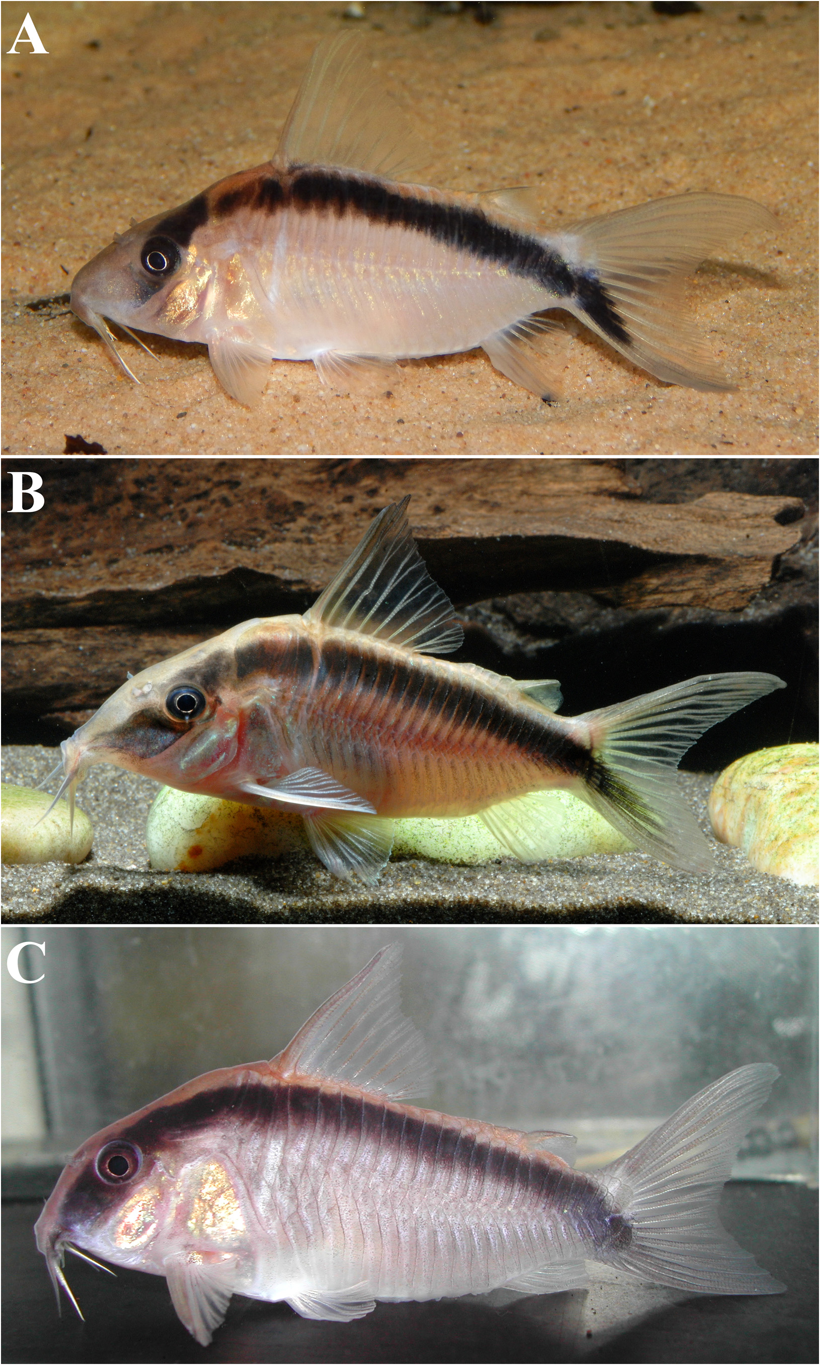
( Figs. 1 View FIGURE 1 , 2A View FIGURE 2 , 3A View FIGURE 3 , 4-6 View FIGURE 4 View FIGURE 5 View FIGURE 6 , Table 1 View TABLE 1 )
Corydoras View in CoL sp. CW006:— Alexandrou et al., 2011: 85–86 (“ C. sp. CW6”; phylogeny; as member of the lineage 8; mimicry).— Tencatt et al., 2019: 459–460 (comparison with C. arcuatus View in CoL ; photo in life (fig. 8) of the specimen designated as holotype herein).
Holotype. MUSM 69403 , female, 51.2 mm SL, Peru, Department of Loreto, Requena Province, Soplin District , río Blanco (close to the confluence with the río Tapiche ), aquarium specimen imported in 2017 by Aquarium Glaser GmbH, Germany.
Paratypes. BMNH 2017.5.25.1-21, 21, 43.7–57.3 mm SL, same data as holotype ; BMNH 2018.7.5.4-5, 2, 56.0–56.0 mm SL; MNRJ 52311 View Materials , 1 View Materials , 53.1 mm SL ; ZUFMS 6470 , 1 of 3, 49.9 mm SL, 2 CS of 3, 55.0– 56.9 mm SL, Peru, Department of Loreto, said to be from the río Ucayali basin, aquarium specimens imported in 2018 by Maidenhead Aquatics , Wigan, UK .
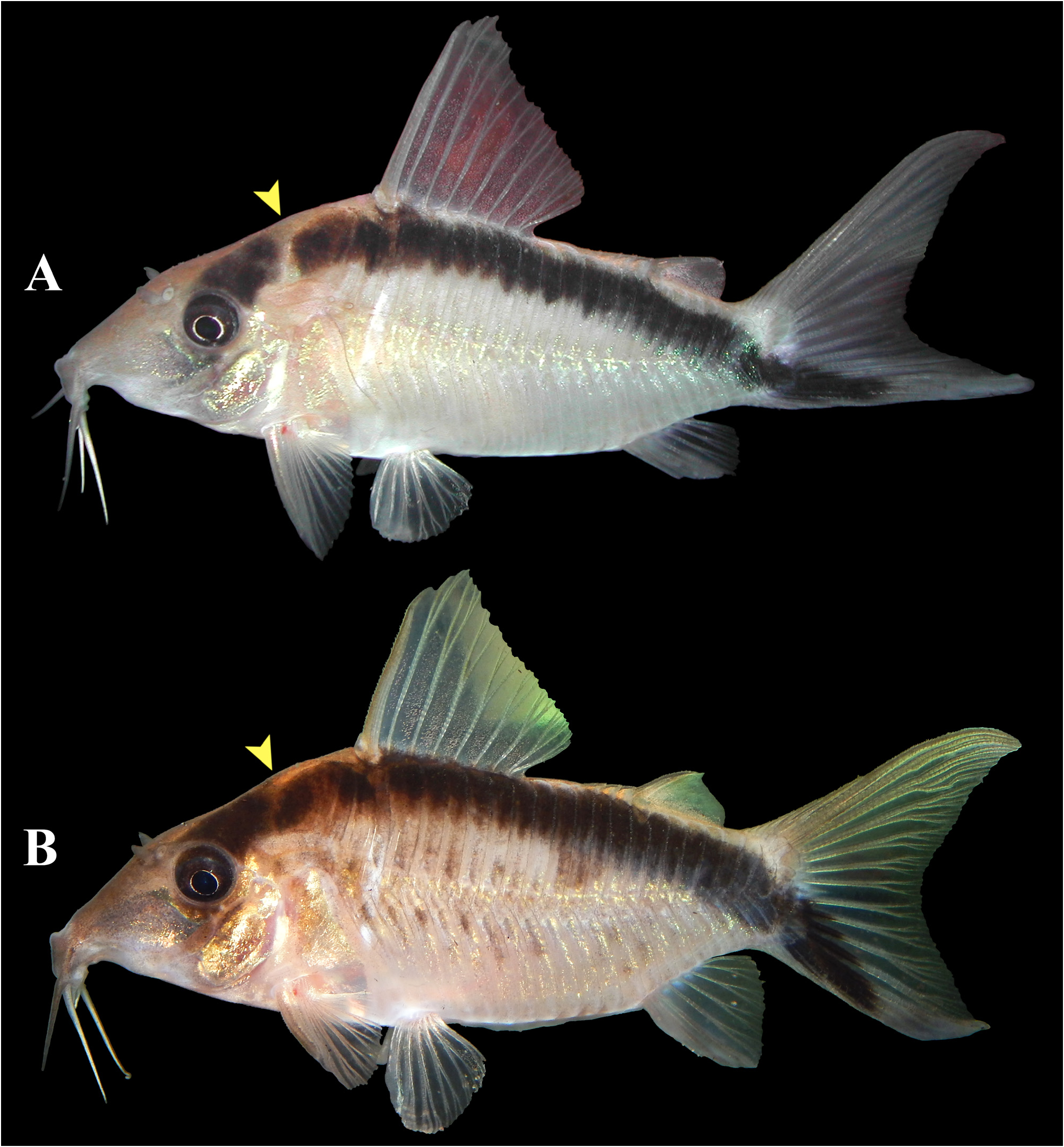
Diagnosis. Corydoras bethanae can be distinguished from its congeners, except for those belonging to the lineage 8 sensu Alexandrou et al. (2011), by having the posterior margin of dorsal-fin spine with laminar serrations directed towards the origin of the spine ( vs. absence of such serration pattern); from the species within lineage 8, except for C. arcuatus , it differs by the presence of a long, wide, arched, and continuous black stripe, which runs parallel to the dorsal profile of the body, extending at least from the region below anterior origin of dorsal fin to the anterior half of the ventral caudal-fin lobe ( Fig. 2A View FIGURE 2 ) ( vs. absence of a similar stripe in remaining lineage 8 congeners); from C. arcuatus ( Fig. 2B View FIGURE 2 ) differs by the color pattern of the head (presence of a black stripe transversally crossing the eye, forming the typical mask-like blotch; mask clearly not fused to arched stripe in most specimens; some specimens with mask separated from arched stripe by a thin line around the suture between neurocranium (on the region composed by the posteroventral margin of parieto-supraoccipital plus the posterodorsal margin of the compound pterotic) and first dorsolateral body plate in C. bethanae ( Figs. 3A View FIGURE 3 and 6 View FIGURE 6 ) vs. mask-like blotch absent; continuous black stripe extending from area at corner of mouth to anteroventral margin of orbit and starting again from posterodorsal margin of orbit; running close and in parallel to dorsum from this point to anterior margin of first dorsolateral body plate, merging with trunk section of the arched stripe in C. arcuatus ( Fig. 3B View FIGURE 3 ), by having a longer snout (53.4–67.4% of HL vs. 42.1–51.5), a smaller HL (30.4–34.7% of SL vs. 41.1–51.4), and a larger least interorbital distance (42.6–49.2% of HL vs. 28.7–36.0). Additionally, the new species differs from C. gracilis Nijssen & Isbr ̹cker, 1976 and C. narcissus Nijssen & Isbr ̹cker, 1980a by having laminar serrations on posterior margin of pectoral-fin spine ( vs. conical); from C. gracilis and C. urucu Britto, Wosiacki & Montag, 2009 by having pointed snout, presenting a long mesethmoid, with anterior tip larger than 50% of the entire length of the bone ( vs. rounded snout, presenting a short mesethmoid, with anterior tip smaller than 50% of the entire bone length); from C. granti Tencatt, Lima & Britto, 2019 ( Fig. 2C View FIGURE 2 ) by having ventral surface of trunk covered by small, non-coalescent platelets ( vs. ventral surface of trunk entirely or partially covered by relatively large and coalescent platelets).
Description. Morphometric data are presented in Table 1 View TABLE 1 . Head laterally compressed with convex dorsal profile, roughly triangular in dorsal view. Snout conical in dorsal view, ranging from pointed to conspicuously pointed. Head profile in lateral view slightly concave from tip of snout to anterior nares, ascending slightly concave from this point to anterior portion of parieto-supraoccipital process; slightly convex or nearly straight from this point to dorsal-fin origin. Profile slightly convex along dorsal-fin base. Postdorsal-fin body profile slightly concave to adiposefin spine, concave from this point to caudal-fin base. Ventral profile of body nearly straight or slightly convex from isthmus to pectoral girdle, and slightly convex from this point until pelvic girdle. Profile nearly straight or slightly convex from pelvic girdle to base of first anal-fin ray, ascending abruptly concave until caudal-fin base. Body roughly elliptical in cross section at pectoral girdle, gradually becoming more compressed toward caudal fin.
Eye rounded, located dorsolaterally on head. Orbit delimited anteriorly by lateral ethmoid, anterodorsally by frontal, posterodorsally by sphenotic, posteriorly by infraorbital 2, and ventrally by infraorbital 1. Anterior and posterior nares close to each other, only separated by flap of skin. Anterior naris tubular. Posterior naris close to anterodorsal margin of orbit, separated from it by distance slightly larger than naris diameter. Mouth small, subterminal, width slightly smaller than bony orbit diameter. Maxillary barbel long in size, reaching anteroventral limit of gill opening. Outer mental barbel slightly longer than maxillary barbel. Inner mental barbel fleshy, base of each counterpart slightly separated from each other. Small rounded papillae covering entire surface of all barbels, upper and lower lips, snout and isthmus.
Mesethmoid long with anterior tip well developed, larger than 50% of bone length (see Britto, 2003: 123, character 1, state 0; fig. 1A); posterior portion relatively narrow, entirely covered by thin layer of skin. Middle portion of mesethmoid with strongly well-developed lateroventral process; region of process with width clearly larger than width of posterior portion of mesethmoid. Nasal capsule delimited anteriorly and ventrally by lateral ethmoid, and posteriorly and dorsally by frontal. Nasal slender, laterally curved, inner margin laminar; mesial border contacting only frontal. Lateral ethmoid conspicuously expanded anteriorly, with anterodorsal expansion contacting frontal and mesethmoid, and anteroventral expansion connected to lateroventral process of mesethmoid. Frontal elongated, ranging from narrow, width less than half of entire length, to relatively wide, with width equal to half of entire length; anterior projection long, size equal to or slightly larger than nasal length. Frontal fontanel large, slender, and somewhat ellipsoid; posterior tip extension entering anterior margin of parieto-supraoccipital. Sphenotic somewhat trapezoid, contacting parieto-supraoccipital dorsally, compound pterotic posteriorly, second infraorbital ventrally and frontal anteriorly ( Fig. 4 View FIGURE 4 ). Compound pterotic roughly pipe-shaped, with posteriormost portion contacting first lateral-line ossicle, posteroventral margin contacting cleithrum, and anteroventral margin contacting opercle and infraorbital 2, and posterior expansion almost entirely covering lateral opening of swimbladder capsule, leaving slender area on its dorsal margin covered only by thick layer of skin ( Fig. 4 View FIGURE 4 ). Parieto-supraoccipital wide, posterior process long and contacting nuchal plate; region of contact between posterior process and nuchal plate covered by thick layer of skin.
Two laminar infraorbitals with minute odontodes. Infraorbital 1 large, ventral laminar expansion moderately developed, anterior portion with well-developed laminar expansion, reaching to or slightly surpassing anterior margin of nasal capsule; inner laminar expansion strongly reduced ( Fig. 4 View FIGURE 4 ). Infraorbital 2 small, widened, with posterior laminar expansion well developed; posteroventral margin contacting posterodorsal ridge of hyomandibula, posterodorsal edge contacting sphenotic and compound pterotic; inner laminar expansion strongly reduced ( Fig. 4 View FIGURE 4 ). Posterodorsal ridge of hyomandibula close to its articulation with opercle relatively slender, exposed, and bearing small odontodes. Dorsal ridge of hyomandibula between compound pterotic and opercle covered by thick layer of skin; covered by posterodorsal edge of infraorbital 2 in some specimens. Interopercle partially exposed, with anterior portion covered by thick skin layer; subtriangular, anterior projection well-developed. Preopercle elongated, ranging from relatively slender to widened; minute odontodes sparse on external surface. Opercle dorsoventrally elongated, width smaller than half of entire length; free margin slightly convex, without serrations and covered by small odontodes.
Four branchiostegal rays decreasing in size posteriorly. Hypobranchial 2 somewhat triangular, tip ossified and directed towards anterior portion, posterior margin cartilaginous; ossified portion conspicuously well developed, its size about three times cartilaginous portion. Five ceratobranchials with expansions increasing posteriorly; ceratobranchial 1 with small process on anterior margin of mesial portion; ceratobranchial 3 with continuous laminar expansion on postero-lateral margin; ceratobranchial 5 toothed on posterodorsal surface, with 34 to 42 (2) teeth aligned in one row. Four epibranchials with similar size; epibranchial 2 slightly larger than others, with small pointed process on laminar expansion of posterior margin; epibranchial 3 with roughly triangular or somewhat trapezoid uncinate process on laminar expansion of posterior margin; uncinate process curved mesially in left side of paratype ZUFMS 6470, 56.9 mm SL. Two wide pharyngobranchials (3 and 4); pharyngobranchial 3 with roughly triangular laminar expansion on posterior margin; variably notched expansion in some specimens. Upper tooth plate oval, 34 to 47 (2) teeth aligned in two rows on posteroventral surface.
Lateral-line canal reaching cephalic laterosensory system through compound pterotic, branching twice before reaching sphenotic: pterotic branch, with single pore, preoperculomandibular branch conspicuously reduced, with single pore opening at postotic main canal; postotic main canal becoming widened just posterior to pterotic branch. Sensory canal continuing through compound pterotic, reaching sphenotic as temporal canal, which splits into two branches: one branch giving rise to infraorbital canal, other branch connecting to frontal through supraorbital canal, both with single pore. Supraorbital canal branched, running through nasal bone. Epiphyseal branch conspicuously reduced; pore opening close to supraorbital main canal, directed towards frontal fontanel. Nasal canal with two or three openings, first on posterior edge, second, when present, on posterolateral portion and generally fused with first pore, and third on anterior edge. Infraorbital canal running through entire infraorbital 2, extending to infraorbital 1 and opening into two pores. Preoperculomandibular branch giving rise to preoperculo-mandibular canal, which runs through entire preopercle with three openings, leading to pores 3, 4, and 5, respectively.
Dorsal fin subtriangular, located just posterior to second or third dorsolateral body plate. Dorsal-fin rays II,7 (1), II,8* (19), I,9 (4), posterior margin of dorsal-fin spine with 18 to 19 poorly to moderately-developed laminar serrations; most serrations directed towards origin of spine; some serrations variably perpendicularly directed; serrations absent close to origin of spine; small odontodes on anterior and lateral surfaces of spine ( Fig. 5A View FIGURE 5 ). Nuchal plate well developed, almost entirely exposed, with minute odontodes. Spinelet short, spine moderately developed, adpressed distal tip slightly surpassing posterior origin of dorsal-fin base, and anterior margin with small odontodes. Pectoral fin roughly triangular, its origin just posterior to gill opening. Pectoral-fin rays I,6*(1), I,7 (2), I,8 (4), I,9 (15), I,9,i (1), posterior margin of pectoral spine with 24 to 28 poorly- to moderately-developed laminar serrations along almost its entire length, absent close to origin of spine; most serrations directed towards pectoral-fin origin; some serrations perpendicularly directed or directed towards tip of spine; small odontodes on anterior, dorsal and ventral surfaces of spine ( Fig. 5B View FIGURE 5 ). Anteroventral portion of cleithrum exposed; posterolateral portion of scapulocoracoid moderately developed, exposed, with anterior portion strongly expanded anteriorly, almost reaching to or contacting anteroventral portion of cleithrum; exposed areas bearing small odontodes; Opening of axillary gland sensu Kiehl et al. (2006) located just posterior to pectoral-fin spine base. Pelvic fin oblong, located just below first ventrolateral body plate, and at vertical through region between first and second branched dorsal-fin ray. Pelvic-fin rays i,5* (24). Adipose fin roughly triangular, separated from base of last dorsal-fin ray by generally six dorsolateral body plates. Anal fin subtriangular, located just posterior to 12 th or 13 th ventrolateral body plates, and at vertical through adiposefin spine base. Anal-fin rays ii,6* (22). Caudal fin bilobed, with dorsal lobe slightly larger than ventral lobe; lobes with similar size in some specimens. Caudal-fin rays i,10,i (1), i,11,i (2), i,12,i* (20), i,13,i (1), generally five or six dorsal and ventral procurrent rays.
Three laterosensory canals on trunk. First ossicle tubular, second ossicle laminar, and third lateral-line canal encased in third dorsolateral body plate. Body plates with minute odontodes scattered over exposed area, conspicuous line of odontodes confined on posterior margins. Dorsolateral body plates 24* (22), 25 (2). Ventrolateral body plates 21 (10), 22* (13), 23 (1). Dorsolateral body plates along dorsal-fin base 6 (2), 7* (22). Dorsolateral body plates between adipose- and caudal-fin 7 (3), 8* (18), 9 (1). Preadipose platelets 4* (3). Small platelets covering base of caudal-fin rays. Small platelets disposed dorsally and ventrally between junctions of lateral plates on posterior portion of caudal peduncle. Anterior margin of orbit, above posterior portion of lateral ethmoid, variably with small, irregular platelets bearing odontodes. Ventral surface of trunk densely covered by small irregular platelets bearing odontodes.
Vertebral count 22 (2). Ribs 7 (1), 8 (1); first pair conspicuously large, its middle portion closely connected to first ventrolateral body plate; its tip connected to anterior external process of basipterygium. Complex vertebra moderately developed.
Color in alcohol. Overall color light whitish, grey, light beige, or tan, with area above dark arched line and mask being slightly darker, sometimes orange-brown; melanophores along, above and below the midline of the lateral scutes, being darker and larger in some specimens; snout area anterior to dark eye mask and opercular region dark grey or diffuse black. Eye mask and body stripe (as set out in diagnosis) black; basal portion of six outermost segmented rays of ventral lobe of caudal fin black, and innermost procurrent ray of same lobe black or dark grey, forming extension of body stripe; some specimens with one dark or black pigmented transverse, diffuse, vertical band in caudal fin. Pale area on middle portion of dorsolateral surface of cleithrum corresponds with axillary gland sensu Kiehl et al. (2006) underneath; holotype similarly colored area on the ventral margin of the base of the adipose-fin spine.
Color in life. See Figs 2A View FIGURE 2 , and 6 View FIGURE 6 of the holotype and two paratypes, respectively, whilst alive for color in life. Overall color same as in alcohol; third dorsolateral body scute usually whitish; green or gold to copper hue on lateral scutes above and below midline, opercular area, cleithrum, and underneath and behind the eye; snout area anterior to dark eye mask is usually light grey. Eye mask, body, and caudal fins dark markings as per preserved specimens. Whitish narrow transversal area from ventral half of fourth dorsolateral body plate, passing through dorsal half of first ventrolateral body plate, and reaching to posterolateral margin of cleithrum. Other fin membranes and rays hyaline, with some of the dorsal and adipose fin membranes with whitish chromatophores (colors in Fig. 6 View FIGURE 6 are background colors). Color of fry can be determined from the spawning in the account by Petersson (2020).
Sexual dimorphism. In addition to a tubular genital papilla (see Spadella et al., 2017), males have more pointed and elongated pelvic fins; the dark arched body stripe can appear proportionately wider; and tend to have the lateral scutes with darker and larger markings along, above and below the midline of flank.
Geographical distribution. Corydoras bethanae is so far only known from the río Blanco system ( río Ucayali basin), Soplin District, Requena Province, Department of Loreto, Peru ( Fig. 7 View FIGURE 7 ). Based on the findings of Corahua et al. (2015) it is likely that it is found in the streams that drain into the main river .
Natural history notes. Corydoras bethanae may be found in blackwater streams that drain into the predominantly whitewater río Blanco ( Fig. 8A View FIGURE 8 ), although the UK aquarist Mark Breeze (pers. comm.) states that in the dry season he found small clearwater tributary streams near the confluence with the río Tapiche ( Fig. 8B View FIGURE 8 ). Those streams had a sand substrate with hardly any leaf litter and just overhanging terrestrial plants. Corahua et al. (2015) found several Corydoras species in shallow, lotic blackwater streams or flooded forests that flowed into the middle reaches of the río Blanco, that were low in pH (5.4–5.5), electrical conductivity (4.8–8.9 μs/cm), temperature of 25 degrees Centigrade, with abundant organic detritus, and white sand. A site in the lower region, closer to the type locality of Corydoras bethanae , was the same except it was lentic, the pH ranged from 4.1 – 5.6, and the electrical conductivity was 29.5–41.0 μs/cm. Corydoras granti was found by Corhua et al. (2015) in this location (misidentified as Corydoras arcuatus ); another species with an arched dark body band. Corahua et al. (2015) list at least eight species from the río Blanco, and in addition to those, information from fish collectors indicates that the following Corydoras can also be found in the río Blanco system: Corydoras coriatae Burgess, 1997 , C138 (which is superficially similar to Corydoras bethanae ), and CW073.
Grant (2020) discussed curious behavior exhibited by Corydoras bethanae when in the aquarium. All corydoradins appear to be able to maintain their position in the water column, above the substrate, by using a combination of body movement and fin movement. They usually to do this when maneuvering while performing definite activities e.g., avoiding another fish or predator, feeding, spawning etc. In the aquarium C. bethanae tend to hover regularly and for long periods of time, apparently not related to any particular activity; a behavior that has not been reported for other lineage 8 Corydoras . The pale transversal bar from fourth dorsolateral body plate to posterolateral margin of cleithrum corresponds with a large ligament attached dorsally to the transverse process of dorsal pterygiophore and ventrally to the posterior laminar expansion of the first rib, which itself is connected to the anterior external process of basipterygium. Such condition may have some effect on the ability to hover in the water column, especially by offering a strong support to pelvic fins, which along with pectoral fins seem to be the main fins responsible for keeping them lifting above the substrate. Videos of the holotype and some BMNH paratypes hovering can be seen at: https://www.youtube.com/watch?v=hcg45PlwHjs and https://www.youtube.com/watch?v=v6p1hUdls54
There is a single record of successful spawning of this species, in which the water parameters were pH 6, Total Dissolved Solids (TDS) 82ppm, and temperature 22.4°C ( Petersson, 2020).
Etymology. Named in honor of Bethan Grant, daughter of SG, who, like her brother, has battled health issues all her life.


| CS |
Musee des Dinosaures d'Esperaza (Aude) |
No known copyright restrictions apply. See Agosti, D., Egloff, W., 2009. Taxonomic information exchange and copyright: the Plazi approach. BMC Research Notes 2009, 2:53 for further explanation.
|
Kingdom |
|
|
Phylum |
|
|
Class |
|
|
Order |
|
|
Family |
|
|
Genus |
Corydoras bethanae
| Bentley, Rebecca Frances, Grant, Steven & Tencatt, Luiz Fernando Caserta 2021 |
C. arcuatus
| Elwin 1938 |
Corydoras
| Lacepede 1803 |