
Menestheus cuneatus Distant, 1899
|
publication ID |
https://doi.org/10.11646/zootaxa.4407.2.8 |
|
publication LSID |
lsid:zoobank.org:pub:1B47F51E-C88F-4637-8092-92B06A7408BB |
|
DOI |
https://doi.org/10.5281/zenodo.5980760 |
|
persistent identifier |
https://treatment.plazi.org/id/03CC041D-FFED-FFEC-66CA-F0782F50DDDB |
|
treatment provided by |
Plazi (2018-04-11 07:37:27, last updated 2024-11-28 08:42:49) |
|
scientific name |
Menestheus cuneatus Distant, 1899 |
| status |
|
Menestheus cuneatus Distant, 1899
Menestheus cuneatus Distant, 1899: 431 (original description).
Menestheus cuneatus: Bergroth (1908): 157 (catalog); Kirkaldy (1909): 181 (catalog); Distant (1910): 469 (redescription); Gross (1975): 177 (redescription); Cassis & Gross (2001): 451 (catalog, distribution).
Type material. ST(S) (♀), Australia, Peak Downs , BMNH.
Material examined. Australia, QLD, Millestone, 24-XII-1964, leg. G. Monteith, 2♀♀, 2♂♂ ; Australia, N. QLD. Kowanyama , 7-I-1977, leg. D. L. Hancock, 1 ♀ [ QUMC] Australia, Bogom, 1♀ [ QUMC] ; Australia, QLD, Bowen , leg. A. Simson, 1♂ [ SAMC] ; Australia , rest illegible, 1♂ [ SAMC] .
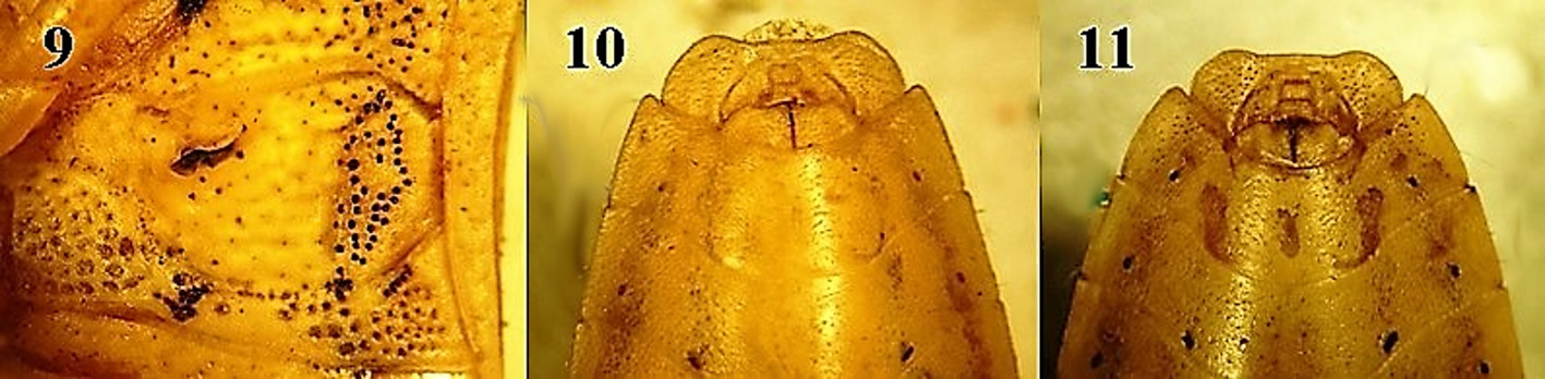
Diagnosis. Body with dark punctures, contrasting with general coloration, body shape elongate, ovate (2.53 times longer than wide) ( Fig. 2 View FIGURES 1–2. 1 ). Head wide, elongate (1.33 times longer than wide) ( Fig. 4 View FIGURES 3–8. 3 ). Basal foveae of scutellum large, well developed. Spiracles black. Emargination of male pygophore wide ( Fig. 8 View FIGURES 3–8. 3 ), male parameres, each with apical lobe elongate, sharp ( Fig. 15 View FIGURES12–16. 12 ). Female paratergites VIII and IX small, elongate ( Fig. 11 View FIGURES 9–11. 9 ).
Distribution. Australia: Queensland ( Gross, 1975; Cassis & Gross, 2002; this contribution), Victoria ( Gross, 1975). Records given by Cassis & Gross (2002) for M. cuneatus from Daly and McArthur rivers and Mornington Island probably belong to M. mcphersoni .
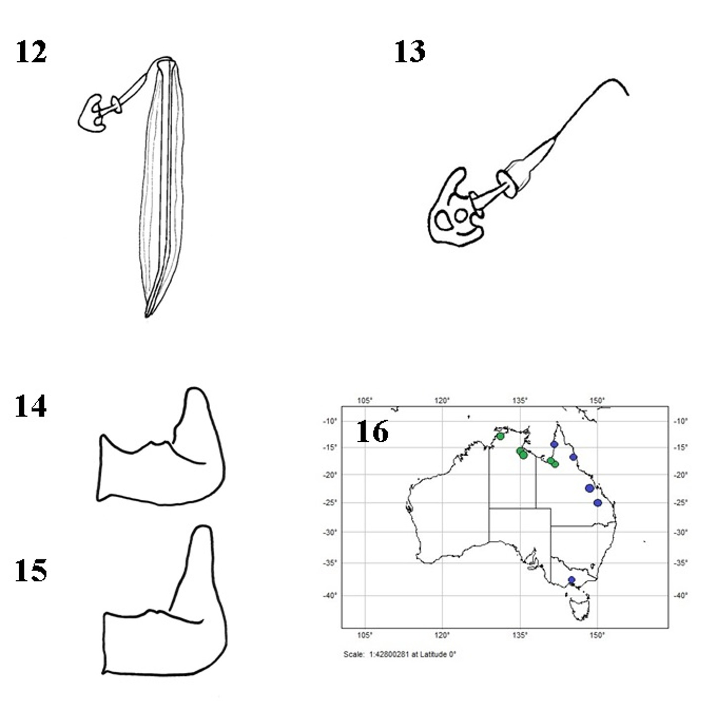
FIGURES12–16. 12. Menestheus mcphersoni sp.nov., spermatheca; 13. M.mcphersoni, spermatheca, pump enlarged;14. M. mcphersoni, left paramere; 15. Menestheus cuneatus Distant,1899, left paramere; 16.Distribution ofthe species of Menestheus (M. mcphersoni in green, M.cuneatus in blue).
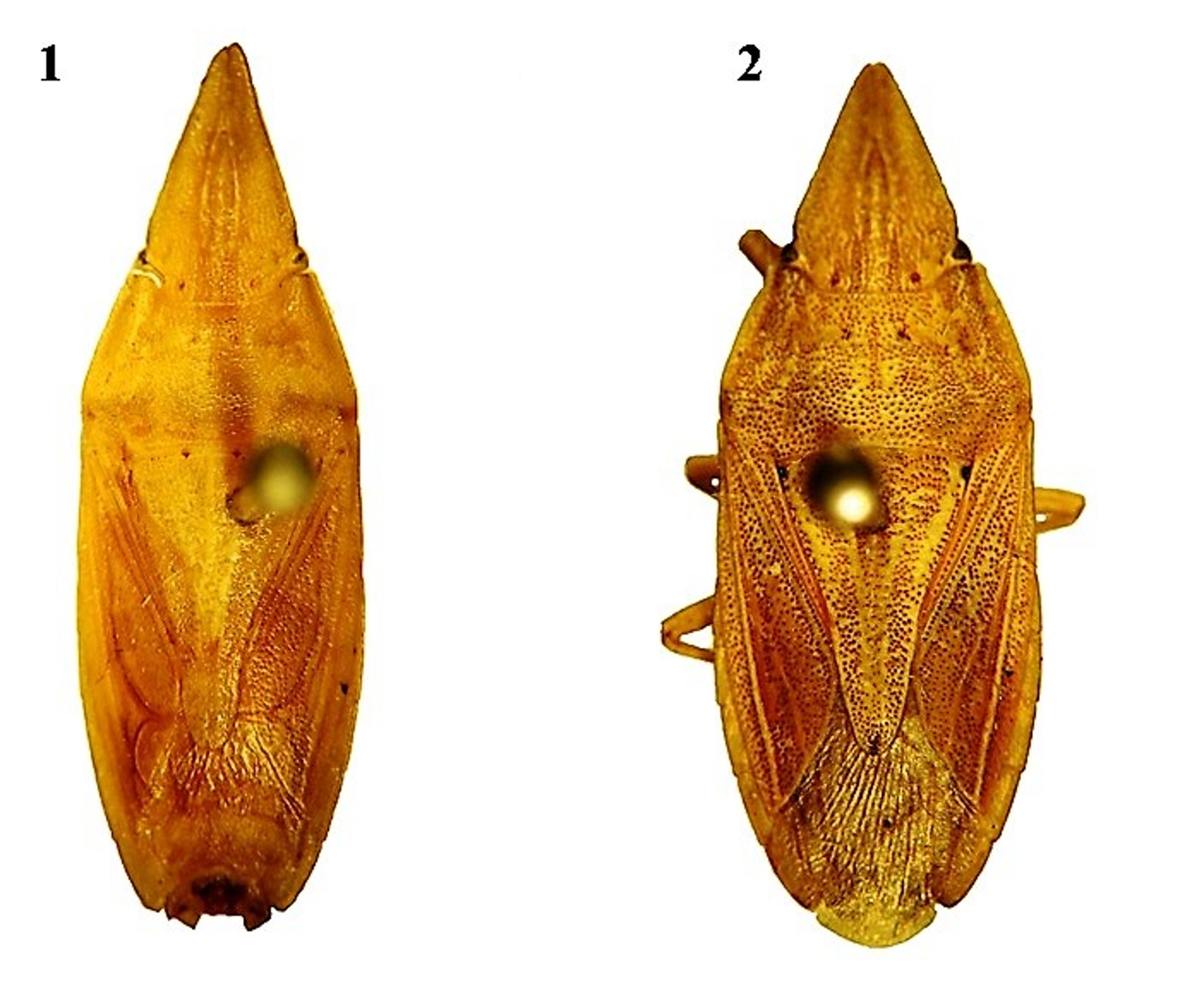
FIGURES 1–2. 1. Menestheus mcphersoni sp. nov., male holotype, habitus; 2. Menestheus cuneatus Distant, 1899, male, habitus.
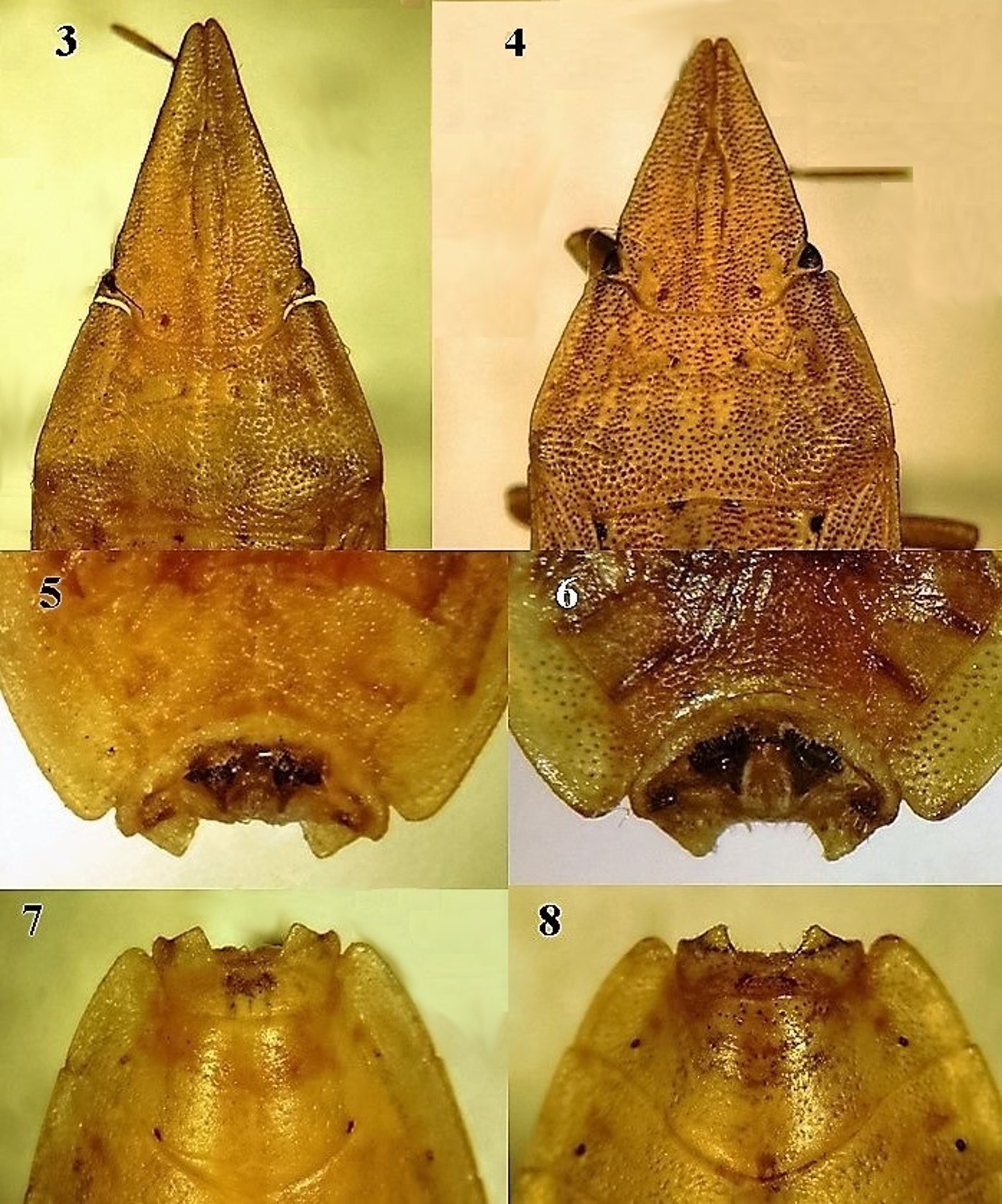
FIGURES 3–8. 3. Menestheus mcphersoni sp. nov. head and pronotum; 4. Menestheus cuneatus Distant, 1899, head and pronotum; 5. M. mcphersoni, pygophore, dorsal view; 6. M. cuneatus, pygophore, dorsal view; 7. M. mcphersoni, pygophore, ventral view; 8. M.cuneatus, pygophore, ventral view.
| SAMC |
Iziko Museums of Cape Town |
No known copyright restrictions apply. See Agosti, D., Egloff, W., 2009. Taxonomic information exchange and copyright: the Plazi approach. BMC Research Notes 2009, 2:53 for further explanation.
|
Kingdom |
|
|
Phylum |
|
|
Class |
|
|
Order |
|
|
Family |
|
|
Genus |
Menestheus cuneatus Distant, 1899
| Faúndez, Eduardo I. & Rider, David A. 2018 |
Menestheus cuneatus
| Distant, 1899 : 431 |
Menestheus cuneatus: Bergroth (1908) : 157
| Bergroth (1908) : 157 |
| Kirkaldy (1909) : 181 |
| Distant (1910) : 469 |
| Gross (1975) : 177 |