
Yanhaoia wynnei ( Reed, 1915 )
|
publication ID |
https://doi.org/ 10.11646/zootaxa.5162.4.1 |
|
publication LSID |
lsid:zoobank.org:pub:DD2279FA-E8F1-4951-A5CA-91082E875580 |
|
DOI |
https://doi.org/10.5281/zenodo.6810372 |
|
persistent identifier |
https://treatment.plazi.org/id/03CB8784-6E7D-1D21-FF3A-C2954BF3F31D |
|
treatment provided by |
Plazi |
|
scientific name |
Yanhaoia wynnei ( Reed, 1915 ) |
| status |
|
Figs 13.4-9 View FIGURE 13
1915 Phacops (Pterygometopus) dagon ; Reed, p. 53–54, pl. 9, fig. 3.
1915 Phacops (Pterygometopus) dagon var. wynnei ; Reed, p. 54–55, pl. 9, figs 7–15.
Material. Lectotype (selected herein): partial exoskeleton from Hwe Mawng, Fig. 13.8 View FIGURE 13 ( Reed, 1915, pl. 9, fig. 10), GSI 11576. Other material: incomplete cephalic shield from Hwe Mawng, Fig. 13.5 View FIGURE 13 ( Reed, 1915, pl. 9, fig. 7), GSI 11573; 2 incomplete cephalic shields from Mong Ha, Fig. 13.6 View FIGURE 13 and unfigured ( Reed, 1915, pl. 9, fig 11,8 respectively), GSI 11577, 11574 respectively; librigenae from Mong Ha, Nawng Yun, and Hwe Mawng ( Reed, 1915, pl. 9, figs 9,13,14 respectively), GSI 11575, 11579, 11580 respectively;_pygidia from Hpawkyi and Nawng Yun, Fig. 13.7 View FIGURE 13 , 9 View FIGURE 9 ( Reed, 1915, pl. 9, figs 12,15) GSI 11578, 11581. All specimens from Hwe Mawng Beds (Katian) at Hpakhi, Hwe Mawng, Mong Ha, or Nawng Yun.
Occurrence. Hwe Mawng Beds, type locality.
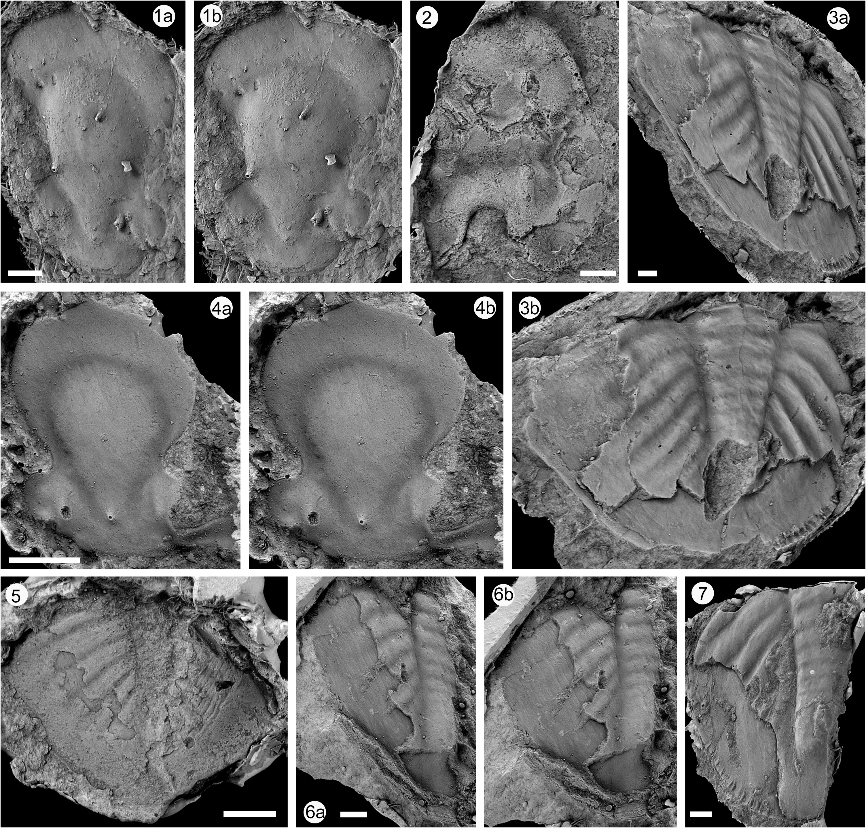
Description. Reed’s (1915) general description can be supplemented by comments on particular features. The lectotype is distorted, but the cast shows the best surface detail. Other cephalic shields are internal moulds for the most part and as a result both the axial and glabellar furrows are deep and wide compared with their expression on the dorsal surface. The cephalic shield was originally somewhat less than twice as wide as long or narrower ( Fig. 13.4 View FIGURE 13 ). Three of the specimens displaying the glabella (Figs 13.4,6,8) show that the short S1 basal glabellar furrow is forked close to its base, and that the posterior fork outlines a basal lateral glabellar lobe –this is clearly seen on the lectotype and in Figs 13.4 View FIGURE 13 , 6 View FIGURE 6 (right). S2 is gently forwardly–directed, and the sigmoid form of gently backwardly-directed S3 is best shown in Fig. 13.6 View FIGURE 13 . Certain species of Calyptaulax show a similar structure. The anterior cranidial border is shown on the lectotype and on the cranidium Fig. 13.4 View FIGURE 13 , where it is narrow and rim-like, especially medially, and defined by a shallow furrow. Eyes are very large, half (exsag.) cranidial length (sag.), the deeply-defined palpebral rims flipped upwards from the level of the intraocular cheeks. The number of dorso-ventral lens files is not precisely determinable but certainly exceeds twenty, on high standing eyes. The free cheek illustrated by Reed (1915, pl. 9, fig. 9) suggests about 15 lenses per dorso-ventral file. The lectotype clearly shows deeply and coarsely pitted sculpture on the intraocular cheeks. The same sculpture extends on to the lateral parts of the fixed cheeks, but more feebly. There is no evidence of a genal spine. The incomplete six thoracic segments on the lectotype show the same kind of sculpture anterior to the deep and narrow epifacetal pleural furrows.
Well-preserved subtrapezoidal pygidium is best shown by cast from external mould ( Fig. 13.9 View FIGURE 13 , right), 75% as long as wide. Eight (?faint ninth) axial rings, but internal mould ( Fig. 13.7 View FIGURE 13 ) would have displayed at least ten. Interpleural furrows much weaker than pleural furrows, defining six or seven ribs fading out on border, but again internal mould certainly shows more ribs extending to the posterior axial rings. Border is gently concave, most noticeably laterally. Posterior termination of pygidium behind axis shows a tendency to come to a median point.
Discussion. Reed (1915) distinguished wynnei as a subspecies of his taxon Phacops (Pterygometopus) dagon from the Upper Naungkangyi Beds. Quite apart from their stratigraphical separation, the cranidium of dagon differs from that of wynnei in having a distinct and wider cephalic border (sag.), relatively wider basal part of the glabella, and the first glabellar furrow S1 has less distinct distal bifurcation. It is regarded as belonging to a different genus. Hence Pterygometopus dagon wynnei is here elevated to species rank, and not regarded as closely related to P. dagon dagon . On the other hand, wynnei is also different from chasmopine pterygometopids with typically inflated and enlarged lateral glabellar lobes. It more closely resembles genera such as Achatella , which, according to Swisher et al. (2016), is a Baltic/Laurentian clade, and one with smaller eyes and generally distinct tuberculate sculpture, unlike wynnei . The presence of three pairs of well-developed lateral glabellar furrows is a plesiomorphic character. Small lateral basal glabellar lobes which become eliminated in more advanced species is pterygometopid, and exceptionally large eyes are typical of several taxa. Although the type species of Yanhaoia is not completely known and is older, Y. wynnei does share with it a narrow glabella posteriorly, and particularly large eyes, and, given the other similarities between Burmese and SW Chinese taxa, is assigned provisionally to this genus pending a revision of the whole group.


| GSI |
Geological Survey of India |
No known copyright restrictions apply. See Agosti, D., Egloff, W., 2009. Taxonomic information exchange and copyright: the Plazi approach. BMC Research Notes 2009, 2:53 for further explanation.