
Pandava Lehtinen, 1967
|
publication ID |
https://doi.org/ 10.5281/zenodo.276087 |
|
DOI |
https://doi.org/10.5281/zenodo.6211645 |
|
persistent identifier |
https://treatment.plazi.org/id/03CB1965-7210-FFE5-FF5B-B63FFB47FC19 |
|
treatment provided by |
Plazi |
|
scientific name |
Pandava Lehtinen, 1967 |
| status |
|
Pandava Lehtinen, 1967 View in CoL View at ENA
Pandava Lehtinen, 1967: 255 View in CoL , figs. 425–426, 440. Type-species by monotypy and original designation: Amaurobius laminatus Thorell, 1878: 168 ; Lehtinen, 1967: 255; Platnick, 2010.
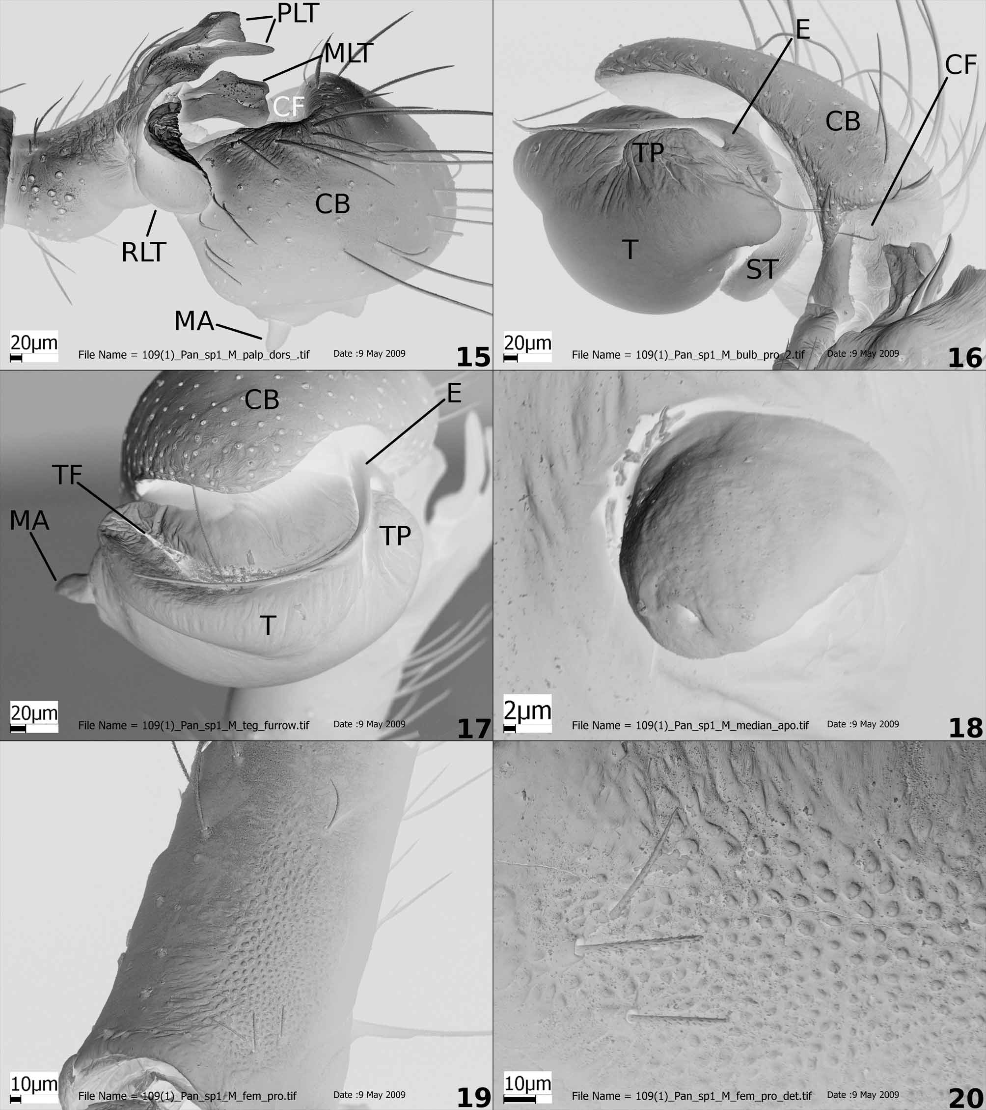
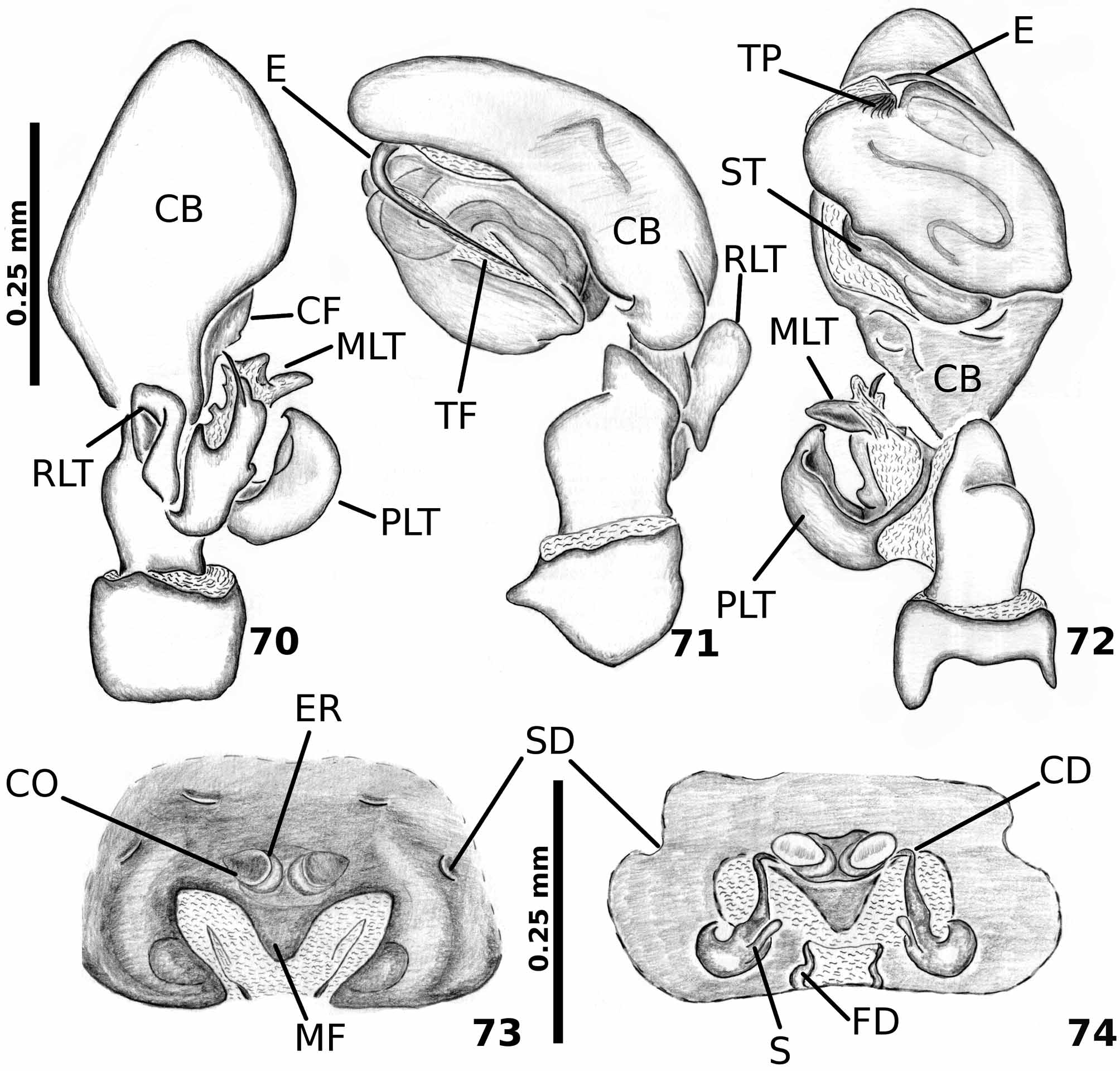
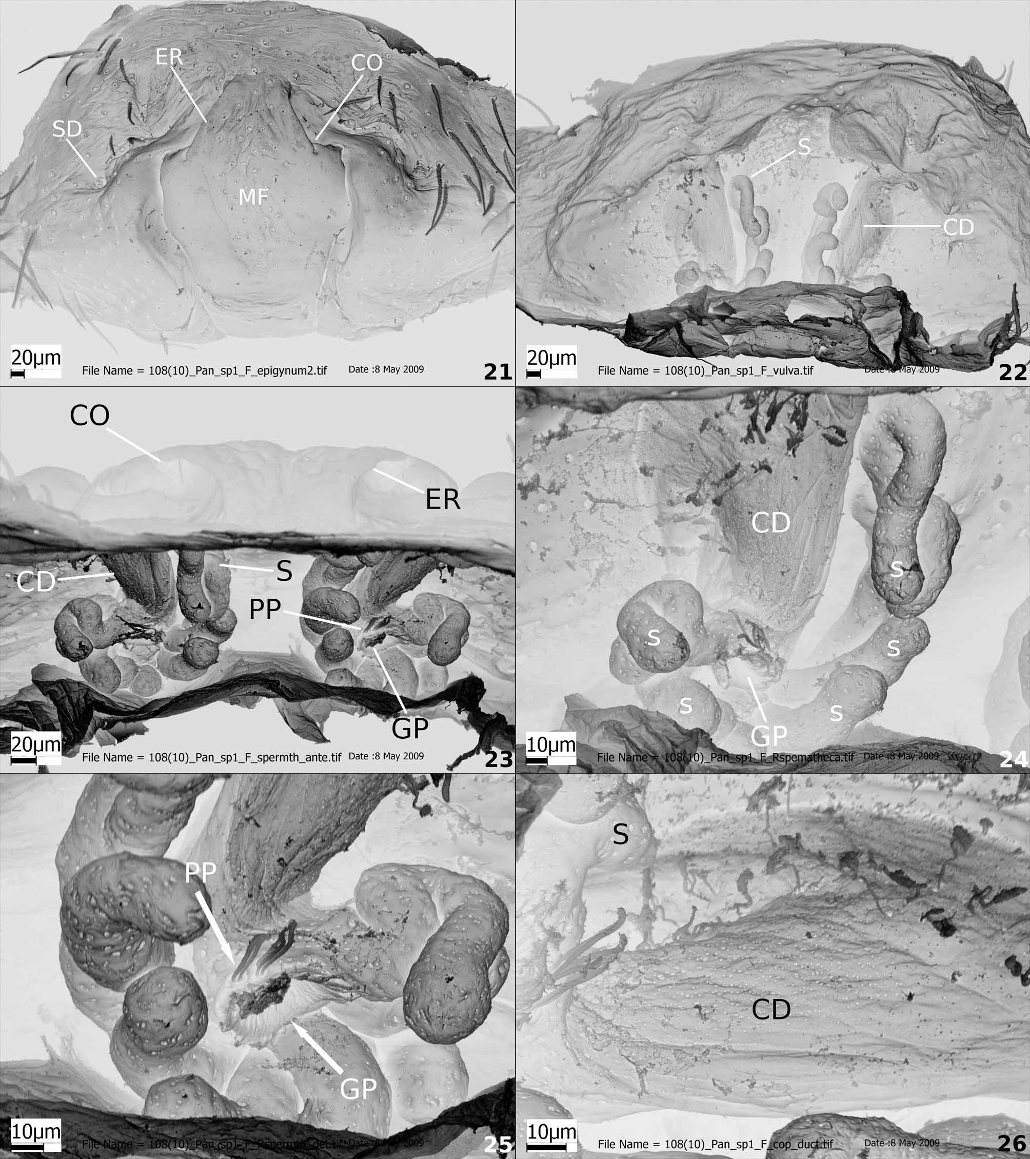
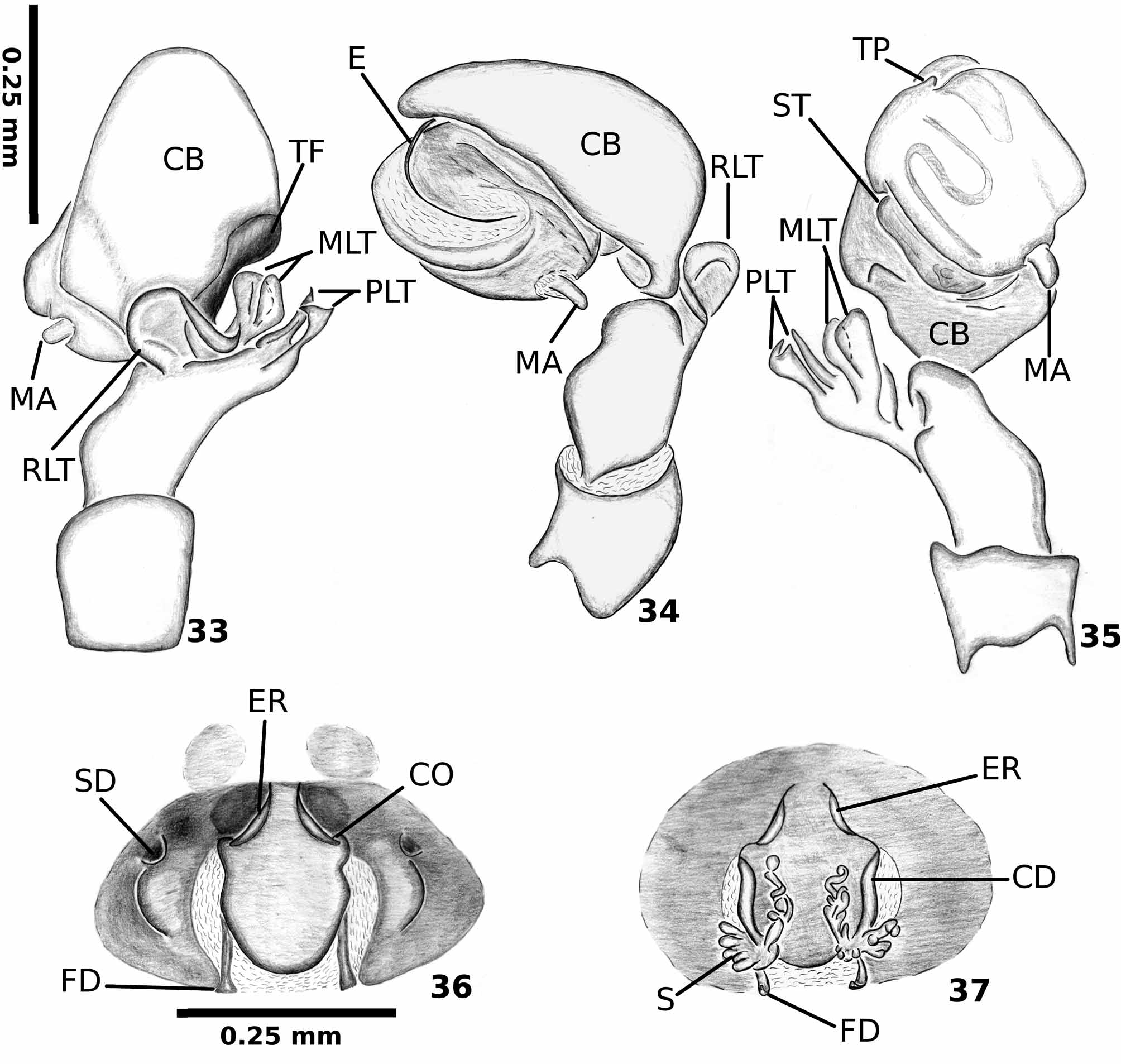
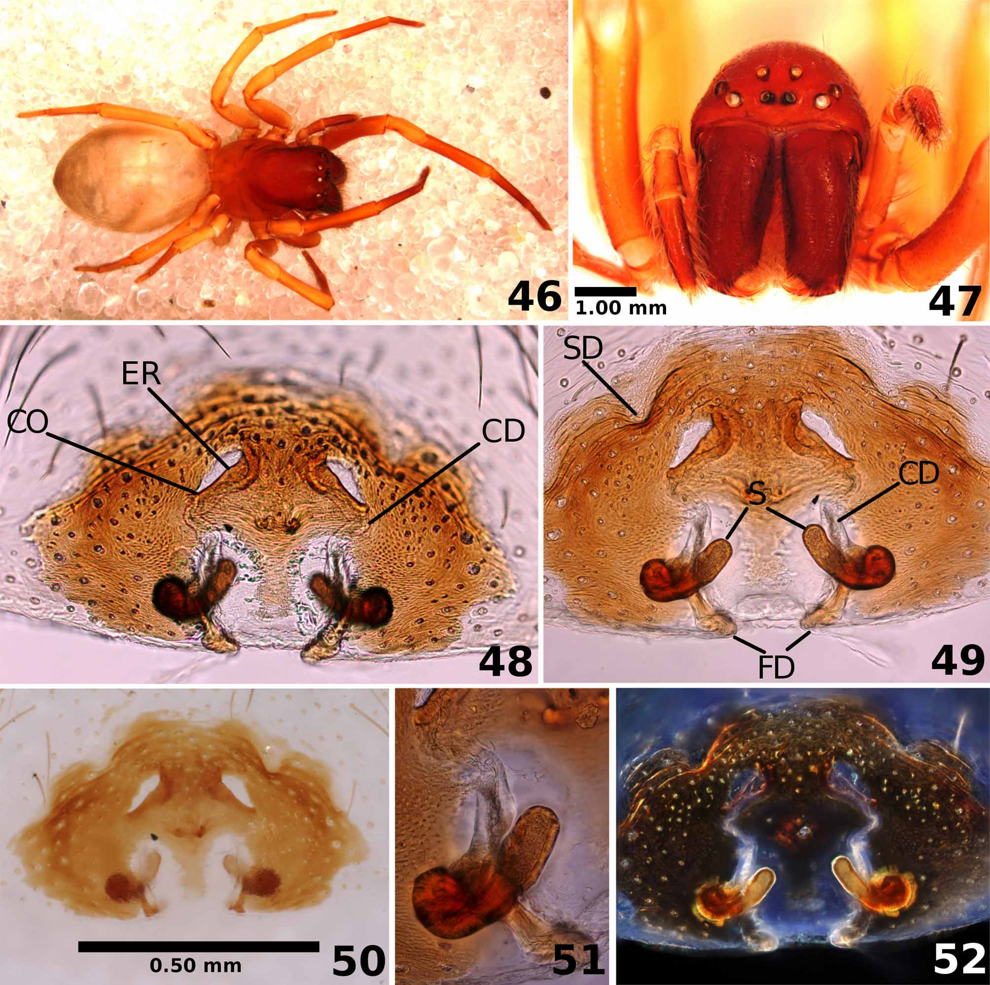
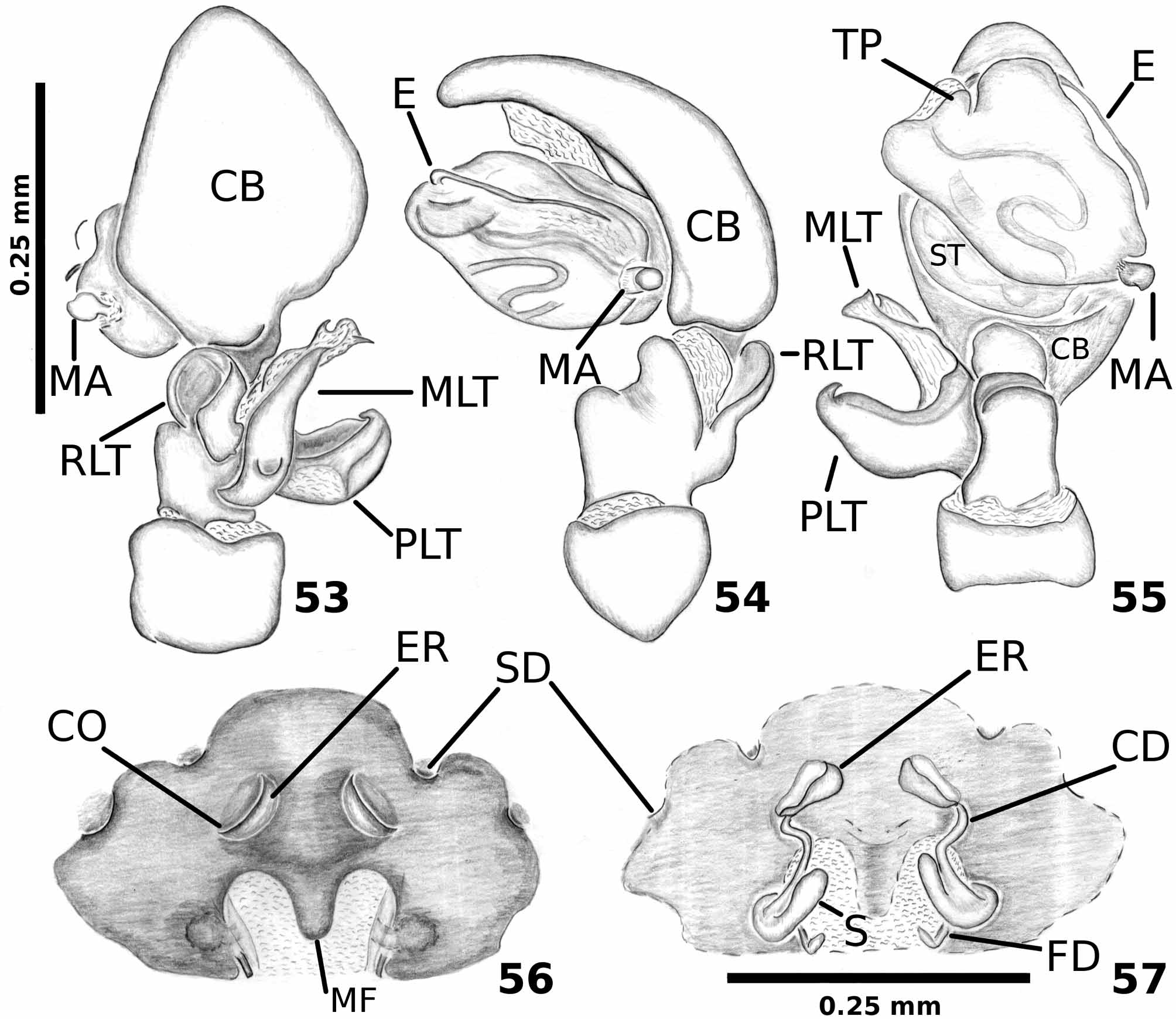
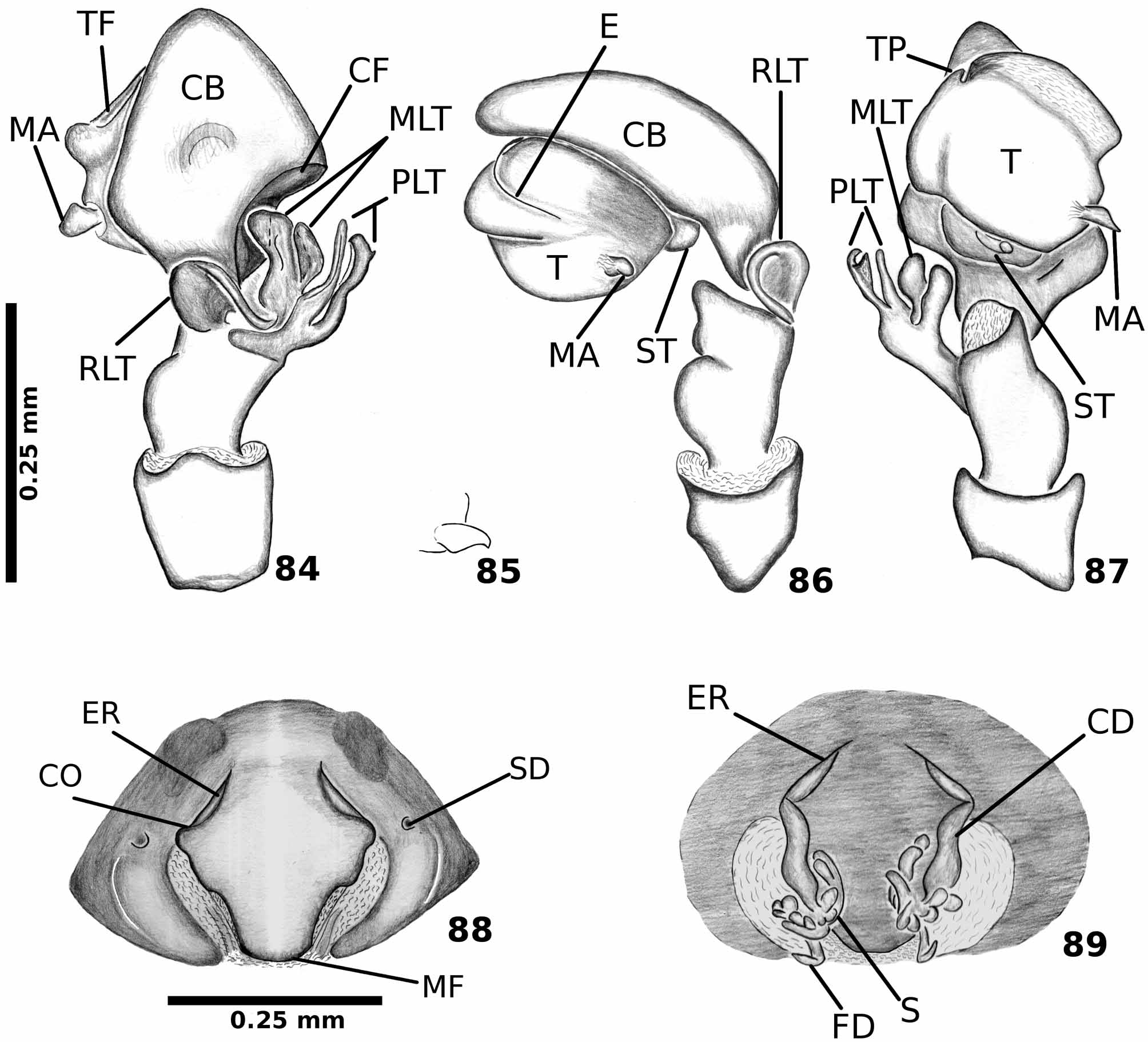
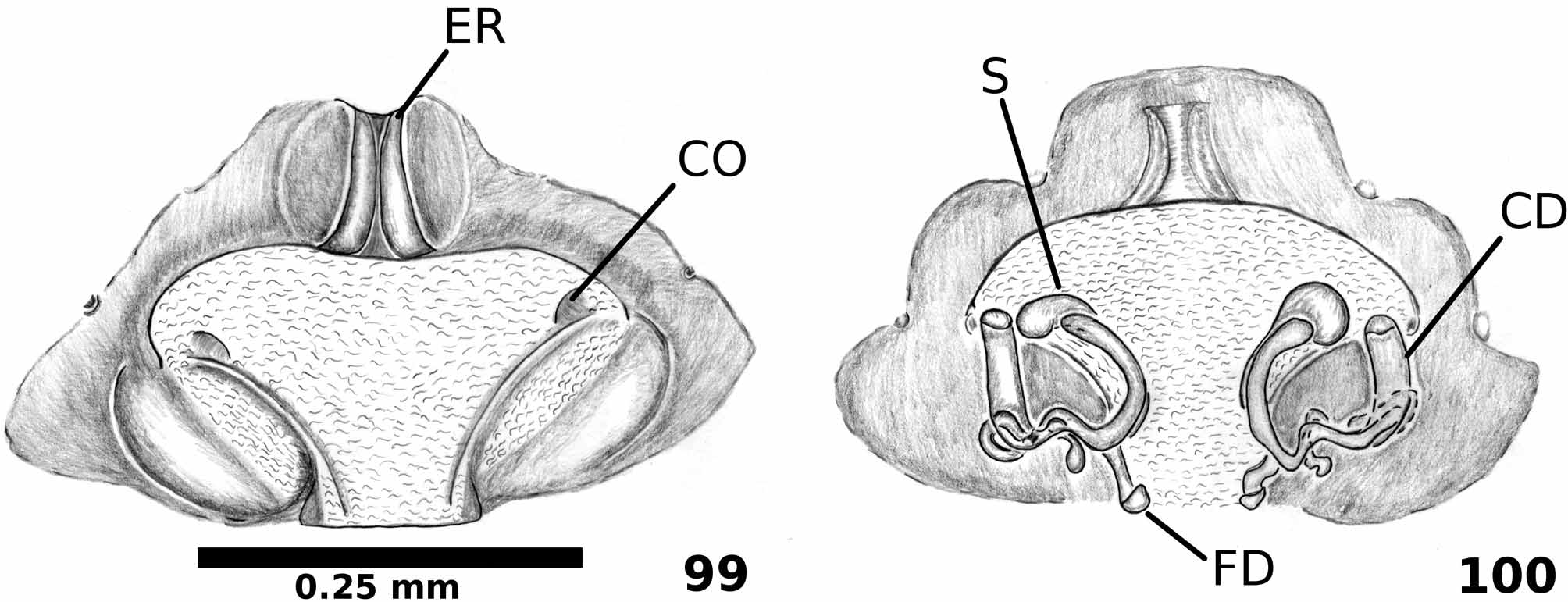
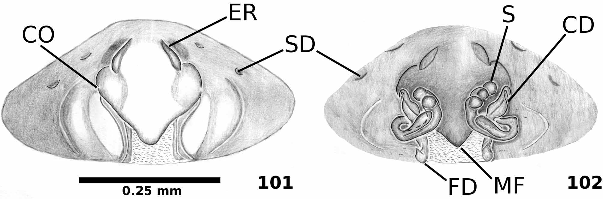
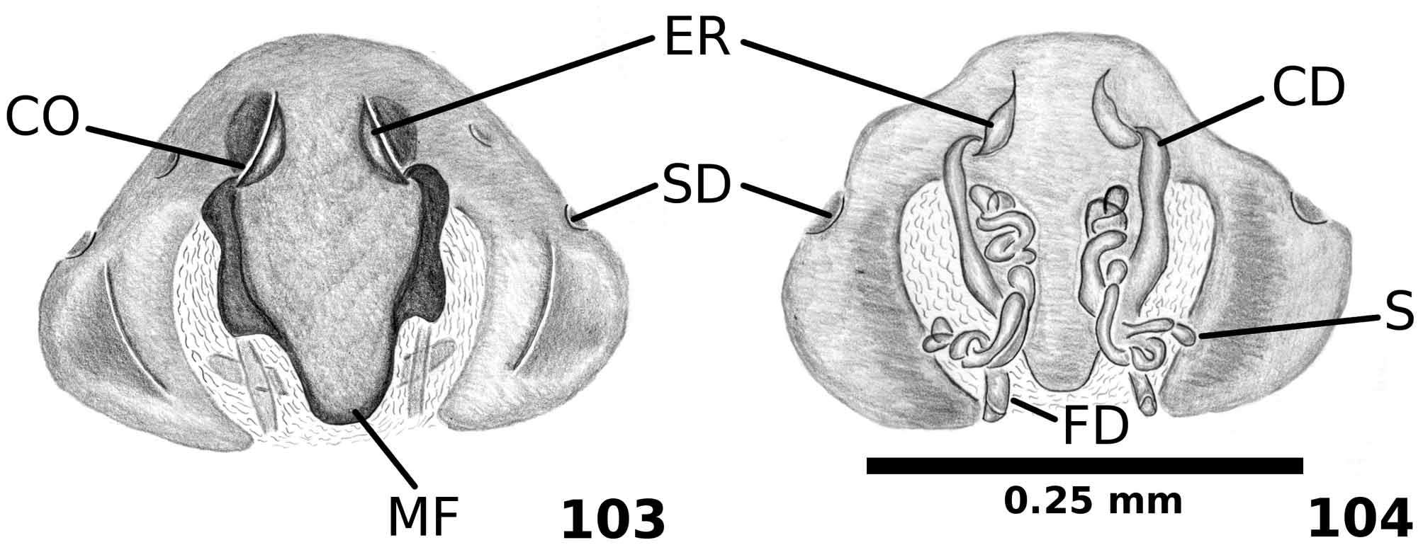
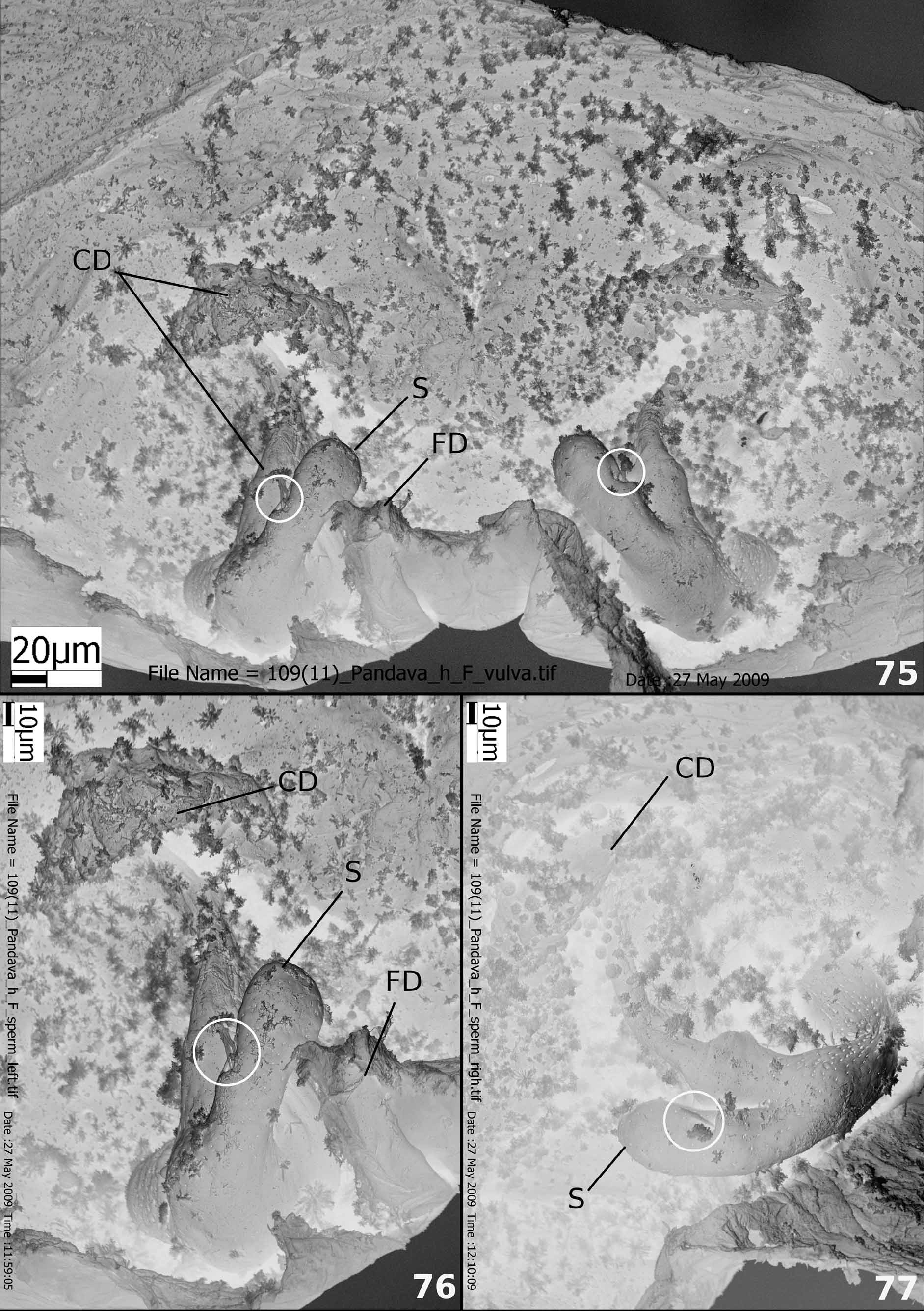
Diagnosis. Males of Pandava differ from Anuvinda , Goeldia , Nurscia and Titanoeca by the reduced tegular process near the base of the embolus ( Figs. 16–17 View FIGURES 15 – 20 ); by the thumb-shaped median apophysis ( Fig. 17 View FIGURES 15 – 20 ), which is absent in P. shiva sp. nov. ( Fig. 72 View FIGURES 70 – 74 ) and divided in all other Titanoecidae ( Griswold et al. 2005: 275, figs. 174B, 174C; Almeida-Silva et al. 2009a: 365, figs. 2, 3; Almeida-Silva et al. 2009b: 63, figs. 1, 2, 4). Females differ from other Titanoecidae by the anteriorly positioned epigynal rims ( Figs. 21 View FIGURES 21 – 26 , 36 View FIGURES 33 – 37 , 48, 50 View FIGURES 46 – 52 , 56 View FIGURES 53 – 57 , 73 View FIGURES 70 – 74 , 88 View FIGURES 84 – 89 , 90 View FIGURES 90 – 96 , 99 View FIGURES 99 – 100 , 101 View FIGURES 101 – 102 , 103 View FIGURES 103 – 104 ), which sometimes cover the copulatory openings; epigynum round and elevated in the median region; spermathecae elongated and varying in number: more than six in P. laminata ( Figs. 22–25 View FIGURES 21 – 26 , 37 View FIGURES 33 – 37 ), P. ganesha sp. nov. ( Fig. 104 View FIGURES 103 – 104 ) and P. sarasvati sp. nov. ( Figs. 89 View FIGURES 84 – 89 , 91–93 View FIGURES 90 – 96 ); three or four in Pandava ganga sp. nov. ( Fig. 100 View FIGURES 99 – 100 ) and P. kama sp. nov. ( Fig. 102 View FIGURES 101 – 102 ), or reduced to a single lobe in P. shiva sp. nov. ( Figs. 74–77 View FIGURES 70 – 74 View FIGURES 75 – 77 ) and P. hunanensis ( Figs. 48–52 View FIGURES 46 – 52 , 57 View FIGURES 53 – 57 ).
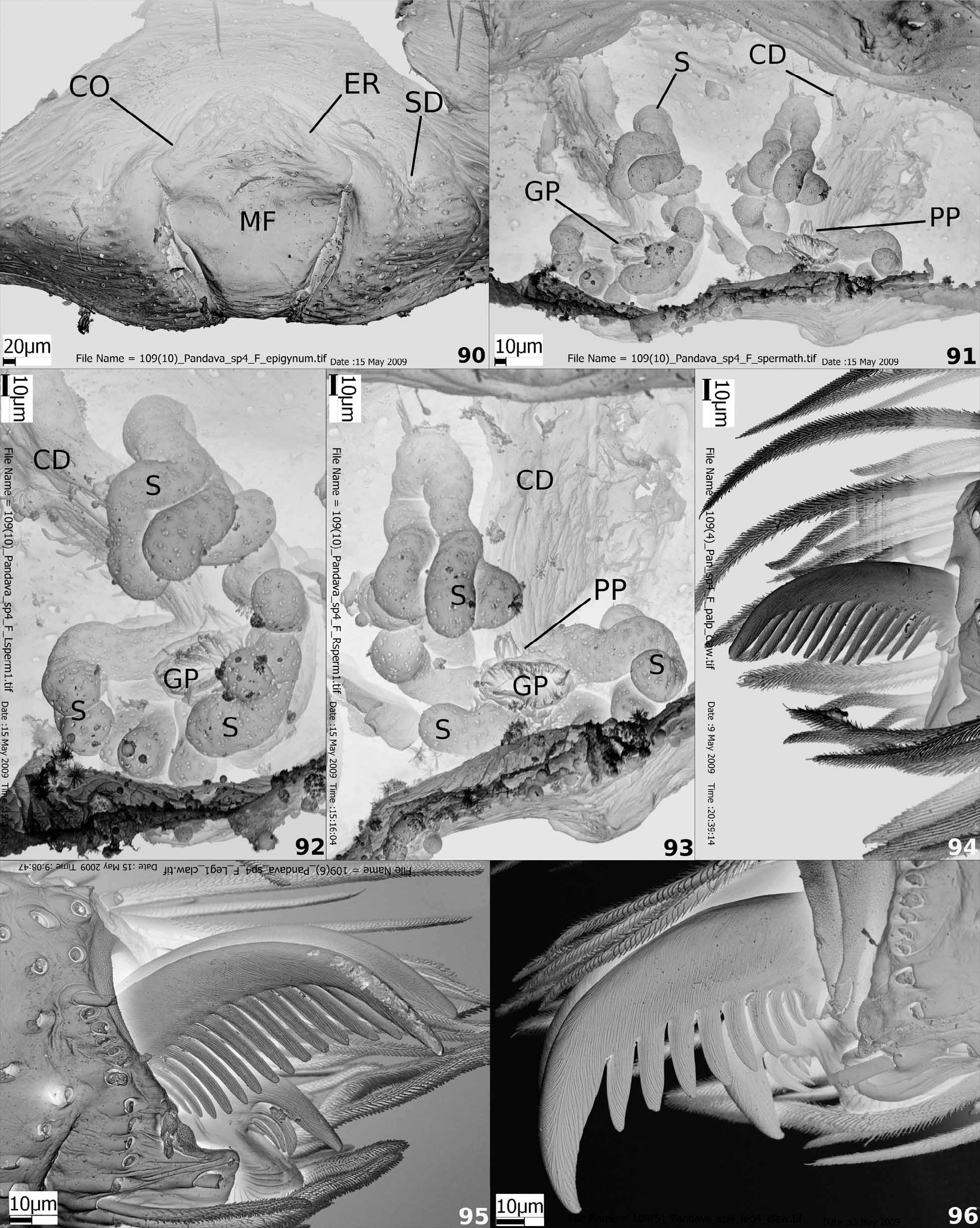
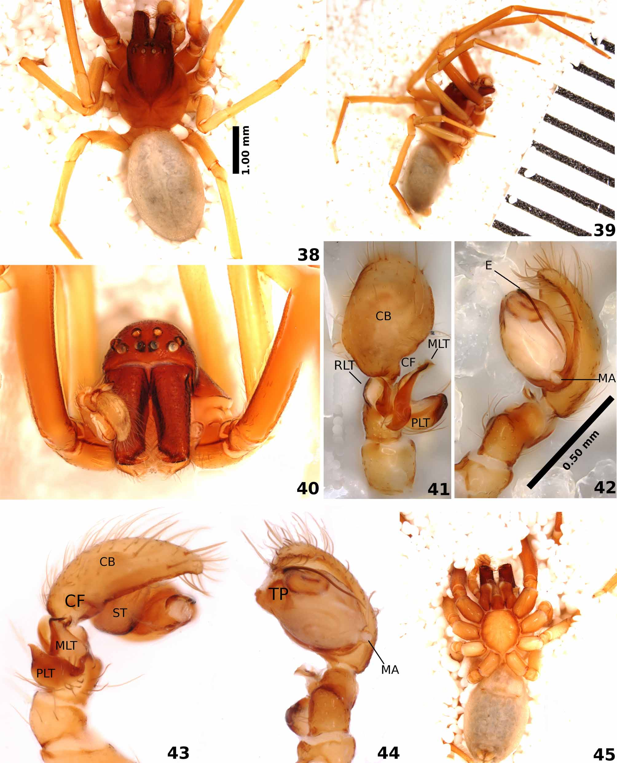
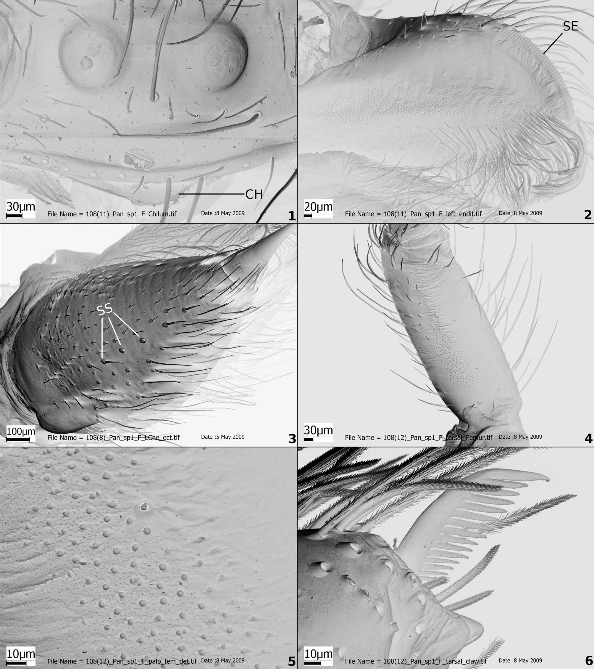
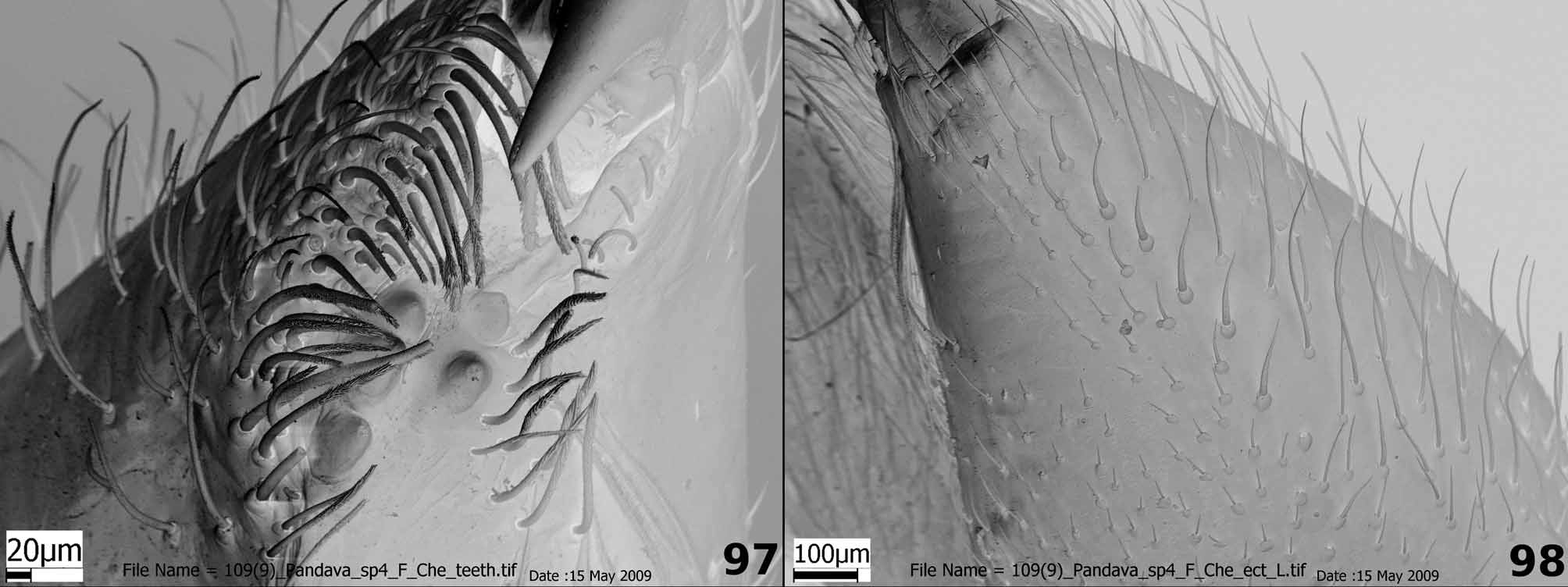
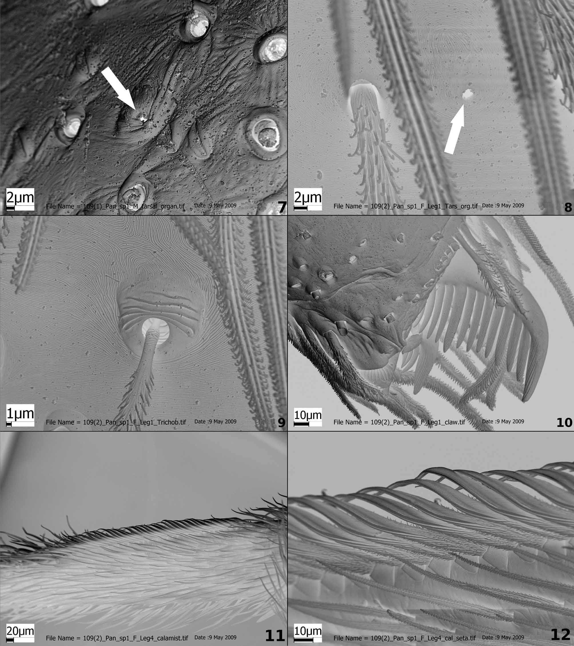
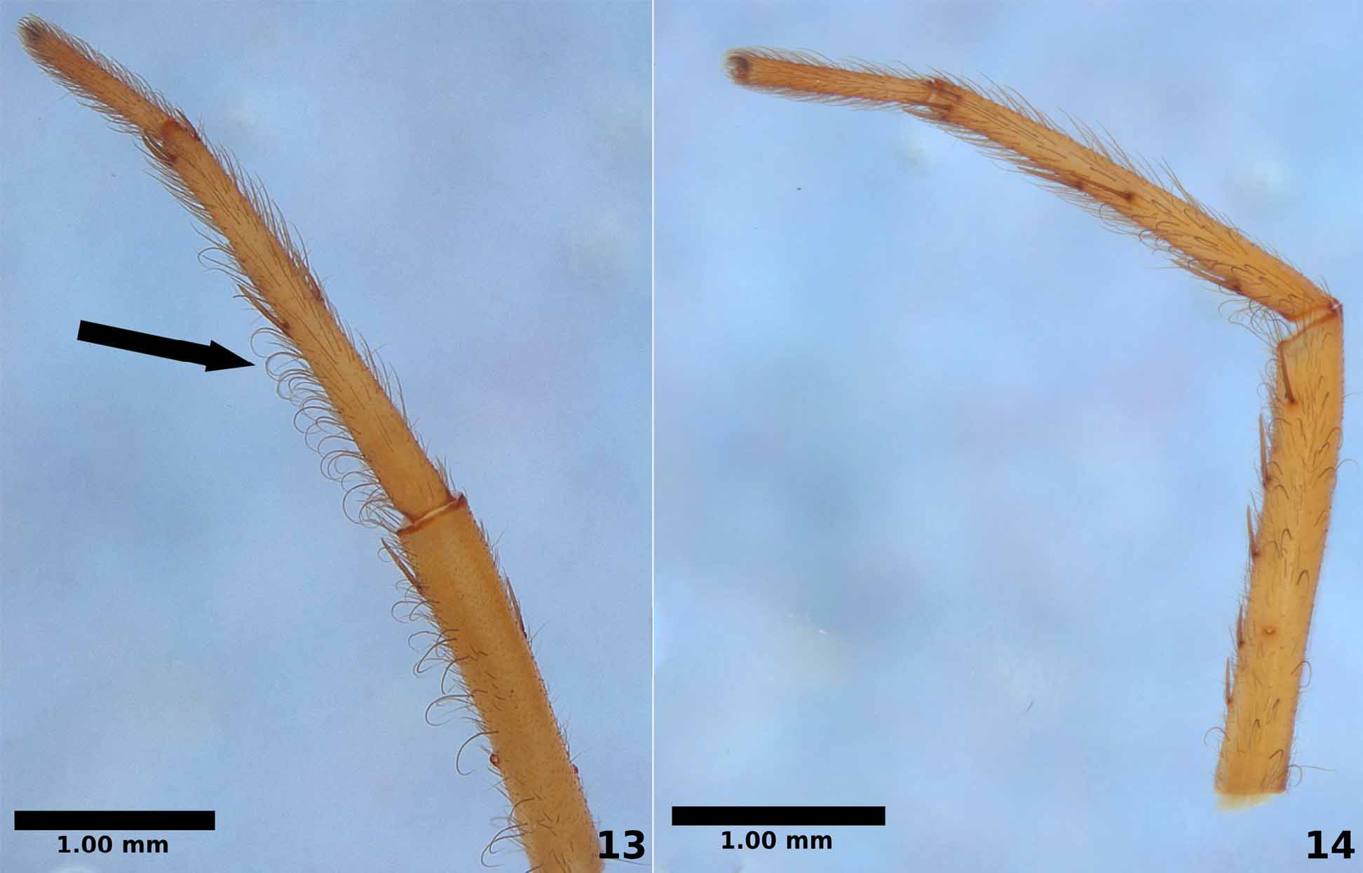
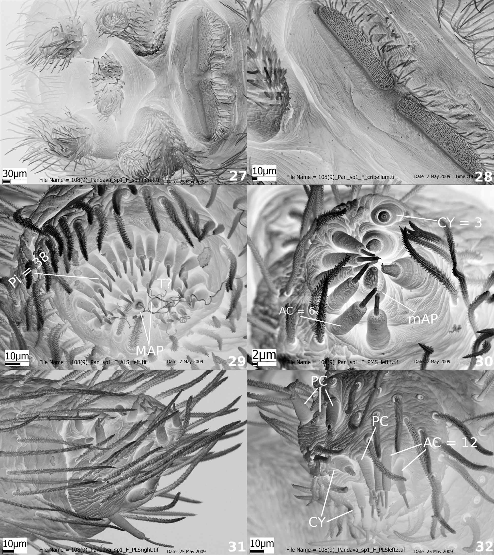
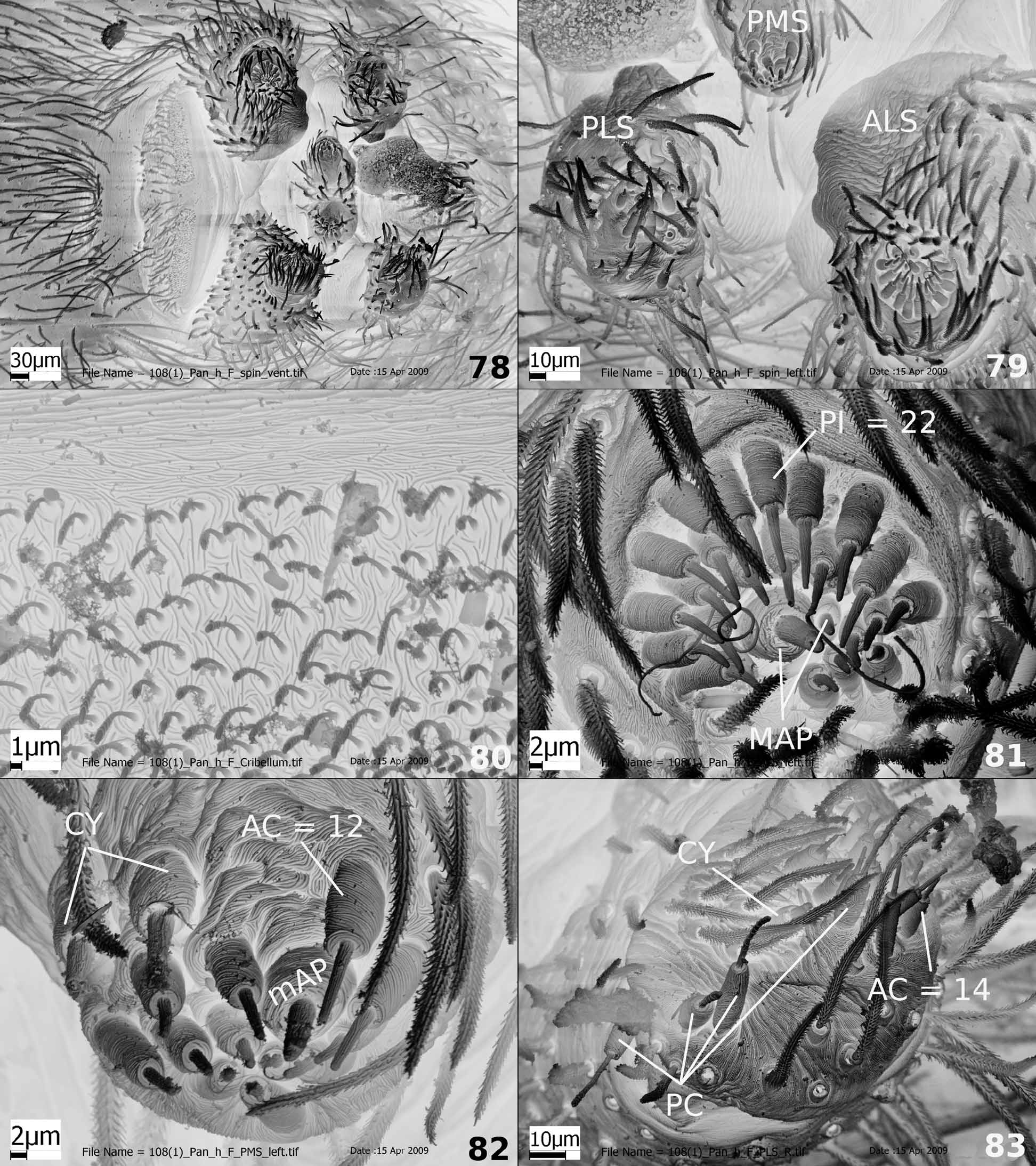
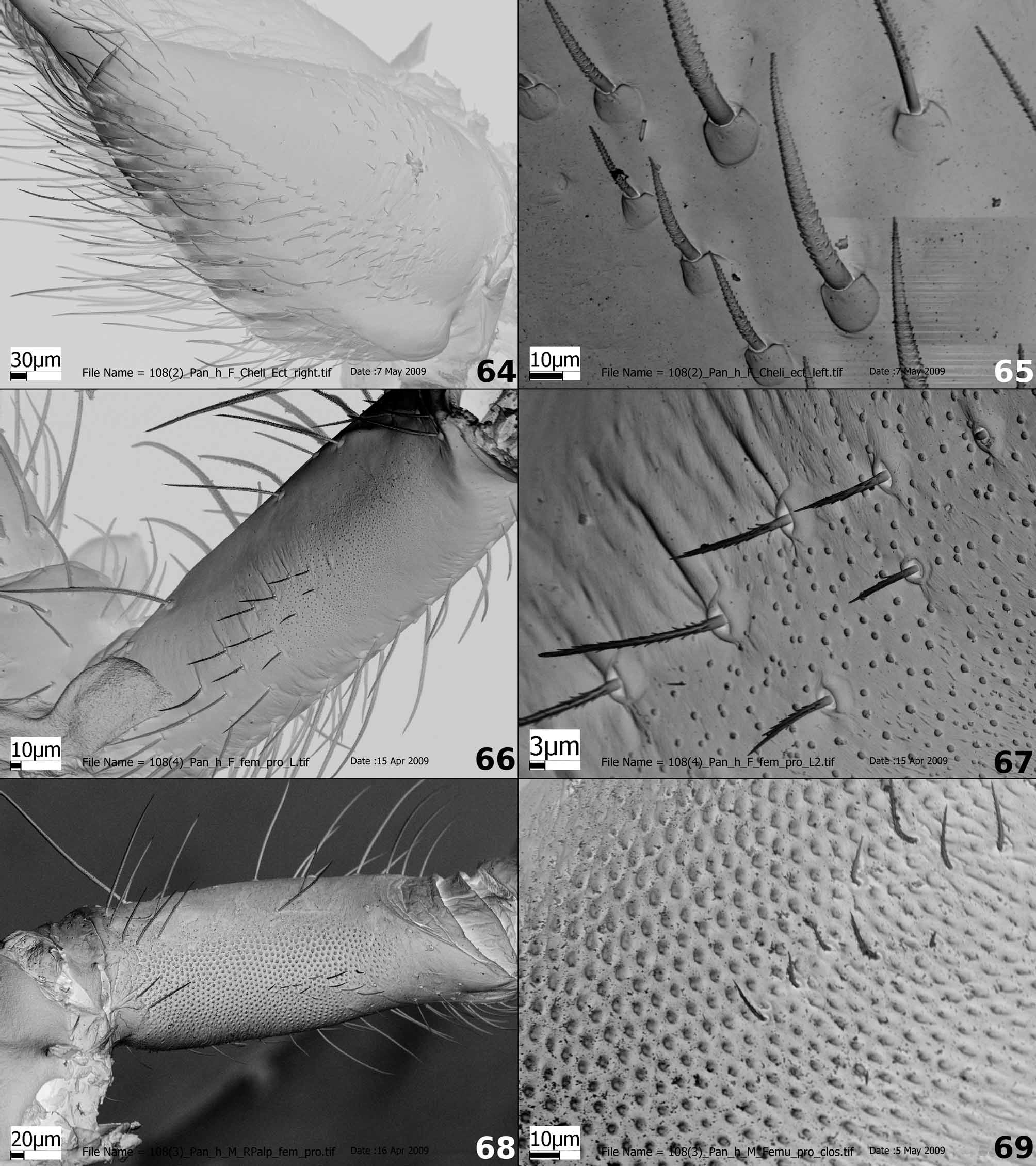
Description. Total length (males and females): 4.00–8.60. Carapace: cephalic area high and thoracic fovea reduced ( Figs. 38, 39 View FIGURES 38 – 45 , 46 View FIGURES 46 – 52 ). Eyes round, AME smaller than ALE, PME and PLE ( Figs. 40 View FIGURES 38 – 45 , 47 View FIGURES 46 – 52 ). Cheliceral fang 1/3 the length of the paturon. Chilum entire, reduced, smaller than outer border of the median eyes and without setae ( Fig. 1 View FIGURES 1 – 6 ). Endites oblong, with regular serrula ( Fig. 2 View FIGURES 1 – 6 ), labium square ( Fig. 45 View FIGURES 38 – 45 ). Cheliceral promargin with three teeth, retromargin with two ( Fig. 97 View FIGURES 97 – 98 ) and retrolateral face with stridulatory setae ( Figs. 3 View FIGURES 1 – 6 , 98 View FIGURES 97 – 98 ). Anterior border of sternum straight, laterally round and posteriorly acute, projecting between coxae IV ( Fig. 45 View FIGURES 38 – 45 ). Leg formula variable but always with leg III shortest. One spine distally positioned on prolateral surface of femur I and II, except in P. s h i v a sp. nov. and P. hunanensis . Distribution and presence of other spines variable. Male tibial crack, when present, positioned after the first spine of tibiae I and II as described by Griswold (1993: 2, figs. 3–4) in Zoropsidae and Zorocratidae ( Griswold et al. 2005: 242, fig. 141F), and by Almeida-Silva et al. (2009b) for Anuvinda . Prolateral surface of the palpal femur with stridulatory files ( Figs. 4–5 View FIGURES 1 – 6 , 19–20 View FIGURES 15 – 20 ). Tarsal claw with 10 to 14 narrow denticles ( Figs. 6 View FIGURES 1 – 6 , 94 View FIGURES 90 – 96 ). Tarsal organ of the male palp on the prolateral distal part of the cymbium, with transverse ridges ( Fig. 7 View FIGURES 7 – 12 ). Tarsal organ of legs and female palp capsulate, with small, round opening ( Fig. 8 View FIGURES 7 – 12 ). One single metatarsal trichobothrium with transverse ridges on basal hood ( Fig. 9 View FIGURES 7 – 12 ), trichobothria absent from tarsi. All leg claws with multiple teeth including three to four denticles on the third claw ( Figs. 10 View FIGURES 7 – 12 , 95–96 View FIGURES 90 – 96 ). Calamistrum uniseriate and extending the entire length of metatarsus IV ( Figs. 11–12 View FIGURES 7 – 12 ). Distally curved setae present on femur, tibia and metatarsus of some males ( Figs. 13–14 View FIGURES 13 – 14 ) as in Anuvinda ( Almeida-Silva et. al. 2009b: 64, figs. 8–9) and also in females of Pandava ganga sp. nov. Abdomen oval. Cribellum divided, as broad as spinneret area ( Figs. 27–28 View FIGURES 27 – 32 , 78, 80 View FIGURES 78 – 83 ). Two major ampullate gland spigots ( Figs. 29 View FIGURES 27 – 32 , 81 View FIGURES 78 – 83 ) and 21 to 38 piriform gland spigots ( Figs. 29 View FIGURES 27 – 32 , 79, 81 View FIGURES 78 – 83 ) on the ALS, MAP recessed into PI spinning field; PMS with one mAP, six to 12 aciniform and two to three cylindrical gland spigots, paracribellar spigots absent ( Figs. 30 View FIGURES 27 – 32 , 82 View FIGURES 78 – 83 ); PLS with four paracribellar, one to two cylindrical and 13 to 14 aciniform gland spigots ( Figs. 31–32 View FIGURES 27 – 32 , 83 View FIGURES 78 – 83 ).
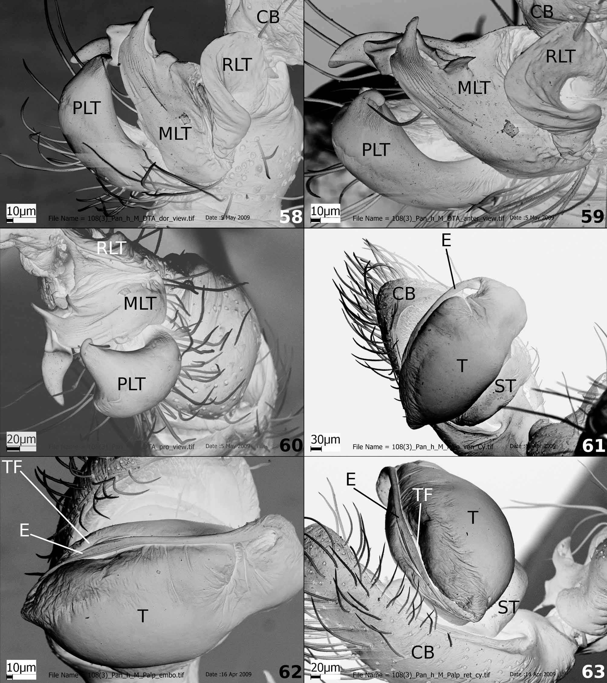
Palpal tibial apophysis with RLT ear-shaped; MLT divided, with an indistinct base; PLT divided as in Pandava laminata and P. sarasvati sp. nov. ( Figs. 15 View FIGURES 15 – 20 , 33, 35 View FIGURES 33 – 37 , 84, 87 View FIGURES 84 – 89 ), or entire and enlarged as in P. hunanensis and P. s h i v a sp. nov. ( Figs. 41, 43 View FIGURES 38 – 45 , 53, 55 View FIGURES 53 – 57 , 58–60 View FIGURES 58 – 63 , 70, 72 View FIGURES 70 – 74 ). Prolateral face of femur with stridulatory files made up of small, round protuberances and with few setae ( Figs. 4–5 View FIGURES 1 – 6 , 19–20 View FIGURES 15 – 20 , 66–69 View FIGURES 64 – 69 ). Cymbium with prolateral furrow as an inverted “L” ( Figs. 15–16 View FIGURES 15 – 20 , 33, 35 View FIGURES 33 – 37 , 41, 43 View FIGURES 38 – 45 , 55 View FIGURES 53 – 57 , 70, 72 View FIGURES 70 – 74 , 84, 87 View FIGURES 84 – 89 ). Median apophysis entire ( Figs. 15, 17, 18 View FIGURES 15 – 20 , 33–35 View FIGURES 33 – 37 , 42, 44 View FIGURES 38 – 45 , 53–55 View FIGURES 53 – 57 , 84–87 View FIGURES 84 – 89 ) or absent ( Figs. 61–63 View FIGURES 58 – 63 ). Tegular process reduced ( Figs. 16–17 View FIGURES 15 – 20 , 35 View FIGURES 33 – 37 , 44 View FIGURES 38 – 45 , 55 View FIGURES 53 – 57 , 72 View FIGURES 70 – 74 , 87 View FIGURES 84 – 89 ). Spermatic duct bent once as an “S” ( Figs. 35 View FIGURES 33 – 37 , 44 View FIGURES 38 – 45 , 55 View FIGURES 53 – 57 , 72 View FIGURES 70 – 74 ). Pars pendula very reduced. Tegular furrow typical of Titanoecidae ( Figs. 17 View FIGURES 15 – 20 , 34 View FIGURES 33 – 37 , 54 View FIGURES 53 – 57 , 62–63 View FIGURES 58 – 63 , 71 View FIGURES 70 – 74 , 86 View FIGURES 84 – 89 ), serving as a resting place for the embolus. Embolus filiform ( Figs. 16–17 View FIGURES 15 – 20 , 34 View FIGURES 33 – 37 , 42, 44 View FIGURES 38 – 45 , 54–55 View FIGURES 53 – 57 , 71–72 View FIGURES 70 – 74 , 86 View FIGURES 84 – 89 ).
Epigynum with copulatory openings and rim anteriorly positioned ( Figs. 21 View FIGURES 21 – 26 , 36 View FIGURES 33 – 37 , 48–50 View FIGURES 46 – 52 , 56 View FIGURES 53 – 57 , 73 View FIGURES 70 – 74 , 88 View FIGURES 84 – 89 , 90 View FIGURES 90 – 96 , 99 View FIGURES 99 – 100 , 101 View FIGURES 101 – 102 , 103 View FIGURES 103 – 104 ). Copulatory ducts with pores ( Figs. 24–25 View FIGURES 21 – 26 , 75– 76 View FIGURES 75 – 77 , 93 View FIGURES 90 – 96 ). Fertilization duct filiform, may be partially covered by cuticle obscuring its connection with the spermathecae, e.g., Fig. 74 View FIGURES 70 – 74 . Spermathecae elongated with lobes forming a bunch ( Figs. 22–25 View FIGURES 21 – 26 , 37 View FIGURES 33 – 37 , 89 View FIGURES 84 – 89 , 91–93 View FIGURES 90 – 96 ) except in P. s h i v a sp. nov. and P. hunanensis , whose spermatheca is limited to a single lobe ( Figs. 49, 51–52 View FIGURES 46 – 52 , 57 View FIGURES 53 – 57 , 74–77 View FIGURES 70 – 74 View FIGURES 75 – 77 ). Spermathecae with two kinds of pores: one enlarged, with the edges covered by a folded tissue, here called giant pore ( Figs. 22–25 View FIGURES 21 – 26 , 91–93 View FIGURES 90 – 96 ), which is located in the intersection among spermathecae and copulatory ducts; the other kind is a “primary pore” according to Bennett (1992: 6 and 17, fig. 32) which in Pandava is commonly located near the giant pore ( Figs. 75–77 View FIGURES 75 – 77 ), except in P. s h i v a sp. nov. The primary pore is also found in other spiders ( Bennett, 1992: 17, fig. 32; Wang, 2002: 16, fig. 41).
Remarks. The homology of the Pandava giant pore with the so-called “dictynoid” pore described by Bennett (1992) is uncertain. The “dictynoid” pore has been described as a single, porous plate in the bottom of a shallow, circular concavity ( Bennett, 1992, figs. 2, 6) or recessed into a deep hole ( Bennett, 1992, figs. 7, 9, 28). The giant pore of Pandava is oblong and does not have an obvious flat poreplate. Notably, the “dictynoid” pore has not been described from Dictyna . Understanding the homology among such giant pores must await optimization of this character on a comprehensive phylogeny of the relevant taxa.
Geographical distribution. Asia: China, Japan, India, Indonesia, Myanmar, Pakistan, Philippines, Sri Lanka, Thailand; Pacific: Marquesas Islands, New Guinea. Introduced in Germany. New records from Africa: Kenya, Tanzania and Madagascar.
Composition. Seven species: Pandava laminata ( Thorell, 1878) ; P. hunanensis Yin & Bao, 2001 ; P. shiva sp. nov., P. sarasvati sp. nov., P. ganga sp. nov., P. kama sp. nov., P. ganesha sp. nov.
No known copyright restrictions apply. See Agosti, D., Egloff, W., 2009. Taxonomic information exchange and copyright: the Plazi approach. BMC Research Notes 2009, 2:53 for further explanation.
|
Kingdom |
|
|
Phylum |
|
|
Class |
|
|
Order |
|
|
Family |
Pandava Lehtinen, 1967
| Almeida-Silva, Lina M., Griswold, Charles E. & Brescovit, Antonio D. 2010 |
Pandava
| Lehtinen 1967: 255 |
| Lehtinen 1967: 255 |
| Thorell 1878: 168 |