
Crettaros valdezi, Cruz-López & Francke, 2015
|
publication ID |
https://doi.org/10.1111/zoj.12299 |
|
DOI |
https://doi.org/10.5281/zenodo.10543455 |
|
persistent identifier |
https://treatment.plazi.org/id/03CA87F9-A27F-FF87-FEA2-FC8EC1BFFB9E |
|
treatment provided by |
Felipe (2021-08-28 21:15:18, last updated 2024-01-21 05:21:29) |
|
scientific name |
Crettaros valdezi |
| status |
sp. nov. |
CRETTAROS VALDEZI View in CoL SP. NOV.
( FIGS 43 View Figure 43 , 44 View Figure 44 , 57 View Figure 57 , 63C View Figure 63 , 64G View Figure 64 )
Type material: MEXICO: San Luís Potosí: ♂ holotype , 1 ♂ and 4 ♀ paratypes [ CNAN-T0799 and CNAN-
T0800 (13.v.2012; J. Cruz, G. Contreras, J. Mendoza, and R. Monjaraz)] (22°0′55.51″N, 100°36′22.21″W). Municipio Villa de Zaragoza, Km 58 Cave.
Material examined: MEXICO: San Luís Potosí: 1 ♂ and 1 ♀ [ CNAN (13.v.2010; O. Francke, A. Valdez, C. Santibáñez, and J. Cruz], same locality data as the types . 1 ♂ and 2 ♀ [ CNAN (13.v.2012; J. Cruz, G. Contreras, J. Mendoza, and R. Monjaraz)] (22°1′0.37″N, 100°35′29.97″W). Municipio Villa de Zaragoza , small cave from Km 58 GoogleMaps . 2 ♂ and 1 ♀ [ CNAN (14.v.2012; J. Cruz, G. Contreras, J. Mendoza, and R. Monjaraz)] (22°4′16.68″N, 100°37′29.96″W). Municipio Villa de Zaragoza , Cueva de la Laguna, Valle de los Fantasmas GoogleMaps .
Etymology: Patronym, dedicated to Alejandro Valdez- Mondragón, friend and colleague, who has contribut- ed to the knowledge of spiders and ricinuleids in Mexico.
Diagnosis: Troglomorphic species. Dorsal tubercles only present in the central portion of mesotergal areas. On male penis the two basal-most pairs of lateral setae very close to each other ( Fig. 44B View Figure 44 ).
Description: Male holotype: Measurements: scutum length 2.6, maximum scutum width 2.2. Legs. I 1.20/ 0.50/0.85/1.10, II 1.50/0.75/1.30/1.35, III 1.30/0.57/1.15/ 1.27, IV 1.80/0.75/1.55/1.97. Dorsum. Rugose, with only small spiniform tubercles on the central portion of mesotergal areas I–IV. Areas II and III with median humps slightly elevated ( Fig. 63C View Figure 63 ). Opisthosoma convex in lateral view. Ocularium in the middle of prosoma, low, slightly projected beyond the eyes, blunt, covered by small tubercles. Lateral clear areas projected from the scutum in triangular shape, apex rounded. Lateral clear areas on apices of area V and free tergites I–II rounded, slightly protruding. Free tergites with one median row of small tubercles. Venter. Entire venter covered by small rounded tubercles, with a few small setae. Stigmatic area an inverted ‘Y’ shape, with lateral margins straight, short ( Fig. 43D View Figure 43 ). Coxa IV with a small dorso-ectal spiniform tooth. Pedipalps. Patella with one mesal spiniform tubercle. Legs. Very similar in ornamentation and size, covered by numerous small rounded tubercles and setae. Ventral ornamentation of femur IV slightly larger. Femora III and IV curved. Metatarsus II without clear annuli. Tarsal count 4(2):6(3):6:6. Male genitalia. Pars distalis elongated, spatular shape; apical margin almost straight. Lateral setae slender and slightly compressed distally, the two basal-most pairs very close to each other. Two pairs of parastylar setae flanking the follis. Spiniform projections not exposed and inconspicuous ( Fig. 44 View Figure 44 ). Female paratype: Measurements: scutum length 2.6, maximum scutum width 2.2. Very similar to male, with the following differences: stigmatic area shorter and narrower than on males. Legs IV slightly thinner ( Fig. 43B, D View Figure 43 ). Tarsal count 4(2):6(3):6:6.
Natural history: All specimens were collected under rocks in the dark zone, never found outside the cave. Generally, one specimen was found under one rock, rarely two or three. When uncovered they showed thanatosis behaviour similar to that of Crettaros santibanezi .

Figure 43. Crettaros valdezi sp. nov. A, habitus, dorsal view. B, legs IV, mesal view. C, habitus, lateral view. D, habitus, ventral view. E, scutum, dorsal view. F, ocularium, frontal view. Scale bars: A = 2.5 mm, B, D, and E = 1.5 mm, C = 1 mm, F = 0.5 mm. The dark lines on (D) indicate the stigmatic area.
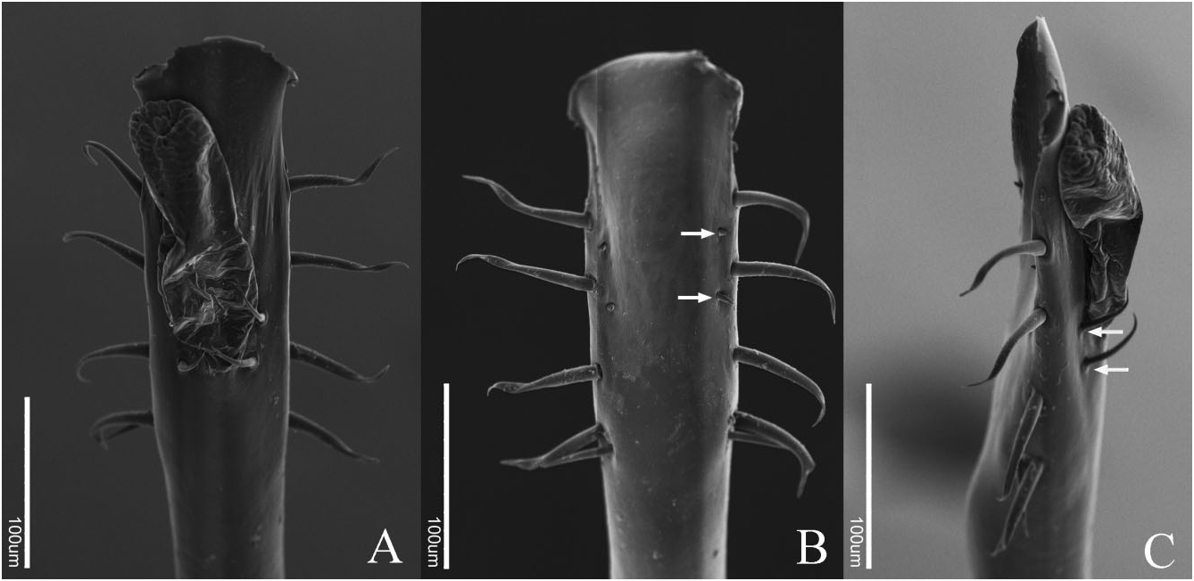
Figure 44. Crettaros valdezi sp. nov. male genitalia. A, dorsal view. B, ventral view. C, lateral view. White arrows indicate ventral microsetae in (B) and parastylar setae in (C).

Figure 57. Distribution map of the species of the Karos genus-group. Black triangles, Monterella tuberculata; black square, Mictlana inops comb. nov. (only locality of material examined was mapped); black circles, Crettaros santibanezi sp. nov.; white circles with black outline, Crettaros valdezi sp. nov.; black crosses, Chapulobunus unispinosus; inverted black triangles, Karos parvus.

Figure 63. A, dorsal view of the anal bulge on Chapulobunus poblano sp. nov.; arrow indicates the bulge. B, lateral view of Huasteca silhavyi sp. nov., line indicates the very high lateral channel area. C, laterodorsal view of scutum of Crettaros valdezi sp. nov., showing the median humps on areas II and III indicated by dotted circles. D, posterodorsal view of scutum of Crettaros santibanezi sp. nov., showing the median humps on areas II and III indicated by dotted circles. E, dorsal view of trochanter IV of Karos barbarikos; arrow indicates the tridentate apophysis. F, anterodorsal view of scutum of Karos barbarikos, the black points mark the tubercles forming a ‘V’. G, dorsal ornamentation of Karos tersum sp. nov. showing the transversal row of tubercles on mesotergal areas; transversal row II indicated by black dots. H, detail of the ventral ornamentation on femur IV of Karos barbarikos. I, detail of the ectal ornamentation on tibia IV of Karos barbarikos; arrows indicate the spiniform tubercles. J, detail of the ventral ornamentation on leg IV in Chapulobunus poblano sp. nov. K, detail of the two apical spines on femur IV in Chapulobunus poblano sp. nov. These images are not at the same scale.
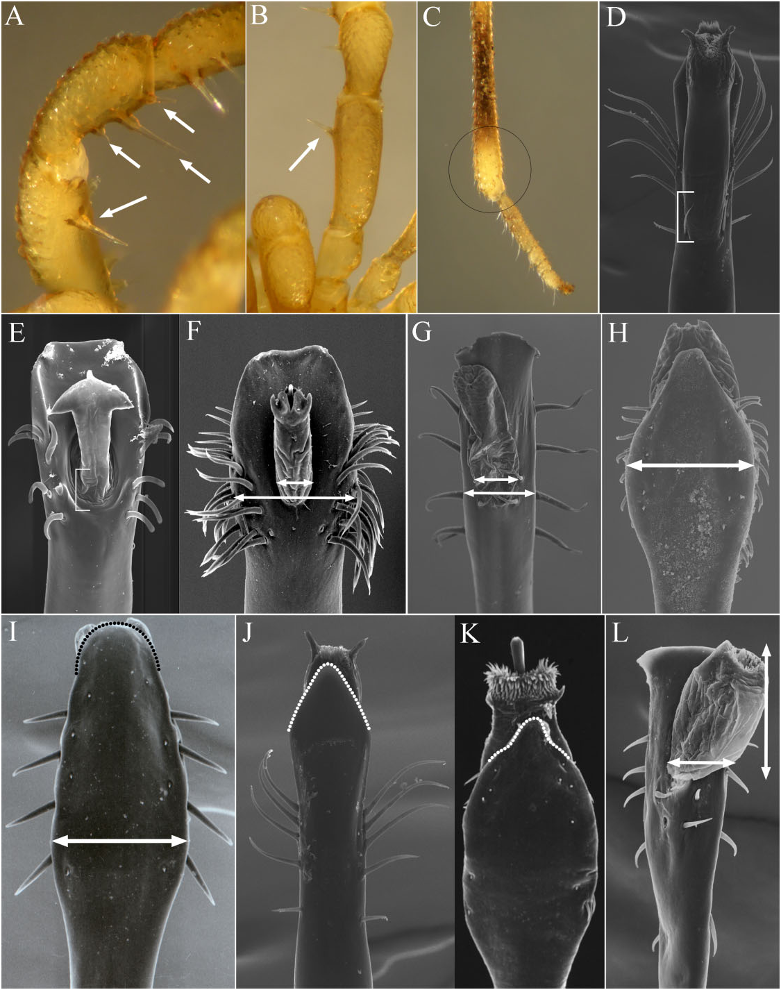
Figure 64. A, mesodorsal view of pedipalp femur and patella of Huasteca silhavyi sp. nov.; arrows indicate the setiferous tubercles on mesal side of these segments. B, dorsal view of pedipalp femur of Karos barbarikos; arrow indicates the mesodistal setiferous tubercle. C, apical portion of metatarsus IV of Karos singularis sp. nov., the circle indicates the swollen area. D, dorsal view of pars distalis of Karos barbarikos; line indicates the exposed base of the follis. E, dorsal view of pars distalis of Philora tuxtlae; line indicates the base of the follis in apical depression. F, dorsal view of pars distalis of Paramitraceras granulatum; arrows indicate the pars distalis width/follis width ratio. G, dorsal view of pars distalis of Crettaros valdezi sp. nov.; arrows indicate the pars distalis width/follis width ratio. H, ventral view of pars distalis of Chapulobunus unispinosus; arrow indicates the width at the middle of the ventral plate. I, ventral view of pars distalis of Mictlana inops the white arrow indicates the slight width in the middle of ventral plate, the black dotted line indicates the apical end of ventral plate. J, ventral view of pars distalis of Karos barbarikos, the white dotted line indicates the apical end of ventral plate. K, ventral view of pars distalis of Chapulobunus poblano sp. nov., the white dotted line indicates the apical end of ventral plate. L, dorsal view of pars distalis of Potosa dybasi; arrows indi-
| R |
Departamento de Geologia, Universidad de Chile |
No known copyright restrictions apply. See Agosti, D., Egloff, W., 2009. Taxonomic information exchange and copyright: the Plazi approach. BMC Research Notes 2009, 2:53 for further explanation.
|
Kingdom |
|
|
Phylum |
|
|
Class |
|
|
Order |
|
|
Family |
|
|
Genus |