
Karos singularis, Cruz-López & Francke, 2015
|
publication ID |
https://doi.org/10.1111/zoj.12299 |
|
persistent identifier |
https://treatment.plazi.org/id/03CA87F9-A242-FFB5-FE88-F9E7C7DEFF36 |
|
treatment provided by |
Felipe (2021-08-28 21:15:18, last updated by Plazi 2023-11-05 10:11:49) |
|
scientific name |
Karos singularis |
| status |
sp. nov. |
KAROS SINGULARIS View in CoL SP. NOV.
( FIGS 18–20 View Figure 18 View Figure 19 View Figure 20 , 59 View Figure 59 , 65F View Figure 65 , 61B, H View Figure 61 , 62C View Figure 62 , 64C View Figure 64 )
Type material: MEXICO: Querétaro: ♂ holotype [CNAN- T0723 (6.v.2011; O. Francke, A. Valdez, C. Santibáñez, J. Cruz, G. Contreras and R. Monjaraz)], (21°11′57.19″N, 99°13′06.78″W) Municipio Landa de Matamoros, Km 7.5 of the road to La Lagunita – Tilaco GoogleMaps . Hidalgo: 1 ♂ paratype [ CNAN-T0724 (6.xi.2010; O. Francke, A. Valdez, E. Miranda and J. Cruz)], (21°09′48.34″N, 98°56′33.86″W). Municipio Chapulhuacán, 1.5 km of insertion to Pisaflores GoogleMaps . 1 ♀ paratype [ AMNH (28.vii.1966; J. and W. Ivie)], (20°33′N, 99°07′12″W). Municipio Zimapán, 8 km N from Encarnación GoogleMaps . San Luís Potosí: 1 ♀ paratype [ AMNH (25.vii.1966; no collector)], (21°14′40.52″N, 98°46′21.28″W). Municipio Tamazunchale, 1.6 km SW from Tamazunchale GoogleMaps .
Etymology: From the Latin singularis (unique), in reference to the unique shape of the scutum.
Diagnosis: Sexual dimorphism in the shape of scutum remarkable, males with scutum broadly rounded, females subrectangular ( Fig. 18B View Figure 18 ). Ornamentation only present on midsection of mesotergal areas III and IV. Spines of the ocularium long, fused at their bases, par- allel, and close to each other ( Fig. 19C View Figure 19 ). Sexual length of femur ratio II: 1.38 and IV: 1.37. Dorsoectal apophyses of coxa IV present, sexually dimorphic. Trochanter II with dorsal apophysis in males ( Fig. 61H View Figure 61 ). Tibia IV in males curved and slightly swollen in the middle, with conspicuous ventral armature ( Fig. 19A View Figure 19 ). Male genitalia: apical margin slightly convex, dentate. Lateral setae cylindrical basally, flattened distally, basal pair shorter than the rest and ventrally displaced. Parastylar setae lateral to follis, basal pair slightly displaced towards follis base. Spiniform projections exposed, noticeably larger than in the other species ( Fig. 20 View Figure 20 ).
Description: Male holotype: Measurements: scutum length 3.0, maximum scutum wide 2.9. Legs. I 1.35/ 0.50/1.07/1.40, II 2.50/0.85/2.07/2.05, III: 1.65/0.60/ 1.45/1.75, IV: 2.75/0.85/2.50/2.77. Dorsum. Scutum Ming vase-shaped, but noticeably broad in the middle ( Fig. 18B View Figure 18 ). Tubercles of the transversal rows present only in mesotergal areas II–IV, increasing in size posteriorly, area II with few central tubercles; tubercles of area III small. Spines of the ocularium long, fused at the bases, parallel and close to each other ( Fig. 19C View Figure 19 ). Lateral clear areas on sides of scutum projected with triangular shape, blunt apically. Lateral clear areas of area V apices and free tergites I and II present, blunt in area V, spiniform in free tergites. Venter. Densely covered by spiniform tubercles, these are larger on coxa IV. Coxa IV only with dorsoectal apophysis, increasing in size distally. Pedipalps. Patella with two mesodistal tubercles. Legs. Legs I and II thinner and less ornate than posterior legs. Femur III curved. Trochanter III globular. Trochanter II with dorsal apophysis ( Fig. 61H View Figure 61 ). Ventral armature of femur IV formed by scattered spiniform tubercles. Distal portion of femur IV with noticeable spiniform tubercles. Tibia IV mesally curved, swollen, and with ventral armature ( Fig. 19A View Figure 19 ). Tarsal count 4(2):6–7(3):6:6. Male genitalia. Apical margin slightly convex, dentate. Lateral setae with bases cylindrical, flattened apically. Basal pair small and displaced ventrally. Ventral microsetae level with distal pair of lateral setae. Two pairs of parastylar setae lateral to follis, basal pair slightly displaced towards base of follis. Spiniform projections exposed. Lateral margins of ventral plate concave ( Fig. 20 View Figure 20 ). Female paratype: Measurements: scutum length 2.7, maximum scutum width 2.2. Similar to male, with the following differences: scutum subrectangular, very different in shape from male; sexual proportion of femora to metatarsi: II 1.38/1.41/1.59/1.32 and IV 1.37/1.29/1.47/ 1.27 ( Figs 18A View Figure 18 , 19A View Figure 19 ). Femur IV thinner, ventral tubercles of leg IV smaller, tibia IV not curved, dorsoectal apophyses of coxa IV reduced. Tarsal count 4(2):6(3): 6:5/6.
Comparisons: Karos singularis sp. nov. is unique among the genus in having noticeable sexual dimorphism in scutum shape, and it is the only species of Karos with dorsal apophysis on trochanter II in males ( Fig. 61H View Figure 61 ).
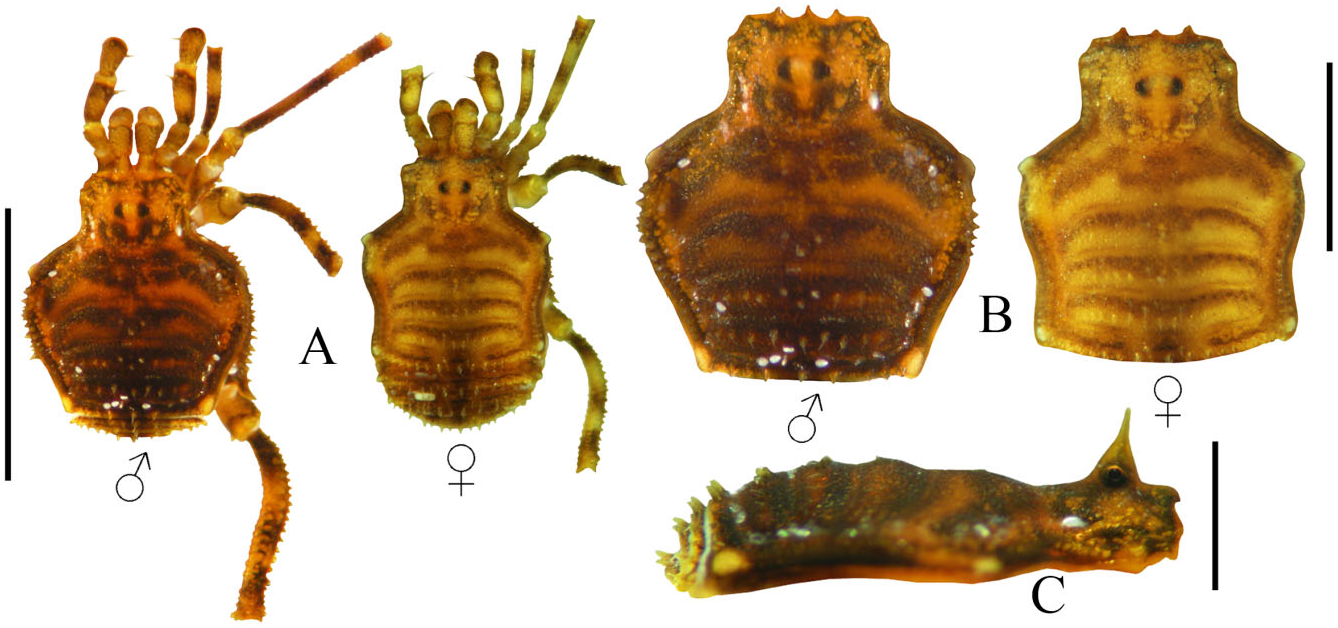
Figure 18. Karos singularis sp. nov. A, habitus, dorsal view. B, scutum, dorsal view. C, habitus, lateral view. Scale bars: A = 3.5 mm, B = 1.5 mm, C = 1 mm.
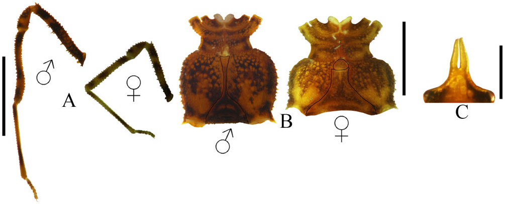
Figure 19. Karos singularis sp. nov. A, legs IV, mesal view. B, habitus, ventral view. C, ocularium, frontal view. Scale bars: A = 2.3 mm, B = 3.5 mm, C = 0.5 mm. The dark lines on (B) indicate the stigmatic area.

Figure 20. Karos singularis sp. nov. male genitalia. A, dorsal view. B, ventral view. C, lateral view. White arrows indicate ventral microsetae in (B) and parastylar setae in (C).

Figure 59. Distribution map of the species of the Karos genus-group, continued. White circles with black outline, Karos tersum sp. nov.; white circles with blue outline, Karos projectus; white circles with red outline, Karos singularis sp. nov.; black squares, Huasteca gratiosa comb. nov.; white square with black outline, Huasteca silhavyi sp. nov.; white square with red outline, Huasteca rugosa comb. nov.; black circles, Karos barbarikos.

Figure 61. A, detail of one of the lateral clear areas forming tubercles on scutum of Crettaros santibanezi sp. nov. B, detail of one of the lateral clear areas projected in tubercles on scutum of Karos singularis sp. nov. C, detail of one of the lateral clear areas projected in tubercles on scutum of Karos tersum sp. nov. D, detail of one of the lateral clear areas projected in tubercles on scutum of Huasteca silhavyi sp. nov. E, detail of one of the lateral clear areas projected in tubercles on scutum of Montabunus foliorum. F, detail of one of the lateral clear areas projected in tubercles on scutum of Monterella tuberculata. G, extra row of pegs in Chapulobunus poblano sp. nov., indicated by arrows. H, dorsal apophyses on trochanter II in male of Karos singularis sp. nov. I, detail of mesotergal areas III and IV in Montabunus foliorum; dotted lines indicate the sulcus between these areas. J, detail of mesotergal areas III and IV on Chapulobunus unispinosus; dotted lines indicate the sulcus between these areas. These images are not at the same scale.

Figure 62. A, dorsal view of lateral clear areas forming tubercles of Karos tersum sp. nov.; arrows indicate the clear areas. B, dorsal view of lateral clear areas projected in tubercles of Huasteca silhavyi sp. nov.; arrows indicate the clear areas. C, lateral view of posterior scutum of Karos singularis sp. nov., showing the spiniform tubercles. D, lateral view of posterior scutum of Montabunus foliorum, showing the rounded tubercles. E, dorsal view of row of pegs in Karos barbarikos; some pegs are indicated by black dots. F, dorsal view of row of pegs in Hoplobunus boneti; some pegs are indicated by black dots. These images are not at the same scale.
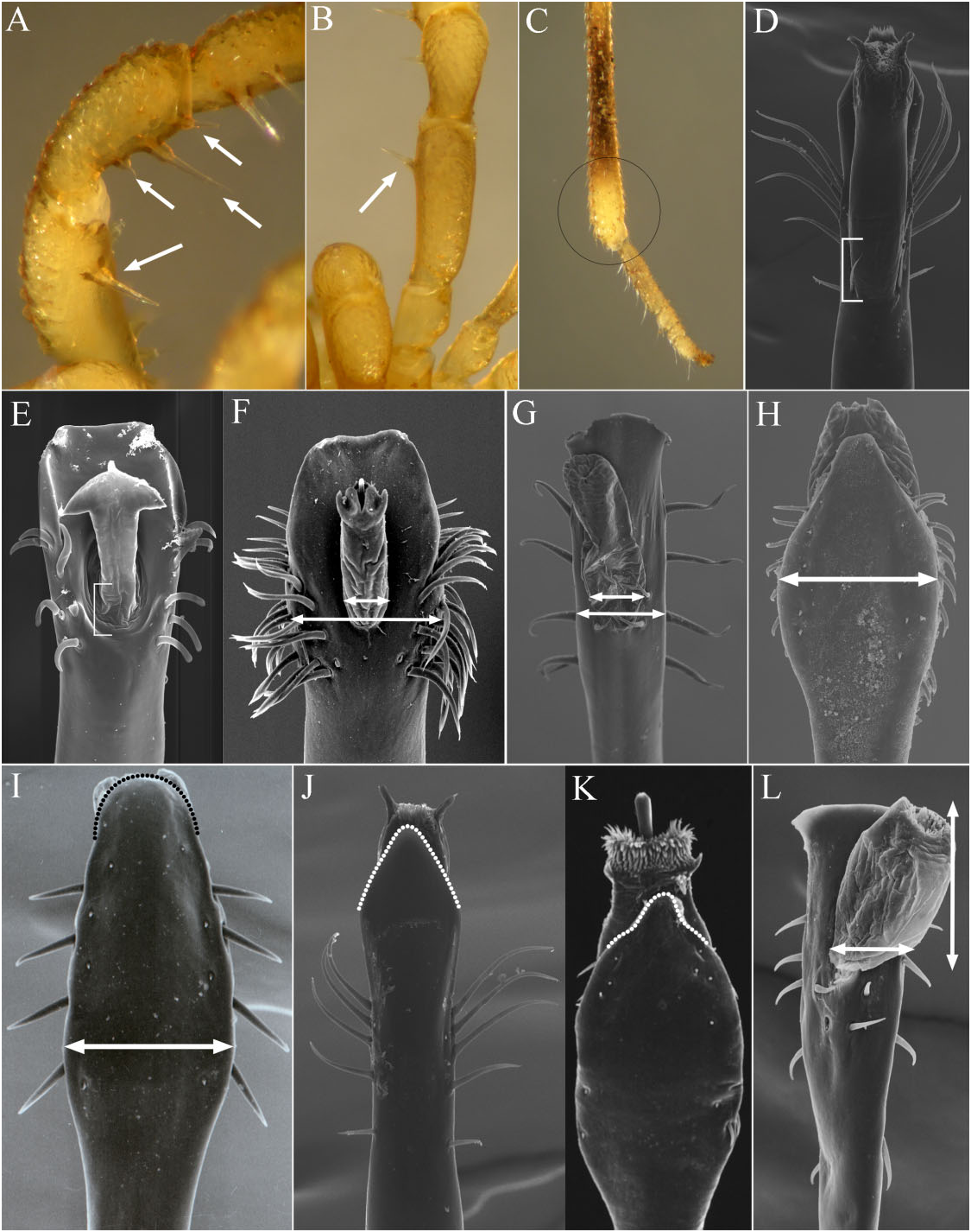
Figure 64. A, mesodorsal view of pedipalp femur and patella of Huasteca silhavyi sp. nov.; arrows indicate the setiferous tubercles on mesal side of these segments. B, dorsal view of pedipalp femur of Karos barbarikos; arrow indicates the mesodistal setiferous tubercle. C, apical portion of metatarsus IV of Karos singularis sp. nov., the circle indicates the swollen area. D, dorsal view of pars distalis of Karos barbarikos; line indicates the exposed base of the follis. E, dorsal view of pars distalis of Philora tuxtlae; line indicates the base of the follis in apical depression. F, dorsal view of pars distalis of Paramitraceras granulatum; arrows indicate the pars distalis width/follis width ratio. G, dorsal view of pars distalis of Crettaros valdezi sp. nov.; arrows indicate the pars distalis width/follis width ratio. H, ventral view of pars distalis of Chapulobunus unispinosus; arrow indicates the width at the middle of the ventral plate. I, ventral view of pars distalis of Mictlana inops the white arrow indicates the slight width in the middle of ventral plate, the black dotted line indicates the apical end of ventral plate. J, ventral view of pars distalis of Karos barbarikos, the white dotted line indicates the apical end of ventral plate. K, ventral view of pars distalis of Chapulobunus poblano sp. nov., the white dotted line indicates the apical end of ventral plate. L, dorsal view of pars distalis of Potosa dybasi; arrows indi-
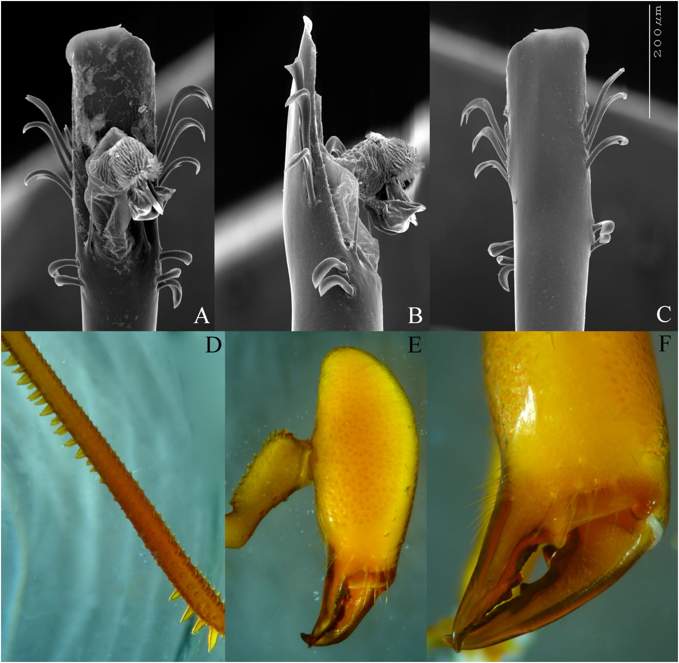
Figure 65. A, dorsal view of the male genitalia of Hoplobunus boneti. B, lateral view of the male genitalia of Hoplobunus boneti. C, ventral view of the male genitalia of Hoplobunus boneti. D, lateral view of femur IV of Hoplobunus boneti. E, lateral view of chelicera of Hoplobunus boneti. F, frontal view of the cheliceral fingers of Hoplobunus boneti. The images in (A–C) are at the same scale indicate on (C). Images (D–F) are not at the same scale.
| R |
Departamento de Geologia, Universidad de Chile |
| AMNH |
American Museum of Natural History |
No known copyright restrictions apply. See Agosti, D., Egloff, W., 2009. Taxonomic information exchange and copyright: the Plazi approach. BMC Research Notes 2009, 2:53 for further explanation.
|
Kingdom |
|
|
Phylum |
|
|
Class |
|
|
Order |
|
|
Family |
|
|
Genus |