
Peltariosilis Wittmer, 1952
|
publication ID |
https://doi.org/10.11606/1807-0205/2020.60.special-issue.16 |
|
publication LSID |
lsid:zoobank.org:pub:3C128243-07E3-4435-A496-D8F50F68389E |
|
persistent identifier |
https://treatment.plazi.org/id/03CA7502-163A-367B-AEAA-FB69DA1736E4 |
|
treatment provided by |
Carolina |
|
scientific name |
Peltariosilis Wittmer, 1952 |
| status |
|
( Figs. 1-17 View Figure 1 View Figure 2 View Figure 3 View Figure4 View Figure 5 View Figure 6 View Figure 7 View Figure 8 View Figure 9 View Figure 10 View Figure 11 View Figure 12 View Figure 13 )
Silis (Peltariosilis) Wittmer, 1952: 203 , Delkeskamp 1977: 286; Constantin 2010a: 41.
Peltariosilis, Constantin 2010b: 15, 2017: 61 .
Type species: Silis (Peltariosilis) scutulata Wittmer, 1952 , by original designation.
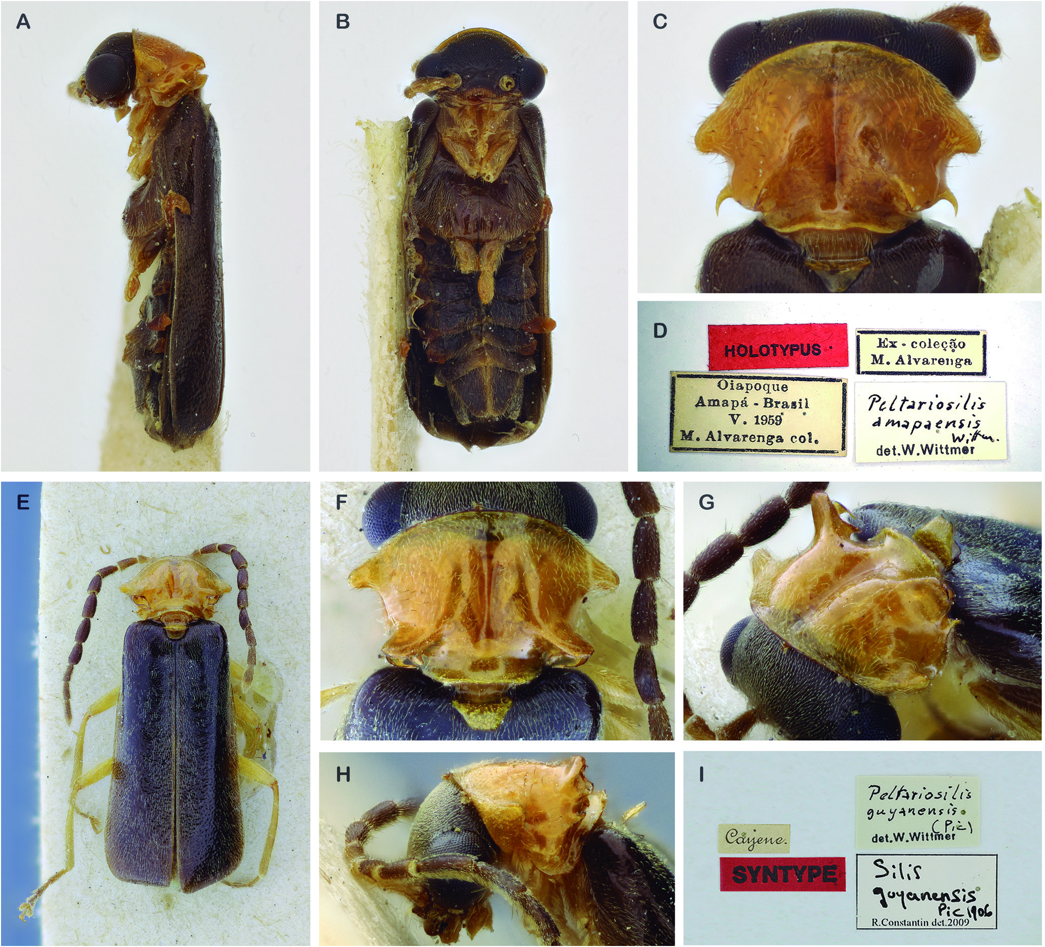
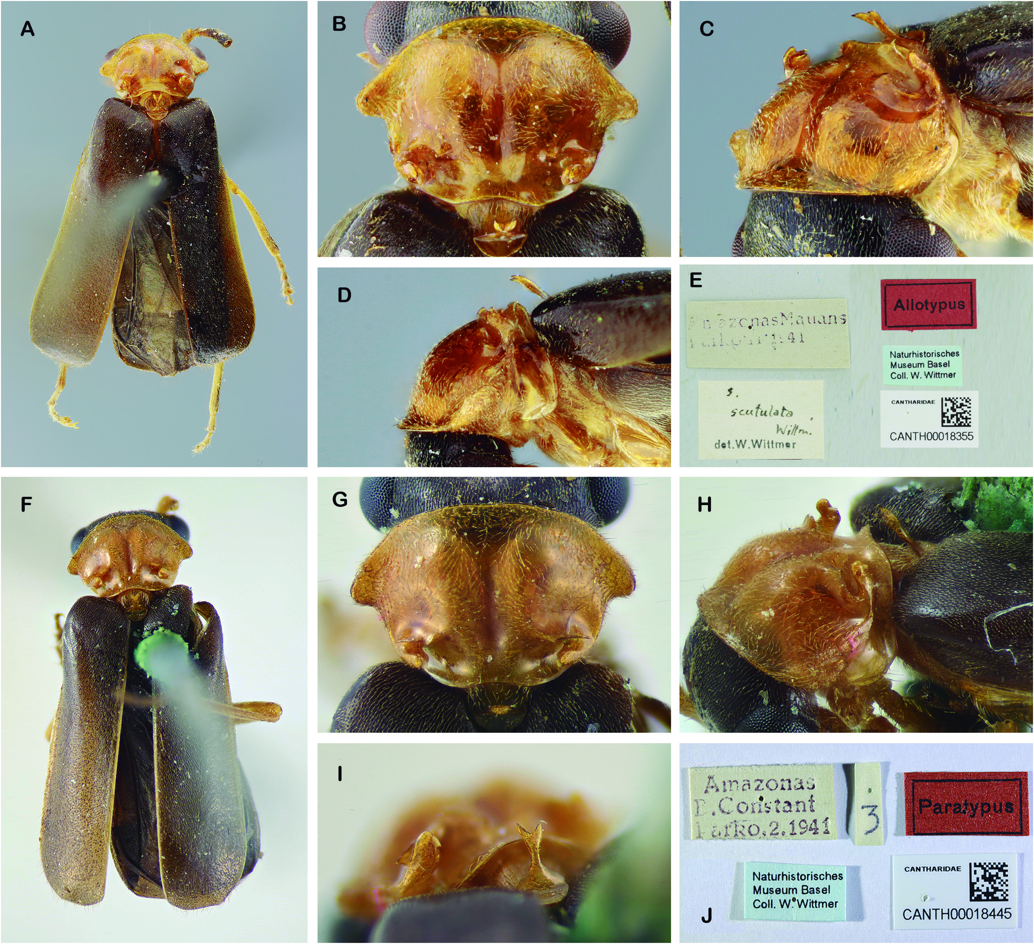
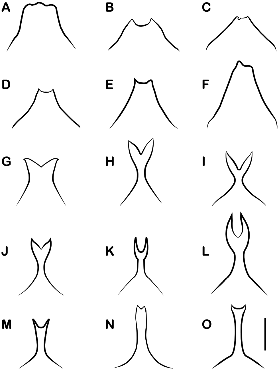
Diagnosis (Males): Clypeus slightly projected, anterior margin with an arched notch; mandibles with a minute tooth near base; pronotum variously modified, anterior margin broadly arched, sides with spines, lobes and projections; scutellum projected anteriorly in a broad lamella or narrow fork; anterior fore tarsal claw with a small rounded basal lobe, mid and hind anterior tarsal claw with small basal cleft. Peltariosilis is distinguished from other genera of Silinae by the unique scutellum, bearing an obliquely or perpendicularly rising lamella, sometimes with a forked apex.
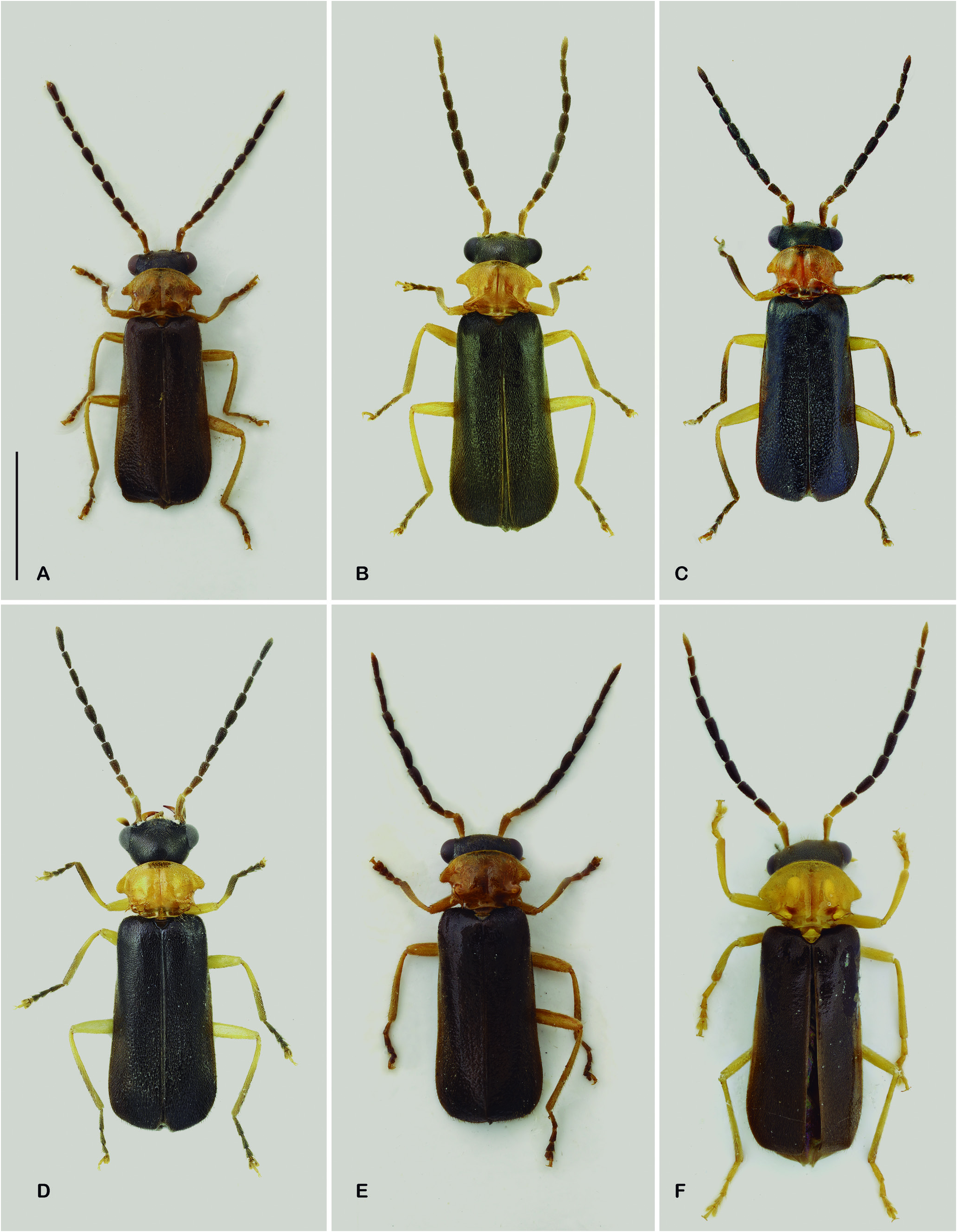
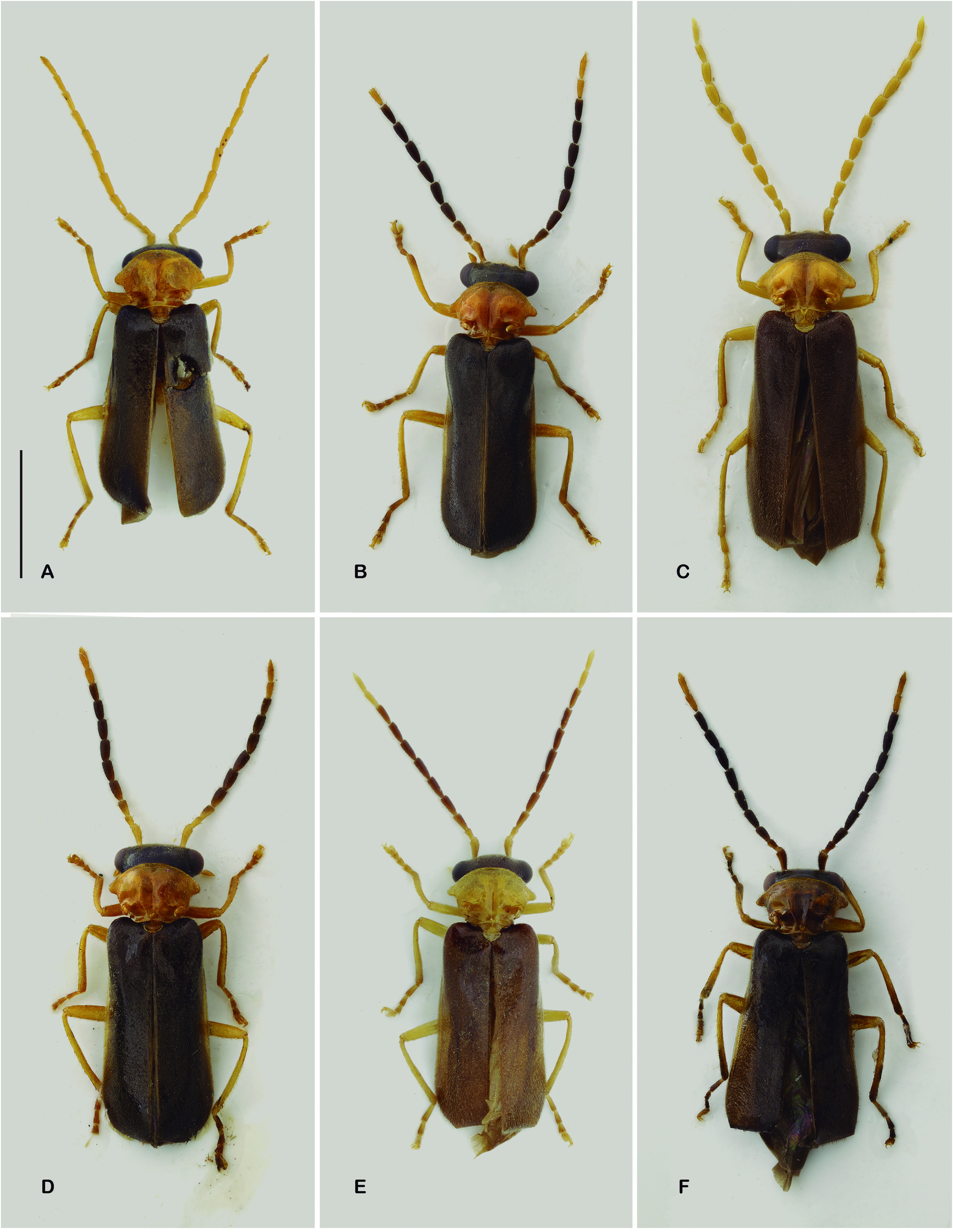
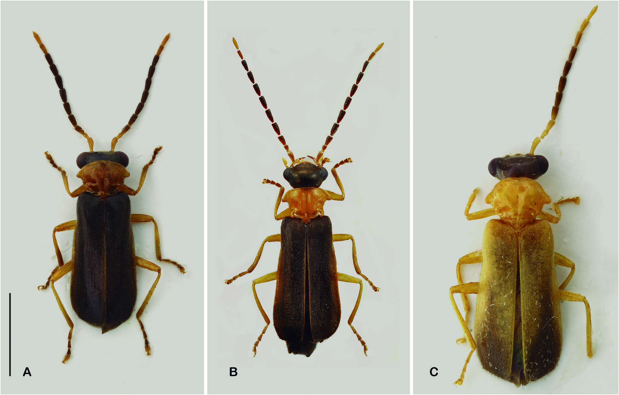
Description: Head, thorax, wings and abdomen dark brown to black; antennae ranging from entirely pale yellow to entirely dark brown; elytra usually dark brown to black, sometimes lighter near lateral margins and suture, or broadly yellowish light brown; pronotum and scutellum pale yellow to light orange brown, sometimes trans- lucent. Body regularly covered with dense very short and fine adpressed pubescence and sparse longer setae on antennae and abdomen.
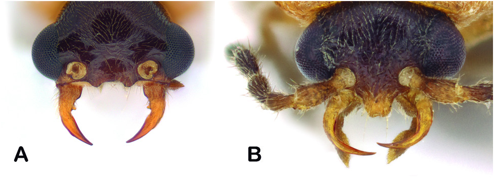
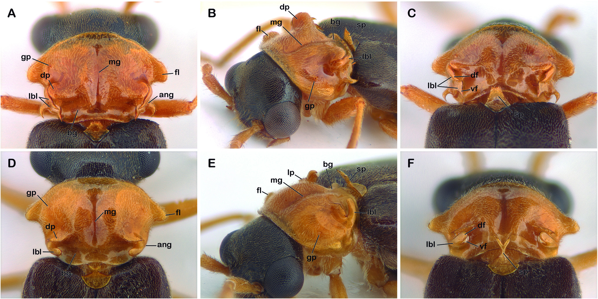
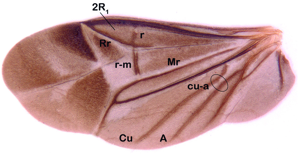
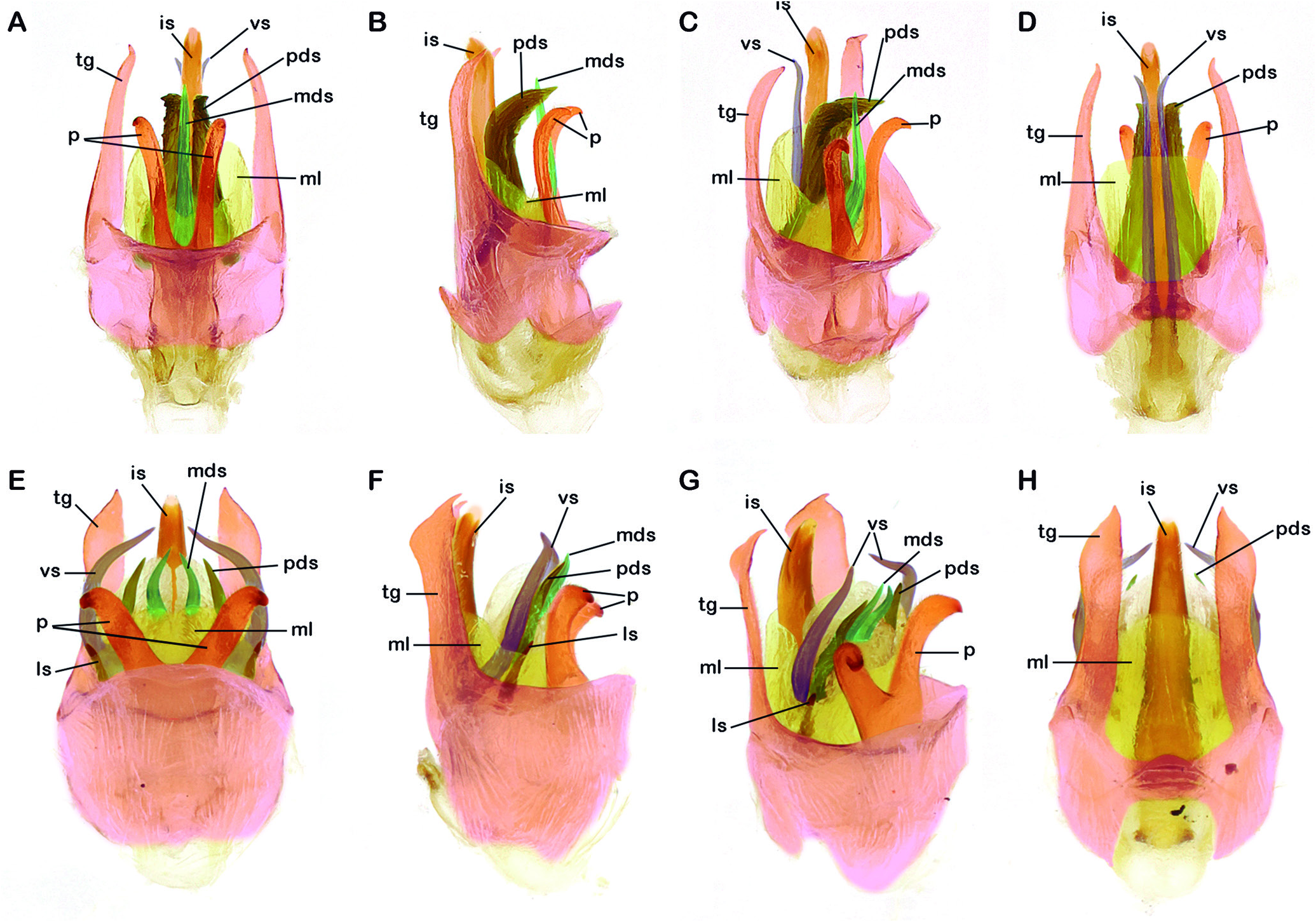
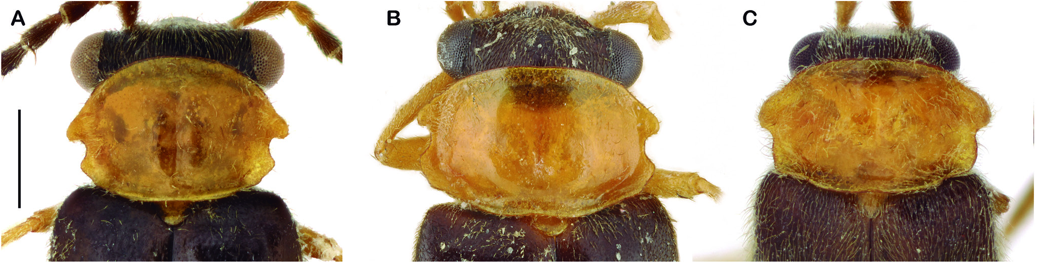
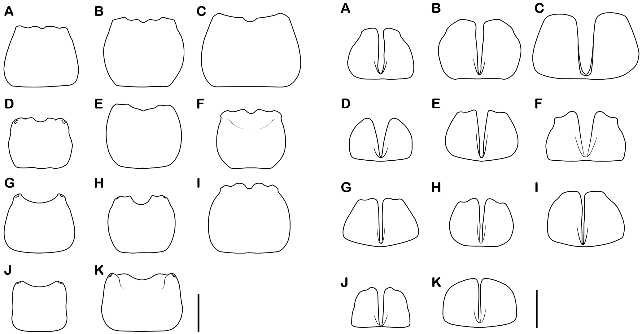
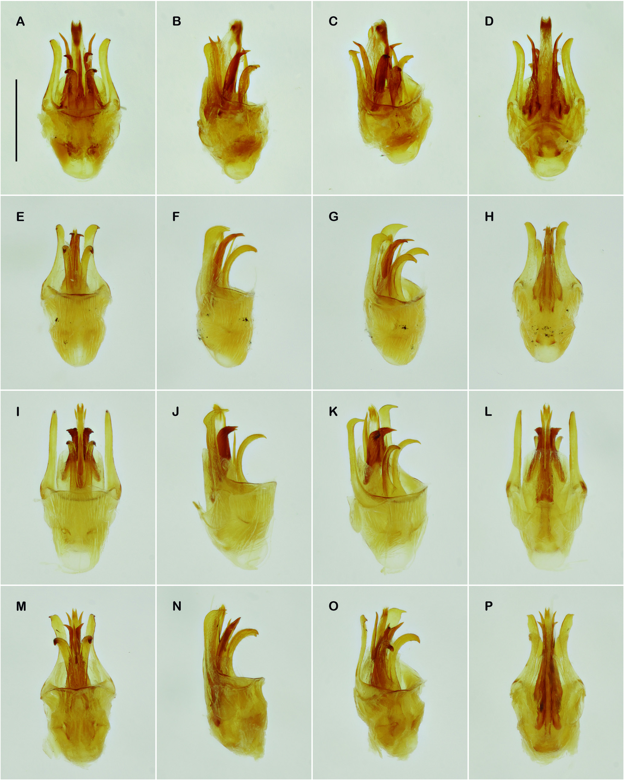
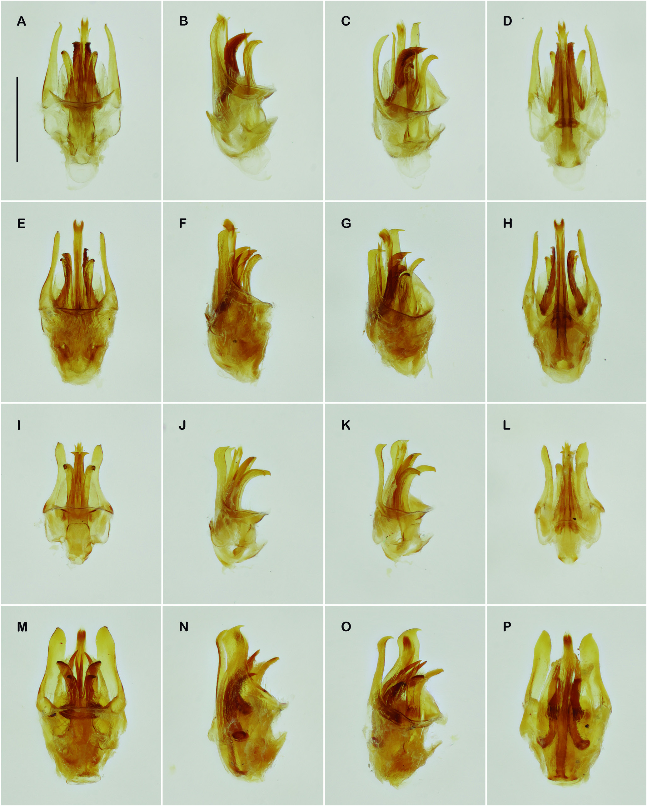
Males: Head ( Fig. 1A View Figure 1 ) (including eyes) broad, nearly as wide as pronotum, ventral part retracted; gula long, gular sutures broadly separated; occipital region and vertex broadly convex, frons flattened; clypeus broadly fused to frons, frontoclypeal suture absent; clypeus broad, slightly prominent, anterior margin with an arched notch; labrum membranous, very reduced, barely distinct in frontal view; genae very short, indistinct in front of eyes. Eyes prominent, bulging, rounded, nearly as long as wide, broadly separated. Antennal insertions broadly separated; antennae usually filiform, sometimes with swollen, rounded antennomeres. Mandibles ( Fig. 1A View Figure 1 ) long, stout, apex acute and sharp, smooth or with a minute tooth near base, lateral margins with rath- er long and dense pubescence. Maxillae weakly sclerotised; stipes short and broad, galea and lacinia membranous, densely setose; maxillary palpi longer than stipes; palpomeres I-III similar in shape, elongate, IV securiform. Labium weakly sclerotised, submentum membranous, palpi 3-segmented, palpomere III securiform. Pronotum ( Figs. 2 View Figure 2 , 10 View Figure 10 ) variously modified; anterior margin broadly arched, sides forming a frontolateral lobe (fl); sides behind frontolateral lobe parallel to slightly convergent posteriorly, distalmost part forming a broad or narrow laterobasal lobe (lbl) directed up and medially; angles (ang) of laterobasal lobes acute, obtuse, round- ed or forming an unciform spine; apex of laterobasal lobe entire or divided into dorsal (df) and ventral flaps (vf). Dorsal surface forming two lateral bumps divided by a median groove (mg), a pair of dorsal projections (dp) that may be broadly rounded or sharp, razor blade shape, sometimes merging with laterobasal lobe; a deep basal groove (bg) behind laterobasal lobes; a pair of glandular pores anteriorly (gp). Scutellum ( Figs. 2 View Figure 2 , 12 View Figure 12 ) projected (sp) upwards and anteriorly in a broad trapezoidal lamella ( Figs. 2C View Figure 2 , 12 View Figure 12 A-F) or in a narrow stem divid- ed apically in a bifid, well-defined fork ( Figs. 2F View Figure 2 , 12 View Figure 12 G-O). Elytra smooth, long, completely concealing the wings and abdomen; lateral sides parallel to divergent, wider posteriorly; apex rounded to nearly truncate; lateral and sutural margins not forming distinct borders. Thorax weakly sclerotised, metaventrite convex. Legs small, slender, without strong modifications in size, shape or pubescence; tibiae with one pair of tiny apical spurs; fourth tarsomere with a transversal slit at base; anterior fore tarsal claws with a small rounded basal lobe; anterior mid hind tarsal claws with a small straight basal cleft. Wings ( Fig. 3 View Figure 3 ) short and broad, radial cell 2R₁ closed, veins r and r-m not coinciding in the meeting point of Rr, Rr prolonged anteriorly beyond the meeting point of r-m, vein cu-a weakly sclerotised, barely visible, anal vein (A) not divided. Abdomen weakly sclerotised, flattened, short and broad; glandular pores very small, barely visible, not prominent, located near the distal margin of tergites; tergite VIII ( Fig. 13 View Figure 13 ) long and broad, with lateral margins straight to arched, glandular pores shallow to rather prominent, distal margin variously notched; ventrites I-VI very wide, distal margin straight; ventrite (i.e., visible sternite) VII ( Fig. 14) with a very deep longitudinal incision, dividing it into two halves. Aedeagus ( Figs. 4 View Figure4 , 15-17 View Figure 15 View Figure 16 View Figure 17 ): ventral plaque of tegmen (tg) lateral, flattened, very elongate, nearly as long as internal sac, variously shaped, straight, curved or sinuous, apex acute, rounded or truncate; parameres (p) dorsally, united at base and moderately to broadly divergent from mid length, elongate, broadly curved dorsally, apex acute; median lobe (ml) thinly membranous, translucent, tubular around internal sac and a series of sclerites; internal sac (is) membranous, very elongate, slen- der, straight to slightly curved dorsally, apex sometimes slightly swollen; a series of sclerites variable in number, position and shape: one pair of median dorsal sclerite (mds) that can be separated, fused medially or totally fused forming one single median sclerite, dorsal, very elongated, straight to curved ventrally, apex acute; one pair of paramedian dorsal sclerites (pds) robust, strongly sclerotised, flattened, usually curved or inclined dorsally, apex acute, gradually or abruptly narrowed, sometimes with scratches or projecting teeth; one pair of ventral sclerites (vs) very long, slender, pressed against ventrolateral wall of internal sac, apex acute, usually directed laterally; one pair of lateral sclerites (ls) present or absent.
Female: Similar to males in colouration and general morphology, differing from males on the base of dimorphic features, such as the antennae not swollen at middle antennomeres, pronotum ( Fig. 11 View Figure 11 ) wider and flattened, without strong modifications, anterior margin less arched, frontolateral and laterobasal lobes rounded, short, not pronounced or recurved; scutellum flattened, not projected anteriorly; pygidium more transverse; abdominal ventrite VII entire, not longitudinally divided, not distinctive among species.
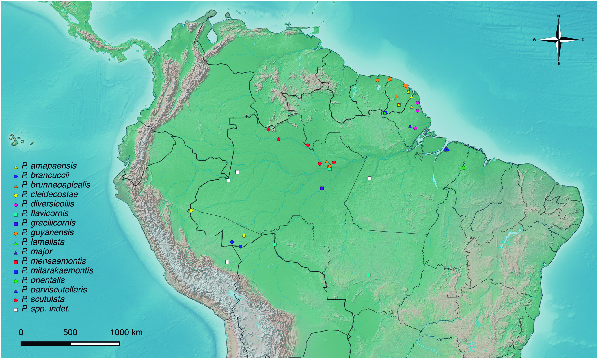
Distribution: French Guiana, Suriname, Brazil, Peru, Bolivia ( Fig. 18 View Figure 18 ).


No known copyright restrictions apply. See Agosti, D., Egloff, W., 2009. Taxonomic information exchange and copyright: the Plazi approach. BMC Research Notes 2009, 2:53 for further explanation.
|
Kingdom |
|
|
Phylum |
|
|
Class |
|
|
Order |
|
|
Family |
Peltariosilis Wittmer, 1952
| Biffi, Gabriel & Geiser, Michael 2020 |
Peltariosilis, Constantin 2010b: 15 , 2017: 61
| Constantin, R. 2017: 61 |
| Constantin, R. 2010: 15 |
Silis (Peltariosilis)
| Constantin, R. 2010: 41 |
| Delkeskamp, K. 1977: 286 |
| Wittmer, W. 1952: 203 |