
Hetereleotris semisquamata Kovačić & Bogorodsky, 2019
|
publication ID |
https://doi.org/10.11646/zootaxa.4608.3.5 |
|
publication LSID |
lsid:zoobank.org:pub:5B31798B-FFA3-42F3-9F3B-DE81B1C318EC |
|
DOI |
https://doi.org/10.5281/zenodo.5696370 |
|
persistent identifier |
https://treatment.plazi.org/id/03C98787-5F74-FFBC-FF78-FF70FE67FEC6 |
|
treatment provided by |
Plazi |
|
scientific name |
Hetereleotris semisquamata Kovačić & Bogorodsky |
| status |
sp. nov. |
Hetereleotris semisquamata Kovačić & Bogorodsky sp. nov.
Semiscaly Goggle goby
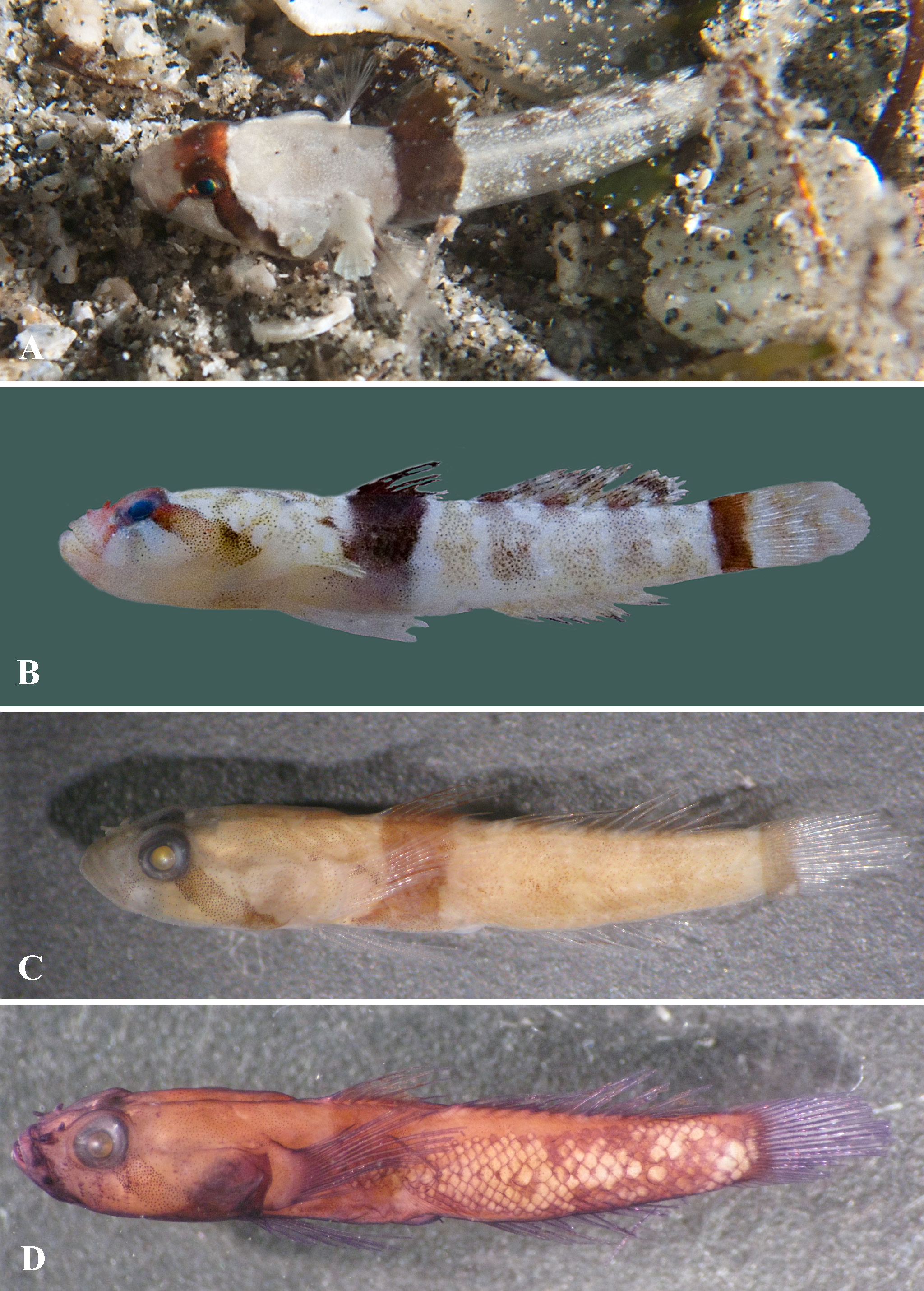
( Figures 3 View FIGURE 3 & 4 View FIGURE 4 )
Holotype. PMR VP3053 View Materials , female, 12.58 mm, caudal fin damaged, Red Sea, Southern Egypt, Shams Alam, 24°41’37.95” N, 35°41’15.80”E, coll. S.V. Bogorodsky, 14 July 2006. GoogleMaps
Diagnosis. Dorsal-fin rays VI + I,11; anal-fin rays I,10; pectoral-fin rays 16, all rays branched; pelvic-fin rays I,5, the fin separated and without frenum, fifth ray unbranched; head length 34.9% of standard length; anterior nostril with a long nasal tube without process from the rim, posterior nostril tube slightly shorter than anterior nasal tube, 0.9 of anterior tube; no tentacle above eye; posterior angle of jaws ending posteriorly to below anterior edge of eye; no opercular spine; no mental frenum; pelvic fin length 23.3% of standard length, pelvic fin shorter than pectoral fin; scales cycloid, along lateral midline tapering from caudal-fin base towards pectoral fin where nearly reaching its base; no head canals; suborbital rows of papillae with four transverse rows; two distinct preorbital rows s present from posterior nostril to above upper lip; row 3 short, extending from lower edge of eye ending near row d, row 4 long, curving at eye extending below of level of row d, row b longitudinal, short, close to middle of transverse row 4 and slightly behind vertical at posterior edge of eye, row d discontinuous; anterior dorsal rows of sensory papillae o and m present; head and body whitish, with brown line from eye to end of upper lip, dark brown band over eye crossing interorbital area and continuing obliquely from eye to corner of opercle, broad dark brown band below first dorsal fin continuing into fin, and vertical dark brown band on caudal-fin base ( Figs. 3A & B View FIGURE 3 ).
Description. Body moderately elongate, its depth at pelvic-fin origin 6.4 in SL, at anal-fin origin 5.8 in SL, laterally compressed posteriorly, caudal-peduncle depth 0.8 of body depth at anal-fin origin, with caudal peduncle moderately deep, caudal peduncle depth 0.7 of caudal peduncle length ( Fig. 3 View FIGURE 3 ). Head large, its length 2.9 in SL, width 4.1 in SL, depth 5.9 in SL, and depressed, its depth 0.7 of width. Snout broad, from dorsal view slightly convex anteriorly, almost rectangular, gently sloping in profile from lateral view, and long, its length 1.0 of eye diameter, 3.9 in head length. Anterior nostril with long tube without process from the rim, posterior nostril tube slightly shorter than anterior nasal tube, 0.9 of anterior nasal tube. Eyes dorsolateral, of moderate size, eye diameter 4.1 in head length, orbit slightly projecting above dorsal profile. Interorbital narrow, 6.0 in eye diameter. No tentacle above eye. Mouth terminal, oblique, jaws ending about equal anteriorly. Mouth small, posterior angle of jaws ending posteriorly below anterior edge of eye. Cheek moderately broad. Upper jaw with single row of recurved pointed teeth; lower jaw with outer row of recurved pointed teeth and smaller teeth behind them as irregular row. Tongue truncated. No mental frenum. Ventrolateral head ridge well-developed, narrowing gular region in “)(“ shape, but distant from fellow at the midline. Branchiostegal membranes fused to isthmus, gill openings restricted to pectoralfin base. Lower limb of first gill arch joined to gill cover by membrane. No spines on preopercle.
Fins. First dorsal fin VI, second dorsal fin I,11; anal fin I,10; branched caudal-fin rays 15, segmented 17. Pectoral-fin rays 16, all rays branched, upper rays not free at tips. Pectoral girdle without flaps on anterior edge. Pelvic fins I,5 + 5,I, left and right fin completely separated and without frenum, fifth ray unbranched, other rays branched, fifth ray longest. Spines of first dorsal fin not elongate or filamentous, second and third spines of first dorsal fin longest, about equal in length, second only slightly longer than third spine, sixth spine short; third to sixth spines of first dorsal fin barely reaching to origin of second dorsal fin when folded down. Origin of first dorsal fin behind vertical at pectoral-fin base. No fin membrane connecting last spine of first dorsal fin and second dorsal-spine base i.e. gap present. Origin of anal fin below vertical of second segmented ray of second dorsal fin. Pectoral fins extending posteriorly to below origin of second dorsal fin. Pelvic fins not reaching anus, 0.9 of distance between pelvic fins and the origin of anus, shorter than pectoral fins, 0.9 of pectoral-fin length. Caudal fin rounded, shorter than head ( Fig. 3B View FIGURE 3 , damaged in preserved specimen).
Squamation ( Fig. 3D View FIGURE 3 ). The squamation reduced, scales cycloid. Scales along lateral midline start from caudalfin base, tapering towards pectoral fin, nearly reaching its base, ending between axil and vertical at first dorsal-fin origin. Head, predorsal area, breast, belly, dorsal- and anal-fin bases, most of anterior dorsal half of body, and narrow area on dorsal and ventral part of caudal peduncle naked. Lateral longitudinal scale count 29 on both sides, transverse scale counts not possible, starting and ending points for the count both scaleless.
Cephalic sensory systems ( Fig. 4 View FIGURE 4 ). No head canals. Rows of head sensory papillae were counted on the both side (left, right), only left side papillae presented on Fig. 4 View FIGURE 4 . Rows of head sensory papillae reduced and additional individual larger papillae present on the position of missing head canals. Preorbital rows: snout with three median preorbital series, upper row r (1, 1) middorsally between eyes above posterior nostrils, s 1 (1, 1) at posterior nostril, row s 3 (1, 1) above upper lip. Lateral series c in four parts: superior c 2 (2, 2) between posterior nostril and anterior nasal tubes, middle c 1 (2, 2) below anterior nasal tube, inferior rows: upper horizontal c 2 (2, 2) above upper lip and lower horizontal c 1 (1, 1) between upper lip and row 1. Suborbital rows: no row a. Row b (2, 2) longitudinal, short, anteriorly beginning close to transverse row 4 and slightly behind vertical of posterior edge of eye. Four transverse suborbital rows ( 1–4) of sensory papillae, row 1 longer, extending from eye reaching upper lip, row 2 short and distant from eye, row 3 short, extending from lower edge of eye ending ventrally close to row d, row 4 long, slightly curving at ventro-posterior edge of eye, then extending vertically below of level of row d ( 1: 6, 5, 2: 4, 4, 3: 4, 5, 4: 10, 10). Row d discontinuous (5 + 4, 5 + 4), anterior part above upper lip at angle of mouth, posterior part below posterior half of eye, high on cheek i.e. above angle of jaw for about 1/3 of distance from horizontal level of angle of jaw to eye, starting anteriorly below row 3 and ending posteriorly nearly transverse suborbital row 4. Preoperculo-mandibular rows: external row e (13 + 11, 13 + 12) longitudinal and uniserial, divided into anterior and posterior sections; internal row i uniserial, divided into anterior and posterior sections (7 + 7, 7 + 8); mental row f longitudinal (4, 4). Oculoscapular rows: three larger papillae longitudinally arranged on position of missing anterior oculoscapular canal behind eye; two larger papillae longitudinally arranged on position of missing posterior oculoscapular canal above opercle (marked aoc and poc on Fig. 4 View FIGURE 4 , respectively); longitudinal row x 1 (3, 3) short; posterior longitudinal row x 2 (1, 1) above two papillae on position of missing posterior oculoscapular canal; anterior lower transverse row z (4, 4) slightly in front from vertical of anterior end of row x 1; transverse row q (2, 2) below and behind posterior end of row x 1; row u (2, 2) between row q and papillae on the position of missing posterior oculoscapular canal; row y absent. Axillary transverse rows as 1 (1, 1), as 2 (1, 1), as 3 (1, 1) present; row la 1 (1, 1) and row la 2 (1, 1) above rows as. Opercular rows: two larger papillae in the position of missing preopercular canal (marked pc on Fig. 4 View FIGURE 4 ); transverse row ot (9, 10); superior longitudinal row os (3, 3); and inferior longitudinal row oi (3, 3). Anterior dorsal rows: single papilla behind eye on position of the missing pore ω of the anterior oculoscapular canal (marked aoc on Fig. 4 View FIGURE 4 ), transverse row n (2, 2) behind it, row o (1, 1) in front and middorsally to row g, row g (2, 2) longitudinal; row m (1, 1) behind and below row g, row h longitudinal (1, 3) in front of the first dorsal fin. Interorbital rows: two pairs of larger papillae present in interorbital, one anteriorly on the position of the missing pore λ and one posteriorly on the position of the missing pore κ (marked aoc on Fig. 4 View FIGURE 4 ).
Color of live female ( Fig. 3A View FIGURE 3 ). In life head white with bright brown line from ventroanterior edge of eye to the end of upper lip and with dark brown band over eye and interorbital continuing obliquely from ventroposterior edge of eye to corner of opercle. Iris and posterior nostril within dark brown bar, pupil dark green. Body transparent white with broad, vertical, dark brown band below entirely base of first dorsal fin and vertical, moderately broad, dark reddish brown bar on caudal-fin base. The first dorsal fin dark brown as band below it, with whitish posterior edge, the second dorsal fin with whitish and brown pigmented areas. Pectoral fin intensively white at base, rest of fin transparent. Other fins and ventral body side not visible on photo.
Color of fresh female ( Fig. 3B View FIGURE 3 ). After death the specimen lost transparency and head and body became white opaque, retaining basic overall coloration pattern of live specimen, including brown line and dark brown band on head, dark brown band below first dorsal fin and vertical dark brown band on caudal-fin base. Head mottled, dorsal half covered with dark brown dots. Body behind band with five indistinct vertical bars which are densely dotted with dark brown. Caudal fin behind vertical dark brown band with weak whitish and pale brown areas. Anal and pelvic fins whitish, the anal fin with blackish margin. Pectoral fin white at base, rest of fin transparent.
Color of preserved female ( Fig. 3C View FIGURE 3 ). Head and body fawn. Head with poorly visible brown stripe from eye to upper lip and with brown bar over eyes and interorbital and from eye back and down to opercular lower edge. Iris dark, pupil grey. Body with brown band below entire base of first dorsal fin i.e. below all six first dorsal-fin spines, and with vertical brown band on caudal-fin base. Diffused coloration pattern from melanophores, head mottled and body with five vertical bars laterally, starting at dorsal-fin origins, even less visible than in fresh condition. First dorsal fin brown as band below it, second dorsal fin with traces of brown pigment poorly visible due to broken membrane. Caudal fin behind vertical dark brown band transparent. Anal, pectoral and pelvic fins transparent.
Etymology. The specific epithet is from the Latin semi, half, and squamata, scaly, and refers to the unique squamation characterized in wedge-shaped pattern retaining dorsal half of anterior body and entirely belly naked.
Habitat. Known only on the basis of the holotype collected from reef flat of seaward reef at Shams Alam, southern Egypt.
Remarks. Based on the shape of urogenital papillae, the holotype could be late juvenile to subadult female or even adult female if species has less prominent urogenital papilla. In this stages the final gobiid squamation is formed and visible, furthermore continuous scales along lateral midline are already present in gobies of similar size even in small juvenile of unidentified sex ( Kovačić et al. 2013). However, in one particular case of gobiid species with semi-reduced squamation the intraspecific variability of area covered by scales was recorded among adults ( Kovačić 2005). Therefore, for this unlikely possibility, the present Hetereleotris key and the new species diagnosis are shaped to be fully functional excluding the squamation data. Regarding the fin meristic characters in diagnosis the same remark can be applied as for Hetereleotris aurantiaca .
The new species is distinct from all 17 described Hetereleotris species and from H. aurantiaca by unique squamation. Hetereleotris semisquamata has cycloid scales, the squamation reduced, along lateral midline, in wedgedshaped pattern, nearly reaching the narrow end the pectoral-fin base anteriorly. Head, predorsal area, breast, belly, dorsal- and anal-fin bases naked, most of dorsal half of anterior body, and narrow naked areas on top and ventrally on caudal peduncle naked (vs. five completely scaleless Hetereleotris species, H. tentaculata with caudal peduncle scaled in some specimens, five Hetereleotris species with only caudal peduncle scaled and seven densely scaled species with continuous scales along lateral midline to pectoral-fin axil and at least caudal peduncle completely scaled).
The new species is different from 14 described Hetereleotris species and from H. aurantiaca by coloration of live and preserved specimen in having dark brown band over eyes and across interorbital continuing diagonally from eye to opercular lower edge and vertical brown band below first dorsal fin, in which it resembles H. diademata , H. kenyae , and H. zonata . In addition to similarity in coloration pattern, the new species shares with H. diademata , H. kenyae and H. zonata general shape of head and body, but also have some firm differences in coloration and morphology. The new species differs from H. diademata in having mainly scaled body vs. body scaleless, by fewer rays in the second dorsal fin (I,11 vs. I,12) and in anal fin (I,10 vs. I,11), in having broad dark band below entire base of the first dorsal fin, i.e. below all six first dorsal-fin spines vs. dark band narrower, below second to fourth spines only; and in having vertical dark band on caudal-fin base vs. no vertical dark band on caudal-fin base. The new species differs from H. kenyae , in addition to squamation, by fewer rays in the second dorsal fin (I,11 vs. I,12), in lacking head canal (vs. anterior oculoscapular head canal present), head length 34.9% of standard length (vs. 27–30%), pelvic fin length 23.3% (vs. 18–20%), and in having vertical dark band on caudal-fin base (vs. no vertical dark band on caudal-fin base). The new species differs from H. zonata , in addition to squamation, by lower count of rays in the second dorsal fin (I,11 vs. I,12–13), in lacking of head canals (vs. anterior oculoscapular head canal present), posterior angle of jaws ending posteriorly to below anterior edge of eye (vs. ending below pupil), head length 34.9% of standard length (vs. 26–28%), 1.0 in snout length (vs. 1.2), in having vertical dark band on caudalfin base (vs. no vertical dark band on caudal-fin base), and in having brown line from eye to upper lip (vs. no line between eye and upper lip).

| PMR |
Prirodoslovni muzej Rijeka |
No known copyright restrictions apply. See Agosti, D., Egloff, W., 2009. Taxonomic information exchange and copyright: the Plazi approach. BMC Research Notes 2009, 2:53 for further explanation.
|
Kingdom |
|
|
Phylum |
|
|
Class |
|
|
Order |
|
|
Family |
|
|
Genus |