
Batagur dhongoka ( Gray, 1832 )
|
publication ID |
https://doi.org/ 10.5852/ejt.2020.652 |
|
publication LSID |
lsid:zoobank.org:pub:DC65C142-53F1-4416-A916-8F78C27DCF93 |
|
DOI |
https://doi.org/10.5281/zenodo.3861105 |
|
persistent identifier |
https://treatment.plazi.org/id/03C7CF0B-C027-1D61-FD8B-A803FEB494F5 |
|
treatment provided by |
Valdenar |
|
scientific name |
Batagur dhongoka ( Gray, 1832 ) |
| status |
|
Batagur dhongoka ( Gray, 1832)
Emys dhongoka Gray, 1832 : pl. 60.
Emys duvaucelli Duméril & Bibron, 1835: 334 .
Kachuga hardwickii Gray, 1869: 202 View in CoL .
Batagur durandi Lydekker, 1885a: 192 .
Batagur dhongoka – Gray 1855 (1856): 36. — Praschag et al. 2007: 439.
Clemmys dhongoka – Strauch 1862: 33.
Dhongoka hardwickii View in CoL – Gray 1870: 56.
Batagur duvaucelli – Anderson 1879: 738.
Kachuga dhongoka – Boulenger 1889: 56.
Type
Unknown ( Iverson 1992).
Material examined
INDIA • 1 specimen, holotype of Batagur durandi ; Siwalik Hills ; Miocene–Pliocene; BMNH 39841 .
COUNTRY UNKNOWN • 1 specimen; IM W19/173 .
Type locality
Not stated originally, restricted by Smith (1931) to “N. India ” ( Iverson 1992).
Occurrence
Miocene/Pliocene – Recent.
Differential osteological diagnosis using shell characters
Batagur dhongoka can be differentiated from other Batagur species by the presence of an elongated fourth vertebral scute that overlaps four neural bones, a second vertebral scute with a posterior protrusion into the third vertebral, a straight humeropectoral sulcus, and a gulohumeral sulcus that forms a right angle.
Description of material examined
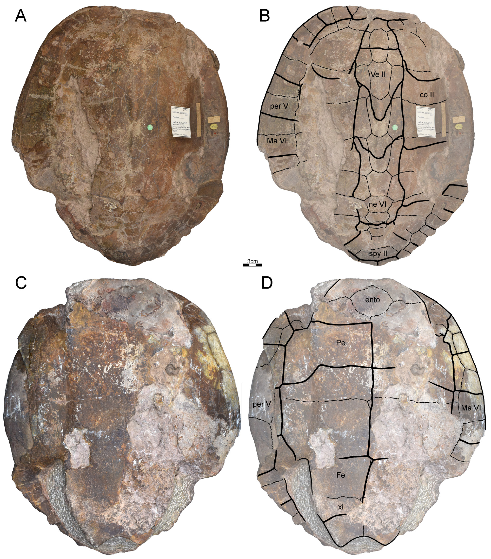
BMNH 39841 ( Fig. 14 View Fig ), holotype of Batagur durandi – This is an almost complete, well-preserved specimen from Miocene/Pliocene of the Siwalik Hills, probably of India, that was presented to BMNH by P.T. Cautley. The original figure by Lydekker (1885a: pl. 24.2) overall compares well to our observations, but we note differences in the shape of vertebrals III and IV and the presence of peripherals, and we document the plastron for the first time ( Fig. 14 View Fig C–D).A portion of the anterior margin of the carapace and some posterior left peripheral bones are missing. The specimen clearly represents an adult female due to its large size (carapace length greater than 40 cm). A median longitudinal carapacial keel is present, which is elevated in the posterior region of the second vertebral scute. All neural bones are hexagonal and anteriorly short-sided. The first to fourth neural bones are about the same size and longer than wide. The fifth to eighth neural bones are wider than long. The seventh neural is anomalously divided into two elements. The first vertebral scute is bell-shaped and has a small anterolateral constriction. The second vertebral scute has a deep protrusion along its posterior margin into the third vertebral. The third vertebral scute has a smaller protrusion into the fourth vertebral scute. The fourth vertebral is twice as long than wide and its anterior margin intersects the fourth neural. The sulcus between the first and second pleural forms a deep anterolateral projection onto the first costal bone. The fifth and sixth marginal scutes overlap part of the costal bones. The anterior plastral margin is not completely preserved. The entoplastron is not intersected by the humeropectoral sulcus. the pectoroabdominal sulcus contacts the fifth marginal scute on one side of the specimen, but the sixth marginal on the other. Both the fifth and sixth marginal scutes overlap the hyoplastron. The hyo-hypoplastral suture and pectoroabdominal sulcus do not overlap or coincide. The xiphiplastra have a small, rounded anal notch.
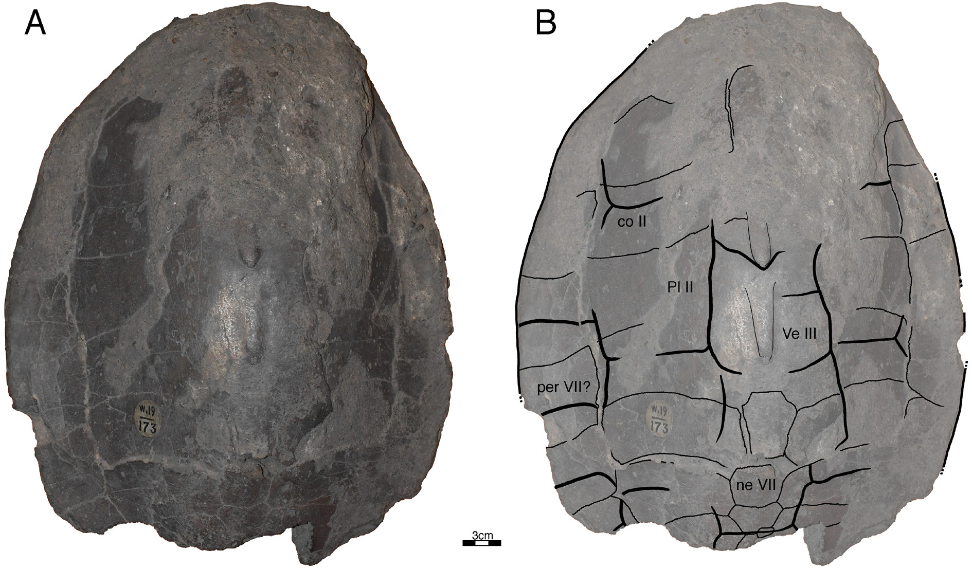
IM W19/173 ( Fig. 15 View Fig ) – This is a well-preserved specimen that lacks provenance data and that appears to be unpublished. The majority of sutures and sulci of the carapace are clearly visible. The specimen likely represents an adult female due to its large size (carapace length greater than 40 cm). The carapace is highly domed at its center. A median carapacial keel is present, with protrusions at the posterior margins of the second and third vertebral scutes. The neural bones, likely the sixth to eighth, are anteriorly short-sided. The sulcus between the first and the second pleurals and the second and third pleurals are positioned over the second and fourth costal bones, respectively. The plastron is damaged and thus not shown here.
Comments
Here, we attribute these two specimens to Batagur dhongoka based on the protrusion of the second vertebral into the third, a medially short third vertebral scute, a fourth vertebral scute that is much longer than wide, and a large plastron with straight humeropectoral sulci that do not cross the entoplastron (noticeable on BMNH 39841). This confirms the synonym of B. durandi with Batagur dhongoka , as first suggested by Boulenger (1889) and later supported by Lydekker (1889a), TEWG (2015) and TTWG (2017).
No known copyright restrictions apply. See Agosti, D., Egloff, W., 2009. Taxonomic information exchange and copyright: the Plazi approach. BMC Research Notes 2009, 2:53 for further explanation.
|
Kingdom |
|
|
Phylum |
|
|
Class |
|
|
Order |
|
|
Family |
|
|
Genus |
Batagur dhongoka ( Gray, 1832 )
| Garbin, Rafaella C., Bandyopadhyay, Saswati & Joyce, Walter G. 2020 |
Kachuga dhongoka
| Boulenger G. A. 1889: 56 |
Batagur durandi
| Lydekker R. 1885: 192 |
Batagur duvaucelli
| Anderson J. A. 1879: 738 |
hardwickii
| Gray J. E. 1870: 56 |
Kachuga hardwickii
| Gray J. E. 1869: 202 |
Clemmys dhongoka
| Strauch A. 1862: 33 |
Emys duvaucelli Duméril & Bibron, 1835: 334
| Dumeril A. M. C. & Bibron G. 1835: 334 |
Emys dhongoka
| Emys dhongoka Gray, 1832 : pl. 60 |
Batagur dhongoka
| Praschag P. & Hundsdorfer A. K. & Fritz U. 2007: 439 |
| Gray 1855 (1856): 36 |