
Kaisquilla laevis, Ahyong, 2002
|
publication ID |
https://doi.org/ 10.5281/zenodo.5392839 |
|
persistent identifier |
https://treatment.plazi.org/id/03C787EC-FFE3-7C12-7771-7C5FB6EC204B |
|
treatment provided by |
Marcus |
|
scientific name |
Kaisquilla laevis |
| status |
sp. nov. |
Kaisquilla laevis n. sp.
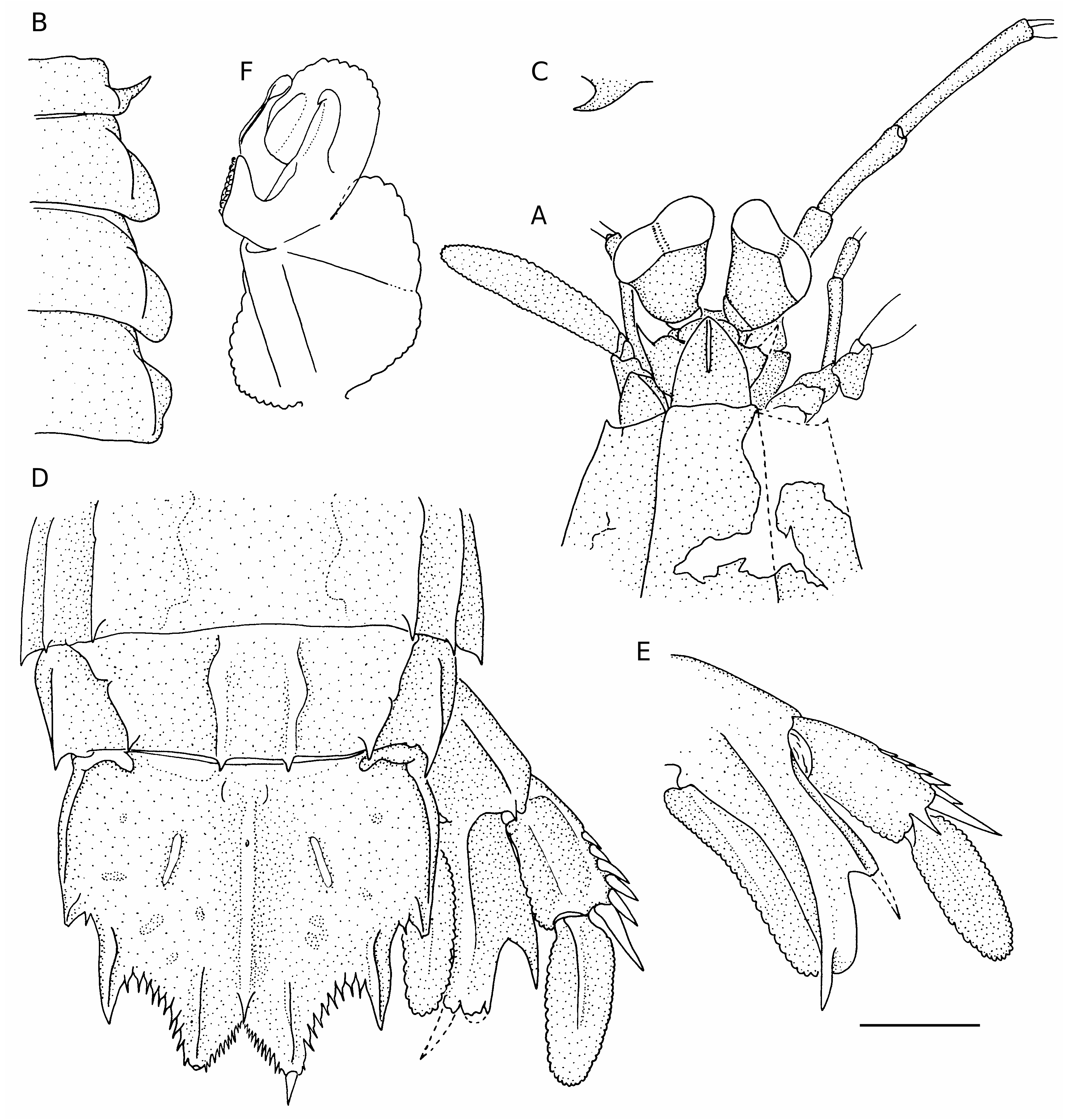
( Fig. 3 View FIG )
TYPE MATERIAL. — Kai Islands, stn DW14, 05°18’S, 132°28’E, 245-246 m, 24.X.1991, holotype tl 28 ( MNHN).
ETYMOLOGY. — Named laevis , meaning smooth, for the absence of median and submedian carina on the abdomen in Kaisquilla n. gen., that are present in species of Anchisquilloides and Anchisquillopsis .
DISTRIBUTION. — Known only from Kai, at 245- 246 m depth.
DESCRIPTION
Dorsal integument smooth, polished.
Eye with cornea bilobed, distinctly broader than and set slightly obliquely on stalk, not extending beyond antennular peduncle segment 1; CI 421. Ophthalmic somite with medially emarginate anterior margin. Ocular scales rounded, separate.
A1 somite dorsal processes with short slender apices, directed anterolaterally; A1 peduncle 1.10cl. A2 scale slender, 0.40cl; entire margin setose.
Rostral plate longer than broad; lateral margins convex; apex blunt; with distinct median carina.
Carapace anterior width less than half median length; anterolateral spines not extending anteriorly to base of rostral plate; with faintly indicated lateral and reflected mg carinae indicated posteriorly only.
Raptorial claw unknown.
Mandibular palp 2-segmented (damaged on left side). MXP1-2 each with epipod. MXP5 basal segment without ventrally directed spine.
Pereiopods 1-3 basal segment unarmed; endopod 2-segmented, distal segment slender.
TS5 lateral process a single slender spine directed anterolaterally; ventral spine absent. TS6-7 lateral process broadly rounded. TS6-8 each with distinct IM carinae. TS8 anterolateral margin rounded; sternal keel produced as a posteriorly directed spine.
AS 1-5 each with IM, LT and MG carinae. AS 6 with SM, IM and LT carinae; with ventrolateral spine anterior to uropodal articulation; sternum posterior margin unarmed, without transverse carinae. Abdominal carinae spined as follows: SM 6, IM 5-6, LT 5-6, MG 4-5.
Telson broader than long, with three pairs of primary teeth, each with dorsal carina; SM teeth with movable apices; prelateral lobe absent; MD carina with proximal pit and posterior spine; dorsolateral surface rugose, with short, low, mid-dorsal carina and a few shallow pits; denticles spiniform SM 12-14, IM 9-10, LT 1; ventral surface without postanal carina; ventrolateral carina extending posteriorly to base of LT denticle.
Uropodal protopod terminating in two slender spines, dorsally and ventrally carinate, inner longer; unarmed dorsally except for dorsal spine above proximal exopod articulation; protopod inner margin smooth, without ventral spine or tubercle anterior to endopod articulation; lobe on outer margin of inner spine rounded and deflected dorsally, broader than adjacent spine, proximal margin faintly concave.
Uropodal exopod proximal segment unarmed dorsally; distal margin with slender ventral spine; outer margin with six movable spines, distalmost not exceeding midlength of distal segment; exopod distal segment longer than proximal segment; endopod unarmed dorsally, entire margin setose.
Colour in alcohol
Largely faded, but with dark pigment around the posterolateral margins of the carapace, lower and posterior margins of the thoracic and abdominal somites. AS 6 with dark submedian patch. Telson dark on posterior half. Uropodal protopod dark proximally, on inner distal margin and apex of outer spine. Uropodal exopod proximal segment with dark pigment distally.
Measurements of holotype
tl 28, cl 5.9, A1 peduncle 6.4, A2 scale 2.5, cornea width 1.4.
REMARKS
Unfortunately, the specimen is badly damaged: both raptorial claws are missing, the right anterior portion of the carapace is fragmented, and the ocular somite fractured and almost fully detached from the cephalon. Therefore, Fig. 3 View FIG shows a partial reconstruction.
The well-developed penes and endopod of PLP1 show that this specimen is sexually mature.
| MNHN |
Museum National d'Histoire Naturelle |
No known copyright restrictions apply. See Agosti, D., Egloff, W., 2009. Taxonomic information exchange and copyright: the Plazi approach. BMC Research Notes 2009, 2:53 for further explanation.
|
Kingdom |
|
|
Phylum |
|
|
Class |
|
|
Order |
|
|
Family |
|
|
Genus |