
Inakayalia cyanea, 2023
|
publication ID |
https://doi.org/10.1093/zoolinnean/zlac072 |
|
publication LSID |
lsid:zoobank.org:pub:010109AB-79F5-4E6D-909B-08BB1803E589 |
|
DOI |
https://doi.org/10.5281/zenodo.7797665 |
|
persistent identifier |
https://treatment.plazi.org/id/03C49B73-697C-FFBE-FF3B-7477B05C746B |
|
treatment provided by |
Plazi (2023-04-04 08:43:21, last updated by Guilherme 2025-02-18 17:46:29) |
|
scientific name |
Inakayalia cyanea |
| status |
sp. nov. |
INAKAYALIA CYANEA ALMEIDA & CARBAYO SP. NOV.
( FIGS 20–23 View Figure 20 View Figure 21 View Figure 22 View Figure 23 )
Zoobank registration: urn: lsid: zoobank. org:act: 3CF36CAC-C035-45EC-B439-79C0EE4DC8E3
Material examined: All specimens collected in Parque Nacional Nahuelbuta, Chile (37°48′′00.0′′S, 073°00′′00.0′′W), coll. F. Carbayo et al., 13 December 2010 .
Holotype: MNHNCL PLAT-15043 (Field code, F4912). Cephalic region : transverse sections on 14 slides; ovarian region: horizontal sections on 100 slides; prepharyngeal region: transverse sections on 30 slides; copulatory apparatus: sagittal sections on 122 slides.
Paratypes: MZUSP PL 2283 (Field code, F4914). Cephalic region : transverse sections on ten slides; ovarian region: horizontal sections on 13 slides; prepharyngeal region: transverse sections on ten slides; pharynx and copulatory apparatus: sagittal sections on 73 slides . MZUSP PL2284 (Field code, F4917). Cephalic region : transverse sections on 15 slides; ovarian region: horizontal sections on 12 slides; prepharyngeal region: transverse sections on 20 slides; pharynx: sagittal sections on 22 slides; copulatory apparatus: sagittal sections on seven slides .
Type locality: Parque Nacional Nahuelbuta, Región de la Araucanía, Chile. The species is only known from this locality.
Etymology: The specific epithet derives from the Greek Κυανός meaning blue, alluding to the colour of the body.
Diagnosis: Species of Inakayalia with a long and widened prostatic vesicle, branched proximally, and provided with a long posterodorsal diverticulum; long unpaired and dilated common ovovitelline duct.
Description
External aspect: The three specimens ( Fig. 20 View Figure 20 ) are mature and measured between 23 and 27 mm in length and 6–8.3 mm in width at rest. Preserved, they measured 28.5–44.5 mm long, 5–6 mm wide and 1.2–1.3 mm high. At rest, the body is lanceolate with undulating margins ( Fig.20D, E View Figure 20 ). The cephalic region narrows to the rounded tip; the posterior narrows abruptly to the pointed tip. The dorsum is flattened except for the convex median region. The ventral side is flat.
With approximately one-eighth of the body length, the cephalic region exhibits two black-blue (RAL 5004) bands, separated from each other by a median pure white (RAL 9010) line ( Fig. 20A–C View Figure 20 ). These two bands are completely ( Fig. 20A, B View Figure 20 ) or incompletely ( Fig. 20C View Figure 20 ) interrupted by a zigzagged, pure white or beige transverse band. Behind the transverse band, the dorsum is black-blue (RAL 5004), darker along the median and paramedian zones to form bands, each with 10–16% of the body width. Additionally, large blue-grey (RAL 7031) haloes mottle the dorsum behind the transverse band except for the median zone.
The ground colour of the ventral side is cream (RAL 9001) ( Fig. 20D, E View Figure 20 ). Numerous small black-blue pigment dots are in the cephalic region. A whitish transverse band continued from the dorsal side separates the cephalic region from the remaining ventral surface, which is covered with numerous dots, either graphitegrey (RAL 7024) or ochre-brown (RAL 8001).
The eyes are of a single-cup type measuring 32–38 µm in diameter. They are organized in a singleto-biserial row around the anterior eighth of the body; behind this body region, they spread onto the dorsal surface and are encircled by the blue-grey haloes.
The sensory pits are simple invaginations 50–57 µm deep and are located ventromarginally in a single row running along an anterior region with 18% of body length. The mouth is positioned at a distance from the anterior extremity equal to 63.4–66.3% of the body length; the gonopore, 83.6–86.7%.
Internal morphology: The creeping sole has 95% of the body width. Abundant gland cells producing coarse (1 µm) xanthophil granules and scarce gland cells producing erythrophil granules discharge their secretion through the entire epidermis of the prepharyngeal region. The cell necks of the xanthophil type are 10–15 µm in diameter and become more abundant toward the body margins of the dorsal side. The glandular margin consists of xanthophil gland cells ( Fig. 21A View Figure 21 ). Rhabdites are discharged through the dorsal epidermis. As the creeping sole narrows toward the anterior extremity of the body, the xanthophil glands become scarce dorsally and abundant ventrally.
The cutaneous musculature comprises three layers, namely, a subepithelial layer of circular muscle (5 µm thick), followed by a double layer (15–40 µm) with decussate fibres and then a well-developed, innermost layer of longitudinal fibres (30–105 µm thick, both dorsally and ventrally) ( Fig. 21B–C View Figure 21 ). The cutaneous musculature thickness relative to the body height is 13–15%. Toward the anterior region of the body, these muscle layers become thinner until they disappear.
There are three strong parenchymal muscle layers, namely, a dense dorsal layer of decussate fibres (30 µm thick), a supra-intestinal layer of transverse fibres (100 µm thick) and a denser subintestinal layer of transverse fibres (100 µm thick; Fig. 21B–C View Figure 21 ). These muscle layers are thinner in the anterior region of the body.
Two of the three main branches of the intestine, namely, the paired ones, may connect to each other at the level of the prostatic vesicle. The oesophagus to pharynx length ratio is 31–33%. The mouth is situated at a distance from the root of the pharynx equivalent to 41–54% of the pharyngeal pouch length ( Fig. 21D View Figure 21 ). The distal portion of the pharyngeal pouch is close to the prostatic vesicle. The pharynx is bell-shaped, with its dorsal insertion slightly anterior to the mouth. The epithelium of the distal portion of the pharynx is pierced by the necks of four types of gland cells, producing xanthophil granules, erythrophil granules, cyanophil granules and amorphous secretion, respectively. The outer pharyngeal musculature consists of a subepithelial layer of longitudinal muscle (5 µm thick), followed by a layer of circular muscle (15 µm thick) and an innermost layer of longitudinal muscle (5 µm thick). The inner pharyngeal musculature consists of a subepithelial circular muscle (75 µm thick), followed by a longitudinal muscle (10 µm thick; Fig. 21E View Figure 21 ).
The rounded-to-irregular testes measure 325– 450 µm in diameter. They are organized into two to four rows in three vertical levels at each side of the body, between the supra-intestinal and subintestinal parenchymal muscles ( Fig. 21A View Figure 21 ). The anteriormost testes lie at a distance from the anterior tip of the body equivalent to 17.3% of the body length; the posteriormost ones, the equivalent to 65% of the body length, i.e. they are lateral to the pharyngeal root.
The sperm ducts run immediately above the subintestinal parenchymal muscle. Laterally to the pharyngeal pouch, each duct opens into the anteroventral region of the respective lateral, short branch of the prostatic vesicle ( Fig. 22A View Figure 22 ). The rest of the prostatic vesicle is unpaired and large, measuring up to 1.3 mm in length. Approximately the anterior-half of the vesicle occupies two-thirds of the body height and exhibits numerous folds filling its lumen ( Figs 22B, C View Figure 22 , 23A–C View Figure 23 ). The posterior-half consists of a dorsal, blind diverticulum (200–250 µm long) and a ventral, widened duct (600 µm long), both running posteriorly. The latter penetrates the anterior section of the penis bulb to communicate with the ejaculatory duct.
The prostatic vesicle is lined with a ciliated epithelium, being cuboidal in the paired portion and columnar in the unpaired one. Abundant gland cells discharge erythrophil granules through the lining epithelium of the prostatic vesicle. The epithelium of this vesicle is underlain by a muscle layer (25–100 µm thick) of decussate fibres.
The ejaculatory duct is wide and opens at the tip of the penis papilla ( Fig. 22A View Figure 22 ). This duct is lined with a cuboidal-to-columnar, ciliated epithelium, surrounded by a circular muscle (50 µm thick). The penis papilla is cylindrical, with a rounded tip and is horizontally located ( Figs 22A–C View Figure 22 , 23B View Figure 23 ). This papilla is covered with a columnar epithelium, which is pierced by the necks of two types of gland cells producing erythrophil and cyanophil granules, respectively. This epithelium is underlain by a 15 µm thick muscle with interwoven circular and longitudinal fibres.
The male atrium is relatively short and not folded. It is lined with a cuboidal-to-columnar epithelium, which is crossed by two types of gland cells producing erythrophil and cyanophil granules, respectively. This epithelium is underlain by a layer of circular muscle, followed by a layer of longitudinal fibres, each layer being 5 µm thick in the proximal region of the atrium and 30 µm in the distal.
The ovaries are ovoid and have a maximum diameter of 400 µm in the longitudinal body axis. These ovaries are located immediately above the ventral nerve plate ( Fig. 21C View Figure 21 ) and at a distance from the anterior tip of the body corresponding to 9.6% of the body length. The ovovitelline ducts emerge from the lateral aspect of the ovaries and run ventrally above the main nerve plate. Close to the mid-region of the prostatic vesicle, each ovovitelline duct opens laterally into the long, dilated common ovovitelline duct ( Figs 22A, C View Figure 22 , 23B–E View Figure 23 ). This long duct is six times wider than the ovovitelline ducts and exhibits longitudinal folds. The common ovovitelline duct ascends gradually to communicate with the common glandular ovovitelline duct. This glandular duct runs posteriorly over the male and female atria to join the female genital canal, which projects dorso-anteriorly from the dorsoposterior region of the female atrium.
The female atrium is elongated to funnel-shaped and is slightly shorter than the male atrium. The common ovovitelline duct is lined with a columnar epithelium, which is crossed by three types of gland cells producing xanthophil, erythrophil and cyanophil granules, respectively ( Fig. 23E View Figure 23 ). This duct is surrounded by a single muscle layer (50 µm thick) comprising circular, diagonal and longitudinal thin fibres. The female genital canal and the female atrium are lined with a 75 µm high columnar, non-ciliated epithelium and the apical side of its cells contains xanthophil granules. Additionally, gland cells discharge erythrophil granules through the epithelium of the female genital canal and that of the female atrium. These epithelia are underlain by a 50–120 µm thick muscle consisting of intermingled longitudinal and circular fibres. The common muscle coat consists of sparse longitudinal and circular muscle fibres.
Remarks on the new tribe Inakayaliini and its genus: The phylogenetic position of Inakayaliini is unstable. It was recovered as sister to Myoplanini ( Fig. 2 View Figure 2 ; Supporting Information, Figs S1 View Figure 1 , S 2 View Figure 2 ) or to Adinoplanini (Supporting Information, Figs S3 View Figure 3 , S 4 View Figure 4 ). Inakayaliini is monogeneric and currently houses four species. Two of them are represented in our phylogenetic trees, namely, I. valdiviana and I. cyanea . These two species are sister to each other. Inakayalia cyanea matches all diagnostic features of the genus, except that the wall of its penis papilla is not irregular but smooth, and the dilated portion of the female ducts does not correspond to ovovitelline ducts, but the common ovovitelline duct. Therefore, the genus is re-diagnosed by omitting the mention of the irregular wall of the penis papilla and the dilation of the female genital ducts. This latter feature is transferred to the diagnosis of the new tribe.
Inakayalia cyanaea is readily distinguished from the other three species in the genus in that it presents a long prostatic vesicle (vs. shorter) and a dilated common ovovitelline duct (vs. paired ovovitelline ducts dilated). Inakayalia cyanea is also the only species in the genus with the testes extending vertically between the supraintestinal and the subintestinal parenchymal muscle (see Fig. 21A View Figure 21 ).
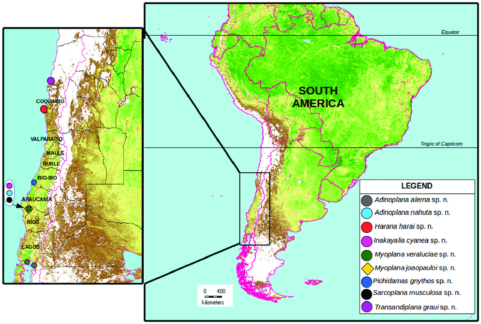
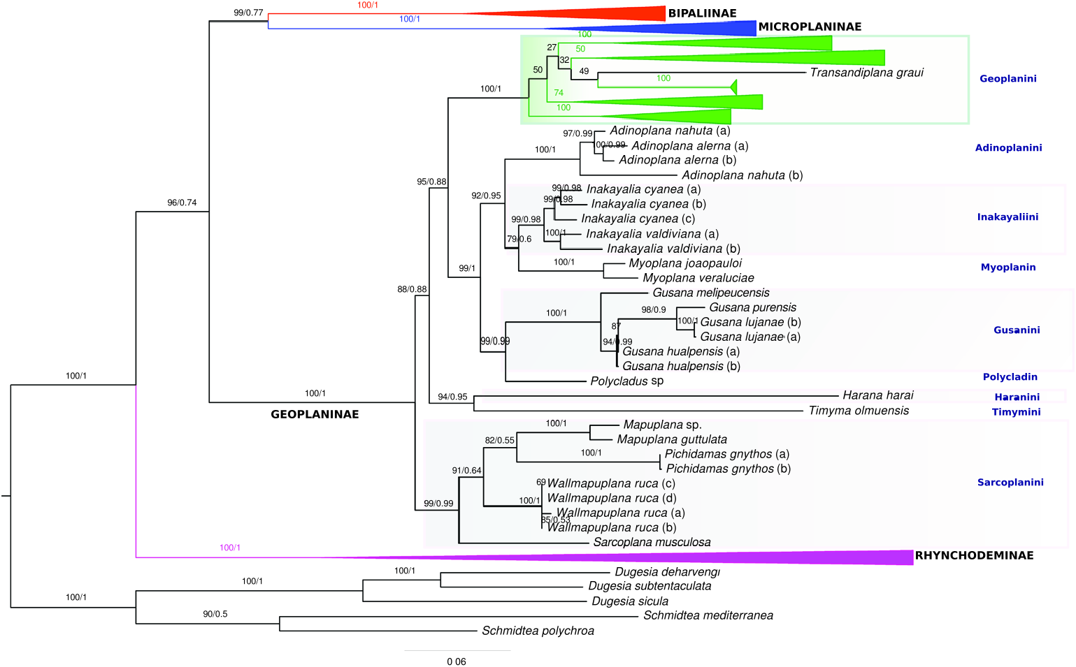
Figure 2. Phylogenetic tree inferred from the concatenated dataset under maximum likelihood as the optimality criterion. Numbers at the nodes correspond to ultrafast bootstrap values. Subfamilies and internal groups of Geoplaninae were collapsed. The same, non-collapsed tree is available in the Supporting Information, Figure S1.
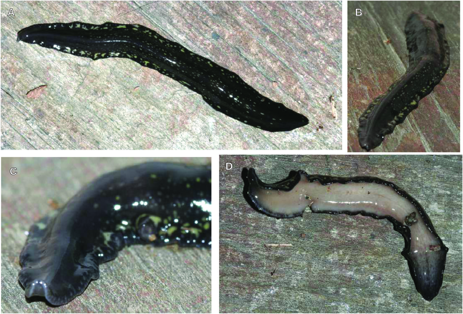
Figure 3. Adinoplana alerna sp. nov. Living holotype in dorsal (A–C) and ventral views (D). Scale not available. The body is approximately 50 mm long.
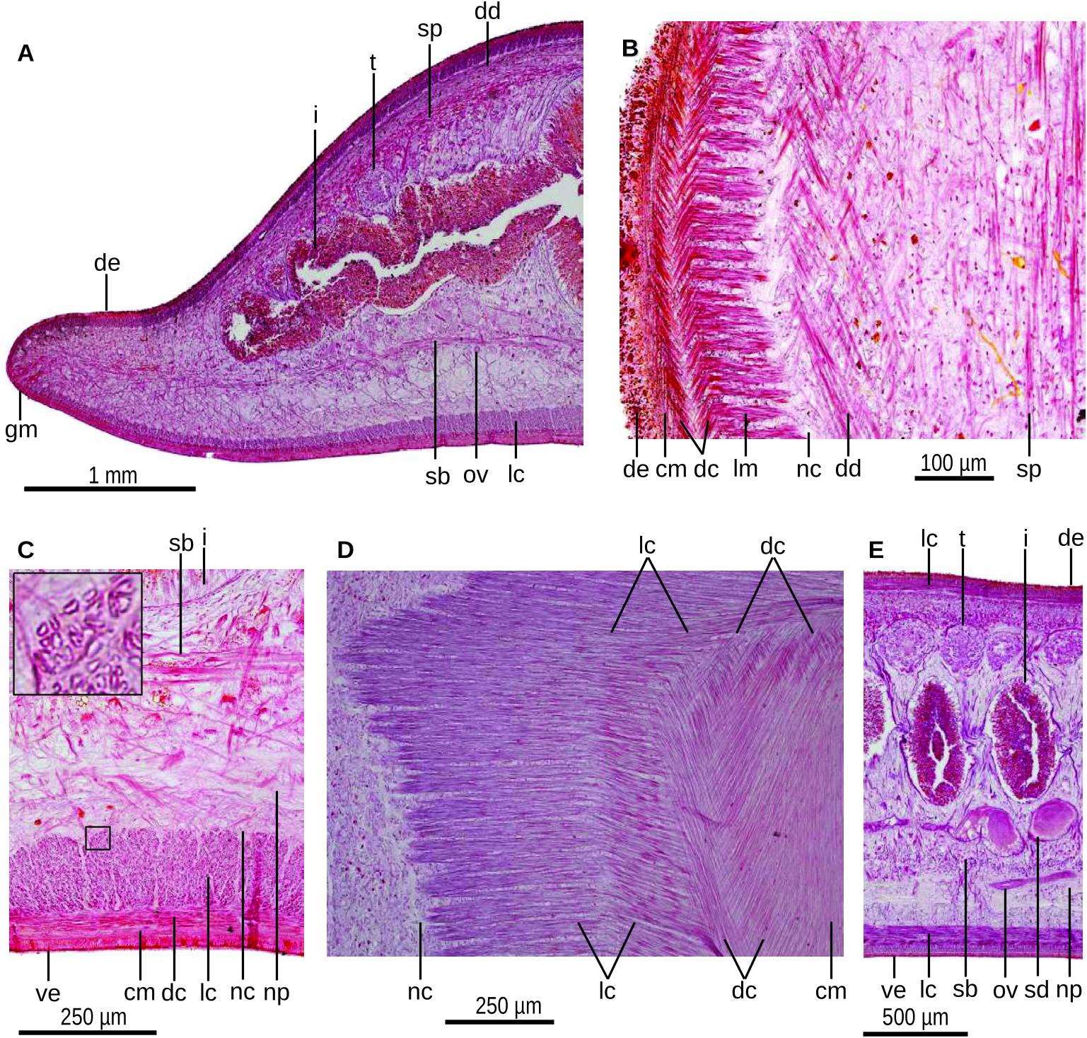
Figure 4. Adinoplana alerna sp. nov. Photomicrographs of the holotype.A, transverse section of the pre-pharyngeal region. B, horizontal section through the dorsal epidermis. C, transverse section of the pre-pharyngeal, ventral region; the inset shows a detail of the hollow longitudinal muscle fibres. D, horizontal section of near ventral epidermis. E, sagittal section of a region close to the penis bulb.
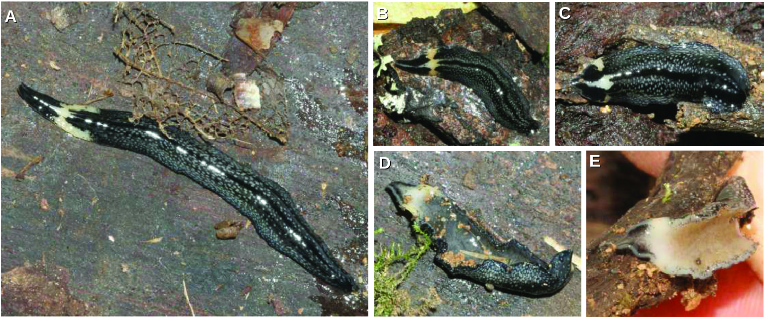
Figure 20. Inakayalia cyanaea sp. nov. Photographs of the live holotype (A, D), paratype MZUSP PL2284 (B) and paratype MZUSP PL2283 (C, E) in dorsal (A–C) and ventral (D, E) views. Scale bars not available.
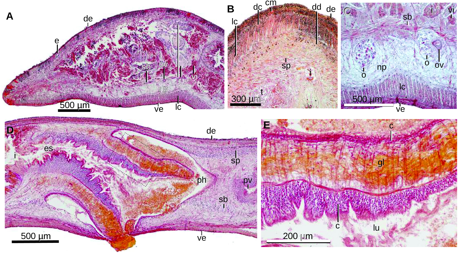
Figure 21. Inakayalia cyanaea sp. nov. Photomicrographs of histological sections of the holotype (A) and paratype MZUSP PL 2283 (B–E). A, transverse section of the pre-pharyngeal region. B, horizontal section of the dorsal portion of the body showing cutaneous and parenchymal muscles. Anterior region to the upper. C, approximately transverse section through the ovaries. D, sagittal section of the pharynx. The region within the rectangle is enlarged in (E). E, sagittal section showing the outer and inner pharyngeal epithelia.
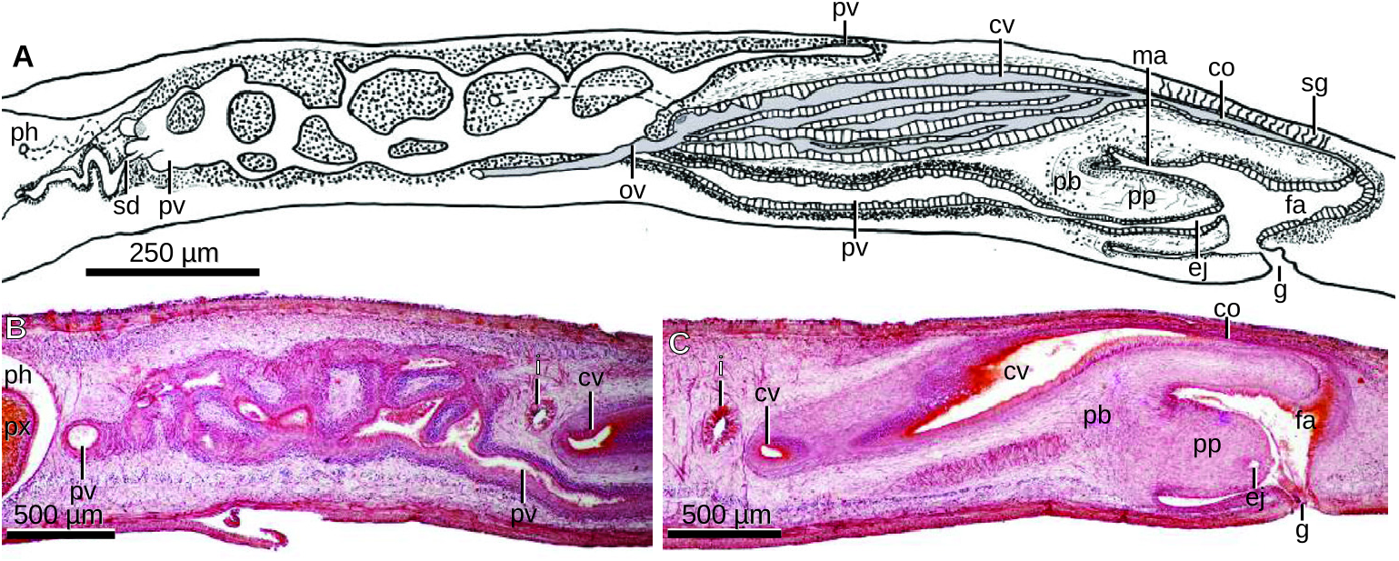
Figure 22. Inakayalia cyanaea sp. nov. A, diagrammatic reconstruction of the copulatory apparatus of the holotype. B, C, photomicrographs of sagittal sections of the copulatory apparatus of the paratype MZUSP PL 2283 showing the prostatic vesicle (B) and the male and female atria (C). Note the instestinal branches connecting both main posterior branches of the intestine (i, B, C).
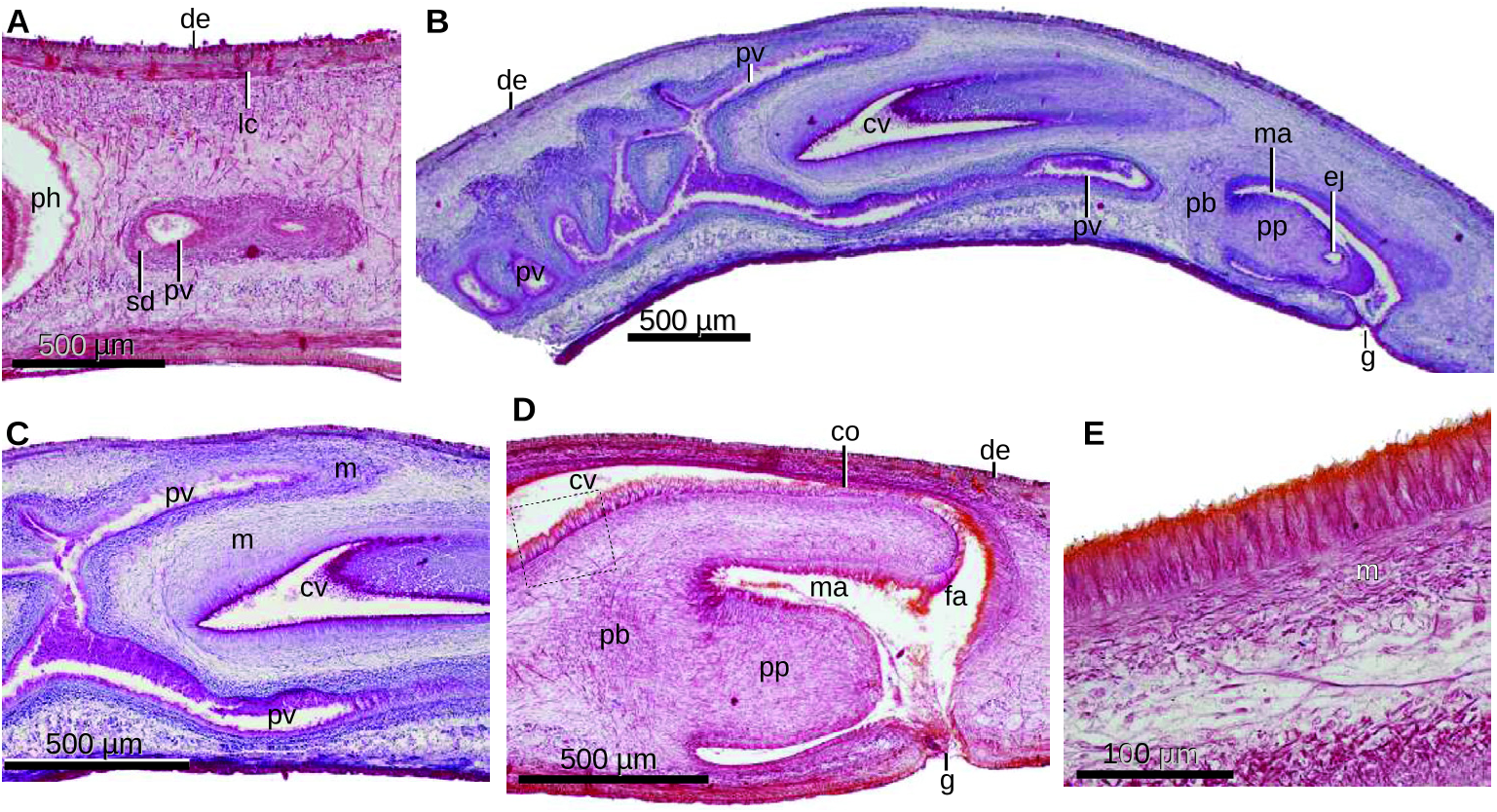
Figure 23. Inakayalia cyanea sp. nov. Photomicrographs of sagittal sections of the paratypes MZUSP PL 2283 (A, D, E) and MZUSP PL 2284 (B, C) showing the anterior region of the prostatic vesicle, the copulatory apparatus (B), the posterior region of the prostatic vesicle, the male and female atria (D) and the epithelium of the common ovovitelline duct.
| MZUSP |
Museu de Zoologia da Universidade de Sao Paulo |
No known copyright restrictions apply. See Agosti, D., Egloff, W., 2009. Taxonomic information exchange and copyright: the Plazi approach. BMC Research Notes 2009, 2:53 for further explanation.
|
Kingdom |
|
|
Phylum |
|
|
Order |
|
|
Family |
|
|
Tribe |
Inakayaliini |
|
Genus |
1 (by plazi, 2023-04-04 08:43:21)
2 (by ExternalLinkService, 2023-04-04 08:52:53)
3 (by diego, 2023-04-10 16:57:04)
4 (by ExternalLinkService, 2023-04-10 17:08:12)
5 (by ExternalLinkService, 2023-04-10 18:34:12)
6 (by plazi, 2023-11-08 22:09:30)
7 (by ExternalLinkService, 2023-11-09 07:03:16)
8 (by ExternalLinkService, 2023-11-09 07:29:14)