
Malagiella vohiparara, Ubick & Griswold, 2011
|
publication ID |
https://doi.org/10.1206/356.1 |
|
persistent identifier |
https://treatment.plazi.org/id/03C2C67F-FFBE-FF98-F2B2-FF7DFE11FD7E |
|
treatment provided by |
Tatiana (2021-08-30 18:44:54, last updated by Plazi 2023-11-06 03:19:09) |
|
scientific name |
Malagiella vohiparara |
| status |
sp. nov. |
Malagiella vohiparara View in CoL , new species
Figures 117–184 View Figs View Figs View Figs View Figs View Figs View Figs View Figs View Figs View Figs , 345–378 View Figs View Figs View Figs View Figs , 464, 468, 469 View Figs , 476, 479 View Figs , 484 View Figs , 487 View Fig ; maps 1–4; table 1
TYPE: Male holotype, female allotype, male paratype, and 3 female paratypes from sifted leaf litter at P.N. Ranomafana, 2.3 km N Vohiparara, 21 ° 12.89S, 47 ° 23.09E, 1100 m, Fianarantsoa Province, Madagascar (28 Apr 1998, C. Griswold, D. Ubick), deposited in CAS (CASENT 9029655, PBI _OON 03078).
ETYMOLOGY: The species is named after the type locality.
DIAGNOSIS: The male of this species differs from other Malagiella by the combination of small size and the absence of lateral spines on the hind legs. The female differs from others in its species group in having a widely sinuous receptaculum, W/L 5 1.0– 1.2 (figs. 182–184, 375–378; map 4).
MALE (PBI_OON 03078) (figs. 117–120, 125–128, 133–139, 142–152, 157–172, 345– 354, 365–372, 476, 484; map 3): Total length 1.32 (1.26–1.32), carapace length 0.64 (0.60–0.66), width 0.55 (0.52–0.56), N 5 9. CEPHALOTHORAX: Carapace orangebrown, broadly oval in dorsal view, pars cephalica strongly elevated in lateral view (figs. 117, 347), anteriorly narrowed to about 0.64 times its maximum width, surface and sides of elevated portion of pars cephalica finely reticulate (fig. 118); lateral margin undulate, smooth; nonmarginal pars cephalica setae dark, present in U-shaped row (figs. 117–120). Clypeus length more than twice eye area length (fig. 117). Eyes medium sized, eye area width 0.36 carapace width, length 0.45 clypeus length (figs. 117, 120). Sternum infracoxal groove with opening only at posterior end (fig. 125) with median longitudinal groove weakly represented (fig. 126); sternal bristles long, straight (fig. 125). Mouthparts: Chelicerae with promarginal tooth and patch of largely retromarginal denticles (figs. 135, 136). Endites distally excavated, serrula present in single row (fig. 128); distomedian part with projection bearing dense scopula (fig. 127), distolateral part with swelling bearing serrula and three stout setae (figs. 127, 128). AB- DOMEN: Cylindrical, slightly compressed (figs. 352, 354). Book lung covers large, ovoid (fig. 370). Dorsal scutum strongly sclerotized, narrowed to 0.6 carapace width; fusion with epigastric scutum a rounded junction (figs. 142–144). Postepigastric scutum broadly rounded, extending to about 0.6 abdomen length. Spinnerets (fig. 149): ALS with three spigots (fig. 150), PMS with two spigots (fig. 151), PLS with three spigots (fig. 152). LEGS: Spination: femur I p0-1-0; tibiae: I v4-4-0, II v4-2-1, IV v0-0-2 (weak). GENITALIA: Epigastric region with sperm pore small, unmodified (fig. 148). Palpal bulb with dorsobasal indentation (figs. 157, 158, 365, 366, 371, 372), embolus sharply bent ventrally (figs. 159, 160); ventral prong long (fig. 169); embolar opening small (fig. 170), closer to retrolateral prong (fig. 172).
FEMALE (PBI_OON 03078) (figs. 121– 124, 129–132, 140–141, 153–156, 173–184, 355–364, 373–378, 464, 468, 469, 479; map 1, 2, 4): Total length 1.36 (1.36–1.48), carapace length 0.66 (0.66–0.70), width 0.58 (0.55–0.59), N 5 4. CEPHALOTHORAX: Carapace similar to male but with lower cephalon, much shorter clypeus (figs. 121– 124), and anteriorly narrowed to about 0.46 maximum width (fig. 124). Eyes medium sized; eye area about 0.45 carapace width, length 0.5 clypeus length (figs. 121, 124). Sternum with infracoxal groove opening only at posterior end (fig. 130). Mouthparts: endites without distal modifications (fig. 132). ABDOMEN: Pedicel tube short (fig. 363); scutum dorsal extension shorter than in male, about 0.4 pedicel diameters (fig. 362). Dorsal scutum covering about 0.7 abdomen length, 0.5 abdomen width. Spinnerets (fig. 153): ALS with three spigots (fig. 154), PMS with three spigots (fig. 155), PLS with five spigots (fig. 156). LEGS: Spination as in male (figs. 468, 469). GENITALIA: Postepigastric scutum weakly sclerotized, externally with round copulatory opening (figs. 176, 177), receptaculum strongly sinuous, W/L 5 1.0–1.2 (figs. 182–184, 375–378; map 4), enclosed in chitinized membrane (figs. 179– 181), rigidly attached to postepigastric scutum (figs. 182–184).
OTHER MATERIAL EXAMINED: MADA- GASCAR: Fianarantsoa Province: Talatakely, P.N. Ranomafana , 21 ° 14.99S, 47 ° 25.69E, pitfall trap, 13–27 Apr 1998 (Griswold et al., CASENT 9029666, PBI _OON 03380), 28 GoogleMaps ; P.N. Ranomafana , 2.3 km N Vohiparara ,
21 ° 12.89S, 47 ° 23.09E, 1100 m, 18 Apr 1998 (Griswold et al., CASENT 9029665, PBI _ OON 03381), 18 GoogleMaps ; pitfall trap, 10–28 Apr 1998 (Griswold et al., CASENT 9029668, PBI _OON 03378), 58 ; sifting leaf litter, 18 Apr 1998 (C. Griswold, D. Ubick, CASENT 9029664, PBI _OON 03382), 2♀ ; 11 Apr 1998 (C. Griswold, D. Ubick (CASENT 9029667, PBI _OON 03379), 1♀ ; Vohiparara , 3.6 km W Ranomafana, 21 ° 14.2439S, 47 ° 23.8429E, 1137 m, evergreen secondary forest, sifting litter, 13 Jan 2009 (D. Andriamalala, C. Griswold, G. Hormiga, A. Saucedo, N. Scharff, H. Wood, AMNH, PBI _OON 35162), 1♀ GoogleMaps ; same data ( AMNH, PBI _OON 35163), 181♀ GoogleMaps .
DISTRIBUTION: Known only from P.N. Ranomafana, Madagascar.
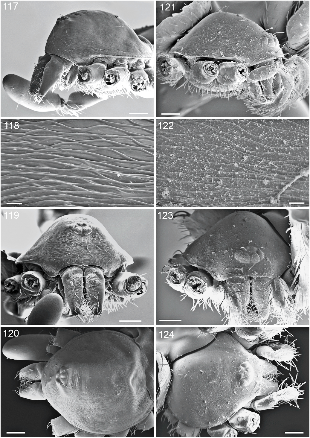
Figs. 117–124. Malagiella vohiparara, new species, carapace. 117–120. Male (PBI_OON 03378); 121– 124 Female (PBI_OON 03078). 117, 121. Lateral view. 118, 122. Same, magnified view of carapace. 119, 123. Same, anterior view. 120, 124. Same, dorsal view. Scale bars are 100 Mm (figs. 117, 119–121, 123, 124) and 10 Mm (figs. 118, 122).

Figs. 125–132. Malagiella vohiparara, new species, sternal region. 125. Male (PBI_OON 03378), sternum, ventral view. 126. Same, sublateral view, arrow shows anterolateral knob of sternum. 127. Same, endites and labium, ventral view. 128. Same, endites, apical view showing serrula (arrows). 129. Female (PBI_OON 03078), sternum, ventral view. 130. Same, magnified view showing sternal grooves and pits (arrows). 131. Same, sternum at coxa IV, posterioventral view, showing sternal pit (arrow). 132. Same, endites and labium, ventral view. Scale bars are 100 Mm (figs. 125, 126, 129) and 50 Mm (figs. 127, 128, 130– 132).

Figs. 133–141. Malagiella vohiparara, new species. 133. Male (PBI_OON 03378), chelicerae, anterior view. 134. Same, lateral view. 135. Same, posterior view. 136. Same, magnified view showing retromarginal denticles and promarginal tooth (arrow). 137. Same, distal tip of chelicerae, anterior view showing flattened setae originating from cheliceral process (arrow). 138. Same, tarsal organ from palp, dorsal view. 139. Trichobothrium from palpal tibia, dorsal view. 140. Female (PBI_OON 03078), claws of tarsus III, prolateral view. 141. Same, tarsus IV, apical view showing large inner prongs (arrow).
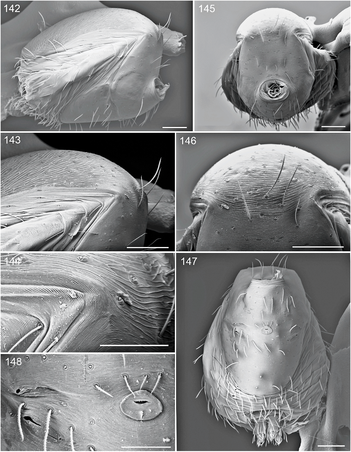
Figs. 142–148. Malagiella vohiparara, new species, male abdomen (PBI_OON 03378). 142. Lateral view. 143. Same, magnified view. 144. Same, detail of fused scutal area. 145. Anterior view. 146. Same, magnification of fused scuta. 147. Ventral view. 148. Same, magnified view of spiracles and sperm pore. Scale bars are 100 Mm (figs. 142, 143, 145–147) and 50 Mm (figs. 144, 148).

Figs. 149–156. Malagiella vohiparara, new species, spinnerets. 149. Male (PBI_OON 03378), spinnerets, apical view. 150. Same, ALS with 3 spigots. 151. Same, PMS with 2 spigots. 152. Same, PLS with 3 spigots. 153. Female (PBI_OON 03078), spinnerets, apical view. 154. Same, ALS, with 3 spigots. 155. Same, PMS, with 3 spigots. 156. Same, PLS, with 5 spigots. Scale bars are 50 Mm (figs. 149, 153) and 10 Mm (all others).

Figs. 157–164. Malagiella vohiparara, new species, male palp, femur to bulb (PBI_OON 03378). 157. Palp, prolateral view. 158. Same, retrolateral view. 159. Bulb, prolateral view with arrow to ventral process (BVP). 160. Same, retrolateral view. 161. Same, dorsal view. 162. Same, retrolateral view showing bulbtarsus junction (arrow). 163. Palp, dorsal view. 164. Same, ventral view. B 5 bulb, F 5 femur, P 5 patella, Ta 5 tarsus, Ti 5 tibia.

Figs. 165–172. Malagiella vohiparara, new species, male (PBI_OON 03378). 165. Embolar region, prolateral view. 166. Same, more dorsal. 167. Same, still more dorsal. 168. Same, fully dorsal view. 169. Same, retrolateral view. 170. Same, apical view. 171. Same, lateroapical view. 172. Same, magnified view showing embolar opening (arrow) and prolateral groove (dash). BVP 5 bulb ventral prong, E 5 embolus, EG 5 embolus groove, EO 5 embolus opening, EPP 5 embolus prolateral process, ERP 5 embolus retrolateral process.
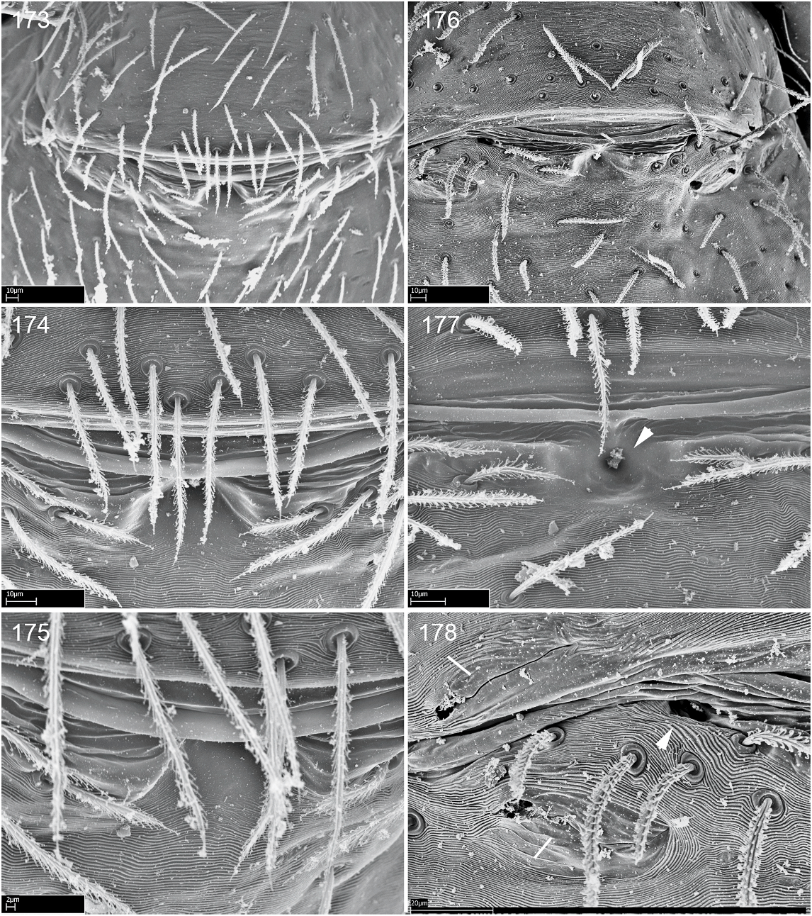
Figs. 173–178. Malagiella vohiparara, new species, female external genitalia (PBI_OON 03078). 173– 175. Ventral views at increasing magnification. 176. Posterior view. 177. Same, magnified view showing copulatory opening (arrow). 178. Same, showing respiratory spiracles (dashes) and apodeme orifice (arrow).

Figs. 179–184. Malagiella vohiparara, new species, female internal genitalia (179–181: PBI_OON 03078; 182–184: PBI_OON 03087). 179. Epigynal region, posteriodorsal view, of partially digested specimen showing membrane enclosing receptaculum. 180. Same, close-up of receptaculum. 181. Same, showing apodeme (arrow). 182. Same, of more fully digested specimen. 183. Same, close-up of receptaculum showing cleaned duct with remains of membrane (arrow). 184. Same, slightly more dorsal view showing attachment of duct (arrow). BL 5 book lung, BLC 5 book lung cover, G 5 gonopore, PA 5 posterior apodeme, R 5 receptaculum, Tr 5 trachea.

Figs. 345–354. Malagiella vohiparara, new species, male (PBI_OON 03078). 345. Habitus, ventral view. 346. Same, dorsal view. 347. Same, lateral view. Scale bar 5 1 mm. 348. Abdomen, ventral view. 349. Cephalothorax, ventral view. 350. Same, anterior view. 351. Same, posterior view. 352. Abdomen, anterior view. 353. Same, lateral view. 354. Same, posterior view.

Figs. 355–364. Malagiella vohiparara, new species, female (PBI_OON 03078). 355. Habitus, ventral view. 356. Same, dorsal view. 357. Same, lateral view. Scale bar 5 1 mm. 358. Abdomen, ventral view. 359. Cephalothorax, ventral view. 360. Same, anterior view. 361. Same, posterior view. 362. Abdomen, anterior view. 363. Same, lateral view. 364. Same, posterior view.
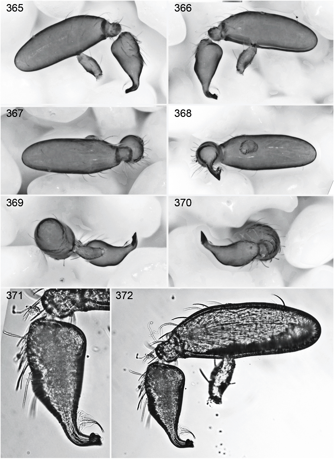
Figs. 365–372. Malagiella vohiparara, new species, male (PBI_OON 03078). 365. Palp, femur to bulb, prolateral view. 366. Same, retrolateral view. 367. Same, dorsal view. 368. Same, ventral view. 369. Same, anterior view. 370. Same, posterior view. 371. Bulb, retrolateral view. 372. Palp, retrolateral view.
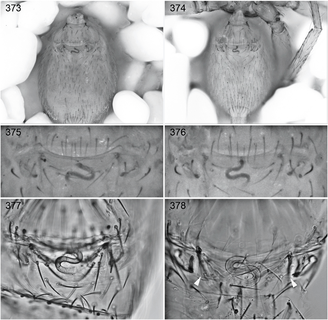
Figs. 373–378. Malagiella vohiparara, new species, female variation (PBI_OON 03078; 373, 375, 377, 378: specimen 1; 374, 376: specimen 2). 373, 374. Abdomen, ventral view. 375, 376. Epigynal area, ventral view. 377. Same, cleared specimen. 378. Same, dorsal view with arrows to apodemes.
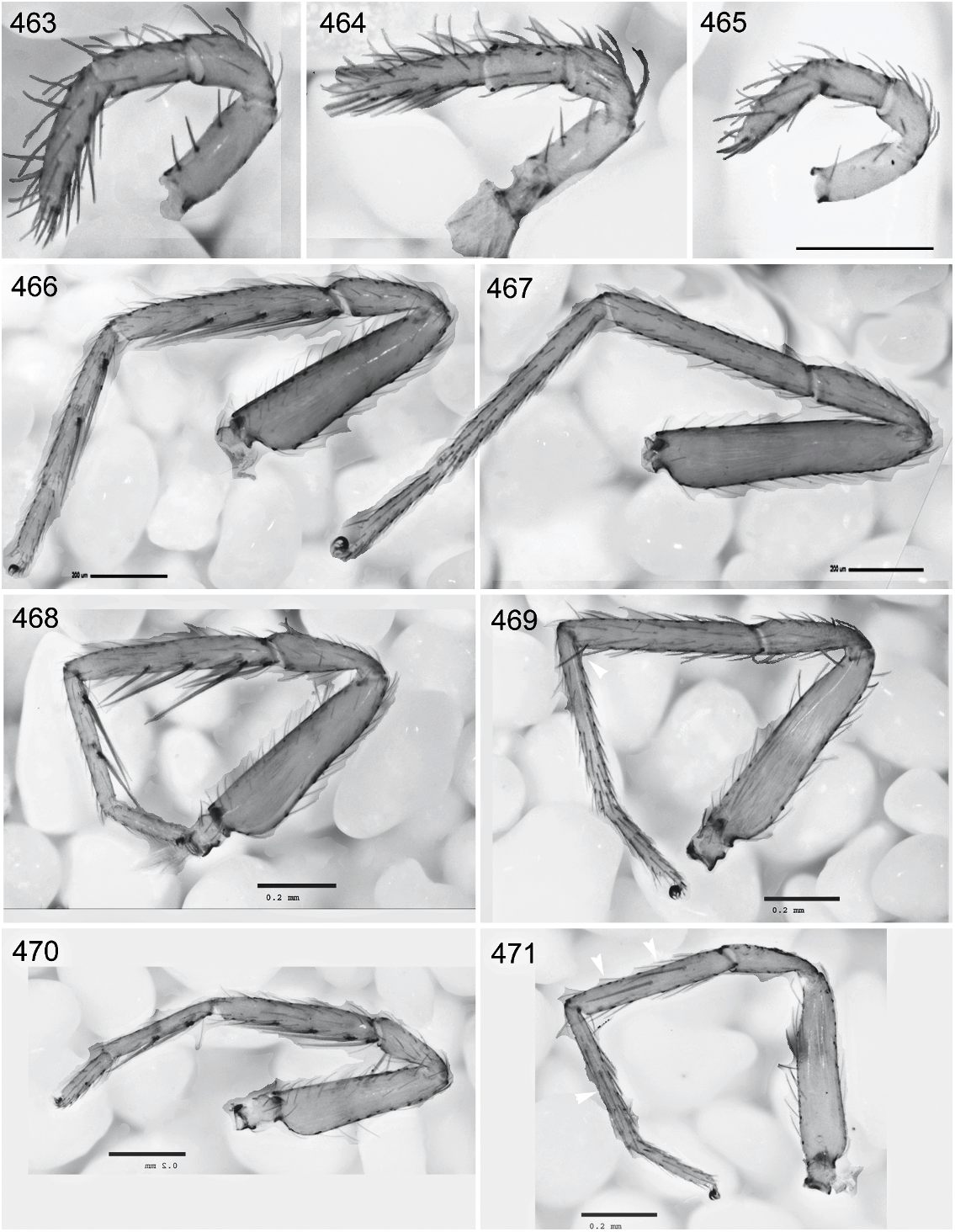
Figs. 463–471. Malagiella species, female palpi and legs. M. ranomafana (PBI_OON 03227): 463, 466, 467; M. vohiparara (PBI_OON 03078): 464, 468, 469; M. toliara (PBI_OON 03221): 465, 470, 471. 463– 465. Palp, retrolateral view. 466, 468, 470. Leg I, retrolateral view. 467, 469, 471. Leg IV, retrolateral view showing leg spines (arrows). Scale bars 5 200 Mm.
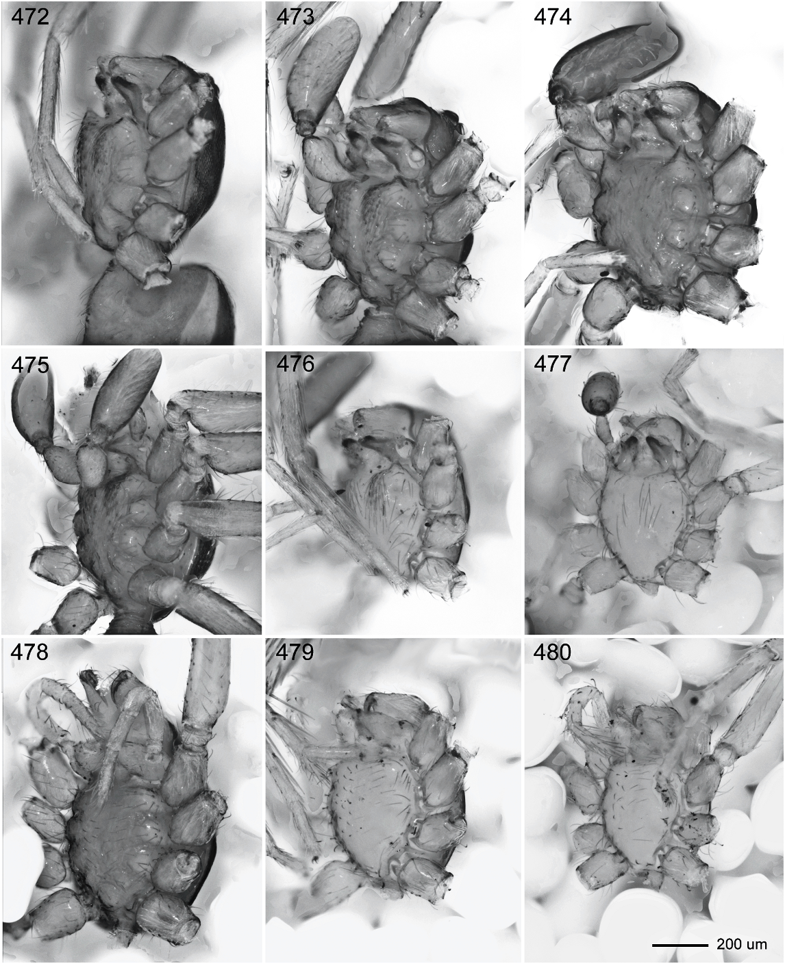
Figs. 472–480. Malagiella species, sternum, ventrolateral view, males (472–477) and females (478–480). 472. M. andringitra, PBI_OON 03234. 473. M. valterova, PBI_OON 03461. 474. M. fisheri, PBI_OON 03452. 475. M. ranomafana, PBI_OON 01999. 476. M. vohiparara, PBI_OON 03078. 477. M. toliara, PBI_OON 03377. 478. M. ranomafana, PBI_OON 01999. 479. M. vohiparara, PBI_OON 03078. 480. M. toliara, PBI_OON 03221.

Figs. 481–486. Malagiella species, male palpus, femur to bulb, prolateral view. 481. M. ranomafana, PBI_OON 03224. 482. M. andringitra, PBI_OON 03234. 483. M. valterova, PBI_OON 03461. 484. M. vohiparara, PBI_OON 03078. 485. M. fisheri, PBI_OON 03452. 486. M. toliara, PBI_OON 03377. Scale bar (200 Mm) applies to all palpi.

Fig. 487. Size variation in Malagiella measured as a function of carapace length to carapace width. Plot is based on measurements of 26 male specimens (‘‘x’’) and 30 female specimens (‘‘o’’). Blue lines enclose male values for a given species and red lines female values; disjunct conspecific readings are connected by straight lines. Green dotted lines enclose the three species groups. Brown dashed lines refer to the size of female DS, with the large dashes (upper right) enclosing species with the largest scutes and small dashes (lower left) those with the smallest. Abbreviations for species names: amba 5 M.ambalavo, andr 5 M. andringitra, fish 5 M. fisheri, good 5 M. goodmani, niki 5 M. nikina, rana 5 M. ranavalona, rano 5 M. ranomafana, toli 5 M. toliara, valt 5 M. valterova, vohi 5 M. vohiparara.
No known copyright restrictions apply. See Agosti, D., Egloff, W., 2009. Taxonomic information exchange and copyright: the Plazi approach. BMC Research Notes 2009, 2:53 for further explanation.
|
Kingdom |
|
|
Phylum |
|
|
Class |
|
|
Order |
|
|
Family |
|
|
Genus |