
Phocoenidae, Gray, 1825
|
publication ID |
https://doi.org/10.4202/app.2012.0127 |
|
DOI |
https://doi.org/10.5281/zenodo.11043689 |
|
persistent identifier |
https://treatment.plazi.org/id/03C287C9-353E-FFED-733D-FD4AA862FA73 |
|
treatment provided by |
Felipe |
|
scientific name |
Phocoenidae |
| status |
|
Phocoenidae View in CoL gen. et sp. indet.
Figs. 2–4 View Fig View Fig View Fig .
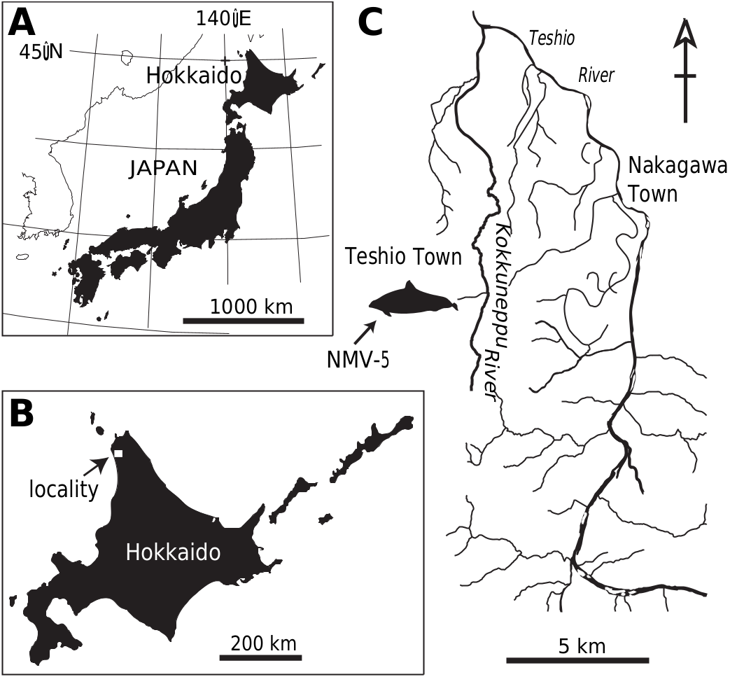
Material.— NMV-5, a partial rostrum and mandible, all cervicals, eight thoracic and three lumbar vertebrae, and several incomplete ribs, from a small, unnamed tributary valley of the Kokuneppu River located in the Sengen district, Teshio, Hokkaido, Japan (44°49′45 N, 141°59′26 E; Fig. 1 View Fig ); Koetoi Formation, early Pliocene (5.5–4.0 Ma) GoogleMaps .
Description
Skull
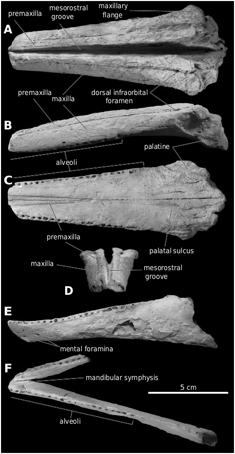
Rostrum.—The relatively well-preserved rostrum (length: 147 mm; Fig. 2A–D View Fig ), missing its distalmost 10–20 mm including the anteriormost alveolus ( Fig. 2B, C View Fig ), is all that remains of the skull. The greatest width of the maxillae is approximately 59+ mm ( Fig. 2C View Fig ), thus resembling the small extant phocoenids Neophocaena phocaeniodes and Phocoena phocoena in size.
Premaxilla.—The premaxilla is slender, although it should be noted that some of the medial portion of the proximal end of the premaxilla is missing. Distally, the premaxilla slopes steeply towards the lateral border of the rostrum ( Fig. 2D View Fig ). As in most phocoenids, the mesorostral groove is moderately open, with the medial edges of both premaxillae being largely parallel. The suture uniting the premaxilla and maxilla is distinct along the entire rostrum.
Maxilla.—In dorsal view, the maxilla steeply slopes away laterally towards its anterior end ( Fig. 2A, D View Fig ). The maxillary flange is flat and narrow. As in Piscolithax longirostris , the anteriormost dorsal infraorbital foramen is located directly adjacent to the premaxilla. In lateral view, the maxilla is slightly curved ventrally ( Fig. 2B View Fig ). The ventral surface of the maxilla is transversely concave ( Fig. 2D View Fig ), and bears a distinct palatine sulcus and greater palatine foramen in its proximal portion ( Fig. 2C View Fig ). The tooth rows are clearly separated, moderately divergent posteriorly, and shortened (as in other phocoenids), with the posteriormost alveolus located 32 mm anterior to the anterior edge of the palatine. 21 alveoli (length of tooth row = 88 mm) are preserved on the left side, and 20 (length of tooth row = 86 mm) on the right, with an average alveolus diameter of 2.5 mm. Six teeth lacking crowns are preserved in situ.
Vomer.—In ventral view, the vomer is narrowly exposed between the premaxillae, unlike in all other phocoenids except Piscolithax longirostris .
Palatine.—Only the anteriormost portions of the palatines are preserved ( Fig. 2C View Fig ), making accurate description impossible.
Mandible.—The left mandible is missing its anteriormost 10–20 mm and the portion posterior to the anterior border of the mandibular foramen. By contrast, only a 40 mm fragment remains of the right mandible ( Fig. 2E, F View Fig ). The mandibular symphysis is at least 15 mm long, and connects both mandibles at an angle of 35°. 20 alveoli are preserved in the left mandible (length of tooth row = 77 mm) and 12 in the right, with an average alveolus diameter of 1.9 mm.
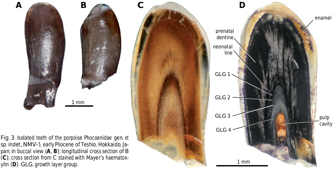
Teeth.—Two small (<3.5 mm long), isolated teeth with spatulate crowns and circular roots (1 mm in diameter) were preserved near the maxillae ( Fig. 3A–D View Fig ). The crowns measure 1 mm buccolingually and 1.25 mm anteroposteriorly, and are covered by smooth enamel with no accessory cusps or ornamentation.
Postcranial skeleton
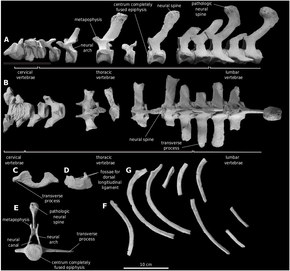
Cervical vertebrae.—The seven cervical vertebrae are unfused and anteroposteriorly compressed, and resemble other extinct phocoenids in their overall morphology ( Fig. 4A–D View Fig , Table 1 View Table 1 ). Weathering has resulted in the loss of most of the neural arches, as well as the erosion of the anterior face of the atlas ( Fig. 4C View Fig ). The ventral transverse process of the atlas is short and moderately robust, and there is no dorsal transverse process. The pedicle of the neural arch of the atlas is attached to the axis. The latter ( Fig. 4D View Fig ) has lost most of its neural arch and transverse processes, but preserves two deep fossae on the dorsal face of the vertebral body, which may represent attachments sites for the dorsal longitudinal ligament.
Thoracic vertebrae.—Eight thoracic vertebrae are preserved, with the posterior five having relatively long centra ( Fig. 4A View Fig ; Table 1 View Table 1 ). In terms of their relative and absolute centrum lengths, these vertebrae are intermediate between those of Numataphocoena yamashitai and extant species. All of the preserved thoracics have flattened anterior and posterior centrum faces, as well as well-developed metapophyses. Unlike in other phocoenids, except Phocoena sinus Norris and McFarland, 1958 and Phocoenoides dalli ( True, 1885) , the neural spines of the posterior thoracics are tall, slender and strongly recurved anteriorly ( Fig. 4A View Fig ). The neural spines of Neophocaena phocaenoides Cuvier, 1829 , are also strongly recurved anteriorly, but differ from those of NMV- 5 in being distinctly shorter dorsoventrally, and longer anteroposteriorly. The neural spines of at least two of the thoracic vertebrae exhibit exostosis, with their distal tips being moderately expanded anteroposteriorly, and slightly swollen laterally.
Lumbar vertebrae.—Three lumbar vertebrae, likely representing L1–L3 are preserved, and, except for the absence of an articular surface for the rib on their transverse processes, resemble the posterior thoracics in their general morphology. The centra of the lumbar vertebrae are distinctly elongated, both in relative (as compared to rostral width) and absolute terms. In dorsal view, the anterior and posterior edges of the transverse process of the first lumbar vertebra and an isolated posterior transverse process are parallel, whereas the transverse process of the second lumbar vertebra slightly widens anteroposteriorly ( Fig. 4B View Fig ). In this, NMV-5 resembles Salumiphocaena stocktoni , Piscolithax longirostris and extant species, and differs from Numataphocoena yamashitai , in which the transverse processes are distinctly widened distally. The neural spines on the first and third lumbars show a remarkable degree of exostosis, making their distal tips appear globular in lateral and dorsal view ( Fig. 4E View Fig ). On the third lumbar, the area affected by exostosis measures 48 mm anteroposteriorly, and 24 mm laterally. The presence of exostosis indicates that this individual suffered from serious disease or injury at some point during its life.
Rib.—At least eight ribs are preserved, including five from the anterior or central portion of the right ribcage ( Fig. 4F, G View Fig ).
No known copyright restrictions apply. See Agosti, D., Egloff, W., 2009. Taxonomic information exchange and copyright: the Plazi approach. BMC Research Notes 2009, 2:53 for further explanation.
|
Kingdom |
|
|
Phylum |
|
|
Class |
|
|
Order |
|
|
SuperFamily |
Delphinoidea |
|
Family |