
Axonchium japonicum, Ahmad & Naz, 2010
|
publication ID |
https://doi.org/10.1080/00222931003690706 |
|
persistent identifier |
https://treatment.plazi.org/id/03C24C64-FFDF-7F79-8E89-FB1379D5FEBF |
|
treatment provided by |
Felipe |
|
scientific name |
Axonchium japonicum |
| status |
sp. nov. |
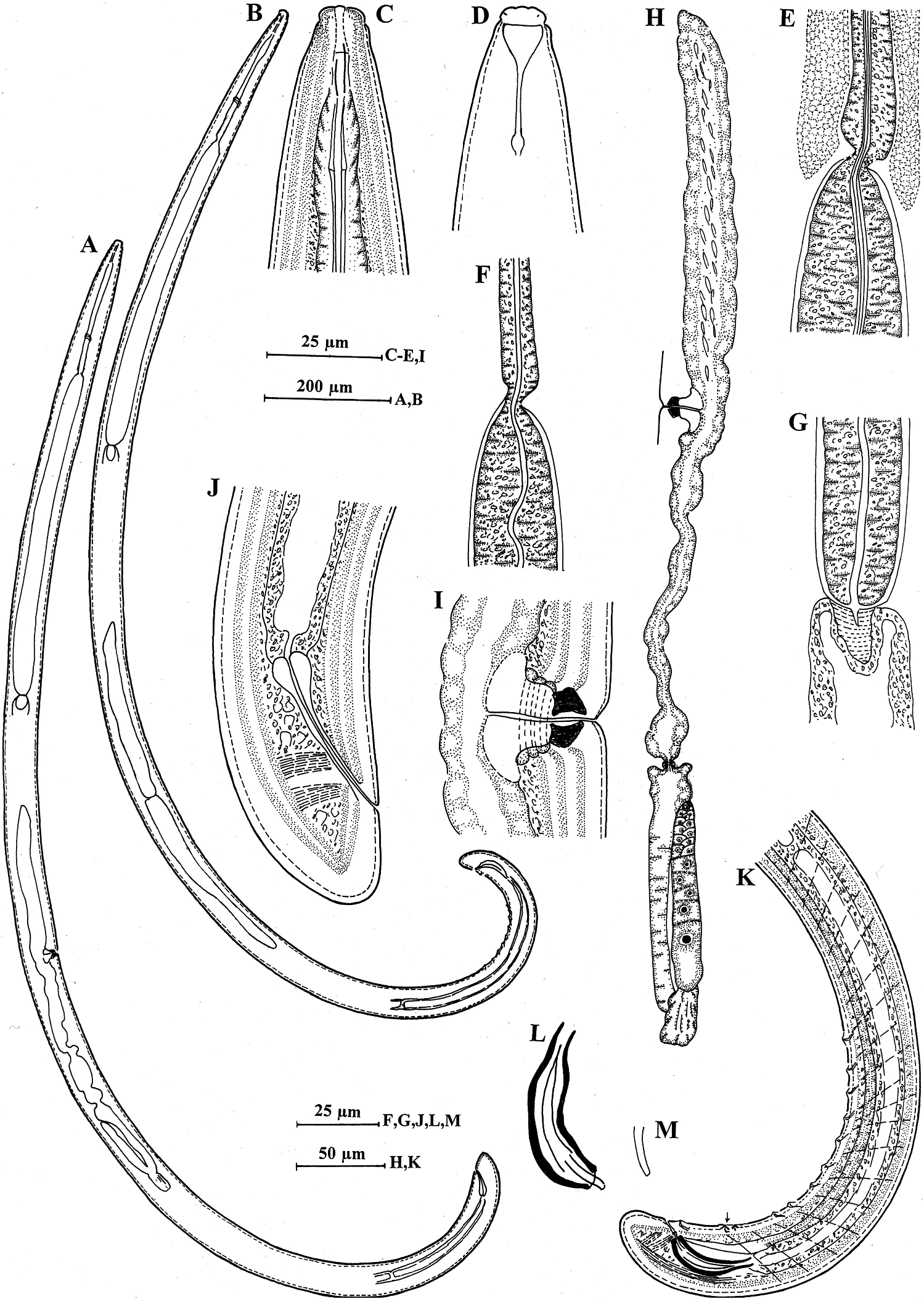
Axonchium japonicum sp. nov.
( Figures 3A–M View Figure 3 , 4A–I View Figure 4 ; Table 2)
Description
Female. Body curved ventrad upon fixation, tapering towards anterior extremity. Posteriorly terminating in a short conoid tail. Cuticle with fine transverse striations, 3–4 µm thick at mid-body and 9–10 µm at tail. Lateral chords about one-fifth of body width at mid-body. Lateral and dorsal pores indistinct; ventral pores 16–17 in the pharyngeal region, 5–6 between pharynx base and vulva and 13–15 between vulva and tail. Lip region offset, about two and half to three times as wide as high or about one-fifth as wide as body width at neck base; lips separate. Amphids cup-shaped, their aperture occupying about three-quarters of lip region width. Odontostyle fusiform, 1.0–1.1 times lip region width long, its aperture about one-third of its length. Guiding ring ‘single’, at 1.1–1.2 times lip region width from anterior end. Odontophore linear, 1.4 times the odontostyle length. Nerve ring encircling the anterior slender part of pharynx at about 17% of neck length from anterior end. Anterior part of pharynx muscular, separated from the posterior expanded part by a short isthmus-like structure, the latter occupying about 69–70% of total neck length and enclosed in muscle sheath with straight bundles. Cardia elongate–conoid, about two-thirds of corresponding body width long. Genital system mono-opisthodelphic; anterior genital branch represented by a simple uterine sac, 3.3–4.1 times the corresponding body width long and filled with spermatozoa. Posterior genital branch well developed; ovary reflexed, not reaching or surpassing the oviduct–uterus junction, measuring 220–320 µm with oocytes arranged in a single row except near tip. Oviduct joining ovary subterminally, measuring 170–180 µm, consisting of a long slender part with prismatic cells and a slightly wider pars dilatata with wide lumen; sphincter between oviduct–uterus junction present. Uterus measuring 200–215 µm, differentiated into three parts, distal and proximal expanded part with wide lumen, intermediate convoluted part with narrow lumen. Vulva transverse. Vagina extending inward, about half of the corresponding body width; pars proximalis vaginae 19–20 µm long with almost straight walls, encircled by circular muscles; pars refringens vaginae with two rhomboid to almost triangular sclerotized pieces, each measuring 6–6.5 × 8 µm (cw = 13 µm); pars distalis vaginae 4–5 µm with slightly concave walls. Prerectum five to six anal body widths long. Rectum about as long as anal body width. Tail short, conical, about 0.7 times anal body width long. Caudal pores two on each side.
Male. Similar to female in general morphology, except for posterior region being more curved ventrad because of the presence of copulatory muscles. Supplements, an adanal pair and 10–11 evenly spaced ventromedians. Two weakly-developed supplements within the range of spicules. Spicules arcuate, thick walled, thickening more pronounced at distal tip, about 1.4–1.5 times anal body width long. Lateral guiding pieces slender, rod-like about one-quarter of the spicule length. Prerectum about nine anal body widths long. Rectum about as long as anal body width. Tail conoid, about 0.7 times anal body width long. Two caudal pores on each side.
Type habitat and locality
From grassland of Ishigaki Island, south Japan, Japan.
Type specimens
Holotype female on slide Axonchium japonicum sp. nov. /1; paratype females and males on slides Axonchium japonicum sp. nov. / 2–3; deposited with the nematode collection of the Department of Zoology, Aligarh Muslim University, India.
Diagnosis and relationships
Axonchium japonicum sp. nov. is characterized by having 2.3–2.4 mm long body; low, offset lip region; 11–12 µm long odontostyle; two parts of pharynx separated by a short isthmus-like structure; anterior uterine branch a simple uterine sac; vagina straight with two distinct almost triangular pars refringens vaginae; 9–10 evenly spaced ventromedian supplements and massive spicules with thickened distal tip.
Among the species with vaginal sclerotization, the new species comes close to Axonchium vaginatum Jairajpuri, 1965 in having short isthmus-like constriction between two parts of pharynx and anterior uterine branch a simple sac without rudiments of ovary or oviduct. However, it differs from it, in the shape of pars refringens vaginae (triangular vs heart-shaped); in the absence of ‘Z’ organ in uterus (vs presence), in the number of ventromedian supplements (10–11 vs 9) and in the shape of spicules (distal end of spicule characteristically thickened vs not thickened), and lateral guiding pieces rod-like with simple tip (vs bifid).
The new species is also close to A. coronatum ( de Man, 1906) Thorne and Swanger, 1936 in having short isthmus-like constriction between two parts of pharynx and spaced ventromedian supplements but differs from it, in having shorter body length (2.3–2.4 vs 2.99–4.18 mm), shorter odontostyle (11–12 µm vs 12–15.5 µm), differently shaped pars refringens vaginae (triangular vs heart-shaped), in the absence of rudimentary ovarian mass and small oviduct at the distal end of anterior uterine branch (vs presence), and in having differently shaped and shorter spicules (robust, 55–60 µm vs cylindrical, 86.5–87.5 µm).

No known copyright restrictions apply. See Agosti, D., Egloff, W., 2009. Taxonomic information exchange and copyright: the Plazi approach. BMC Research Notes 2009, 2:53 for further explanation.