
Eurytion yungarum, Pereira, Luis A., 2005
|
publication ID |
https://doi.org/ 10.5281/zenodo.170535 |
|
DOI |
https://doi.org/10.5281/zenodo.5631344 |
|
persistent identifier |
https://treatment.plazi.org/id/03C24866-FF94-B459-6377-FE67332AFD89 |
|
treatment provided by |
Plazi |
|
scientific name |
Eurytion yungarum |
| status |
sp. nov. |
Eurytion yungarum View in CoL n. sp.
( Figs. 1–41 View FIGURES 1 – 4 View FIGURES 5 – 9 View FIGURES 10 – 15 View FIGURES 16 – 25 View FIGURES 26 – 33 View FIGURES 34 – 35 View FIGURES 36 – 41 )
Diagnosis: An Eurytion species with circumforaminal rim of coxosternum of second maxillae not elongate and coxal organs arranged in 2 clusters in each coxopleuron. Among the Neotropical species included in the genus Eurytion , only the present species and Eurytion lethifer Crabill, 1968 share these particular traits. Characters in table 1 separate these two species.
Type material examined: Holotype ɗ, 49 pairs of legs, body length 19 mm, from ARGENTINA: Jujuy: 900 m a.s.l., ca. 50 Km W of Fraile Pintado, close to rivers Candelaria and Normenta, 17.IV.1993, L. A. Pereira & S. Coscarón leg. ( MLP). (First maxillae and mandibles in a slide, rest of the body in alcohol).
Etymology: This species is named after the biogeographical Province of the 'Yungas' in which the holotype has been collected.
Description of Male holotype: 49 pairs of legs, body length 19 mm, maximum body width 0.5 mm. Colour (of preserved specimen in alcohol) yellowish with cephalic shield and forcipular segment darker (pale ochreous).
Antennae: relatively short, ca. 2.4 times as long as the cephalic plate, distally slightly attenuate, all articles longer than wide. Setae on antennal articles IV of different length and few in number; those of remaining articles progressively shorter and more numerous towards the tip of the appendage ( Fig. 1 View FIGURES 1 – 4 ). Terminal antennal article (a.a.) with ca. 9 claviform sensilla on the external border and ca. 11 on the internal border. Distal end of this a.a. with ca. 5 very small sensilla apparently not split apically ( Fig. 2 View FIGURES 1 – 4 ). Ventral and dorsal surface of a.a. II, V, IX ( Figs. 3–4 View FIGURES 1 – 4 ) and XIII with very small specialised sensilla. On the ventral side these sensilla are restricted to an internal lateroapical area and are represented by two different types: a and b. Type a sensilla are very thin and not divided apically, type b sensilla are very similar to those of the apex of the terminal article (a, b, Fig. 3 View FIGURES 1 – 4 ). Specialised sensilla on dorsal side are restricted to a lateroapical area and are represented by two different types: a and b, similar to a and b of the ventral side (a, b, Fig. 4 View FIGURES 1 – 4 ). Distribution of type a and b sensilla as in table 2.
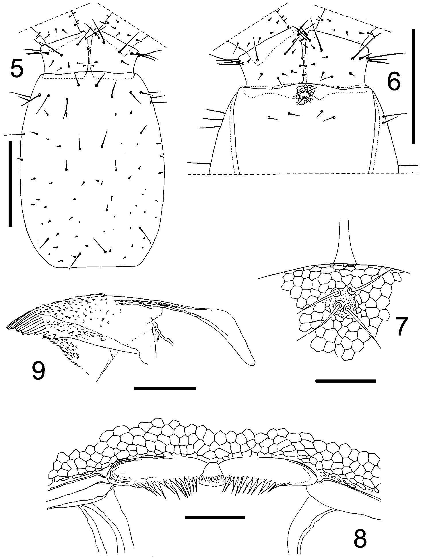
Cephalic plate: nearly subrectangular with sides curved, distinctly longer than wide (ratio 1.35: 1). Shape and chaetotaxy as in Fig 5 View FIGURES 5 – 9 .
Clypeus: with 4 setae located on the clypeal area and 2+2 setae on the middle: remaining clypeal surface without setae ( Fig. 6 View FIGURES 5 – 9 ). Clypeal area relatively small, minutely punctate or granulate, not areolate ( Figs. 6, 7 View FIGURES 5 – 9 ).
Labrum: midpiece trapeziform, well developed and sclerotized, with 7 small tuberculate teeth. Sidepieces with 13+13 long hyaline filaments ( Fig. 8 View FIGURES 5 – 9 ).
Mandible: pectinate lamella with ca. 14 hyaline teeth with shape as in Figure 9 View FIGURES 5 – 9 .
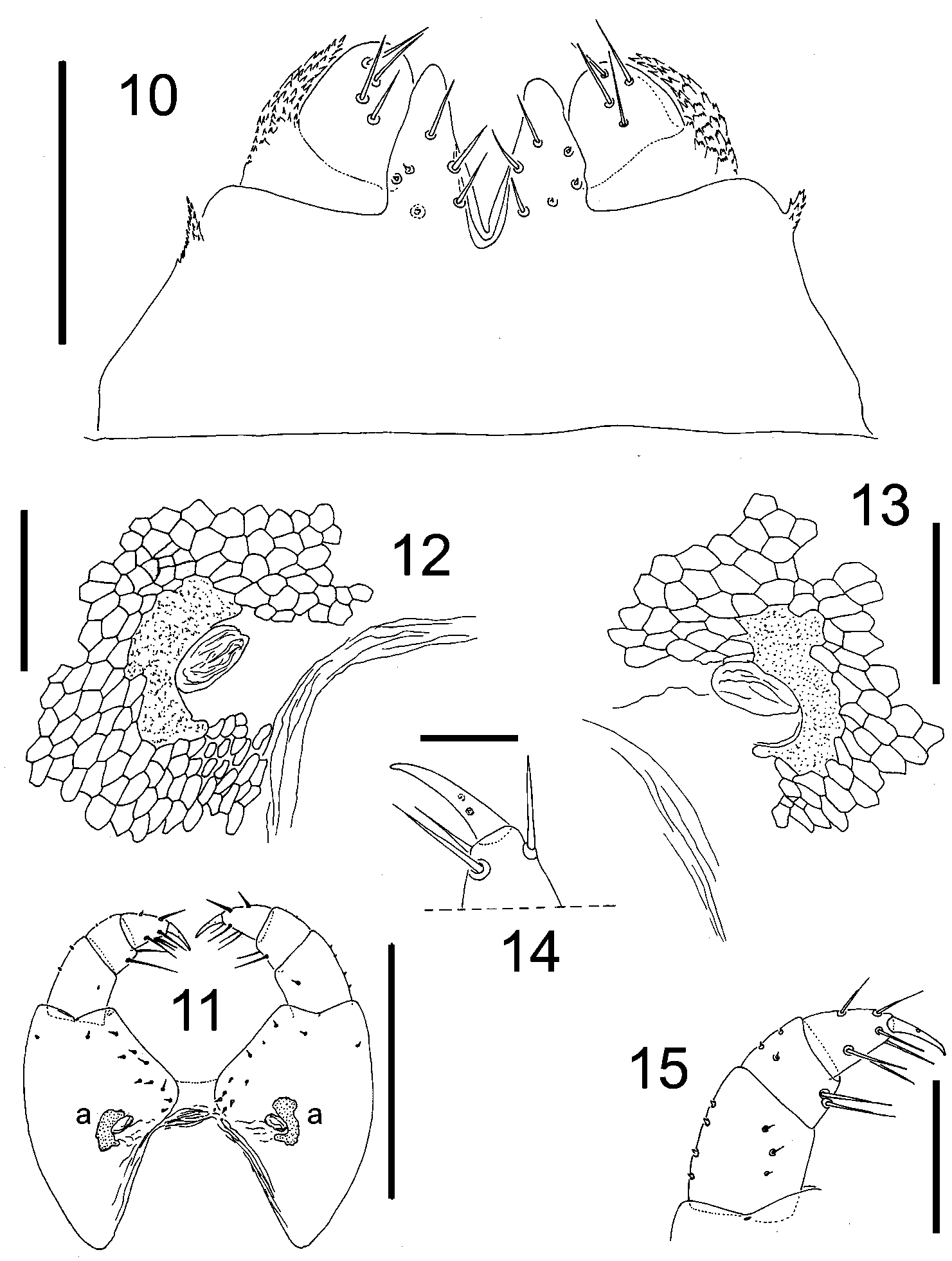
First maxillae: with small lappets on coxosternum; telopodites with well developed lappets almost as long as the telopodite ( Fig. 10 View FIGURES 10 – 15 ). Coxosternum without setae; median projections of coxosternum subtriangular, well developed and provided with 3+3 large setae and 3+3 small sensilla. Article II of telopodite with 4+4 ventral setae ( Fig. 10 View FIGURES 10 – 15 ), dorsal surface apparently without sensilla.
Second maxillae: coxites with 11+11 setae, medially joined through a narrow, hyaline and nonareolate membranous isthmus only ( Fig. 11 View FIGURES 10 – 15 ). Pore surronded by sclerotized rim ( Figs. 12–13 View FIGURES 10 – 15 ). Apical claw of telopodite well developed slightly curved internally at the tip ( Fig. 14 View FIGURES 10 – 15 ). Chaetotaxy of coxosternum and telopodites as in Figs. 11, 15 View FIGURES 10 – 15 .
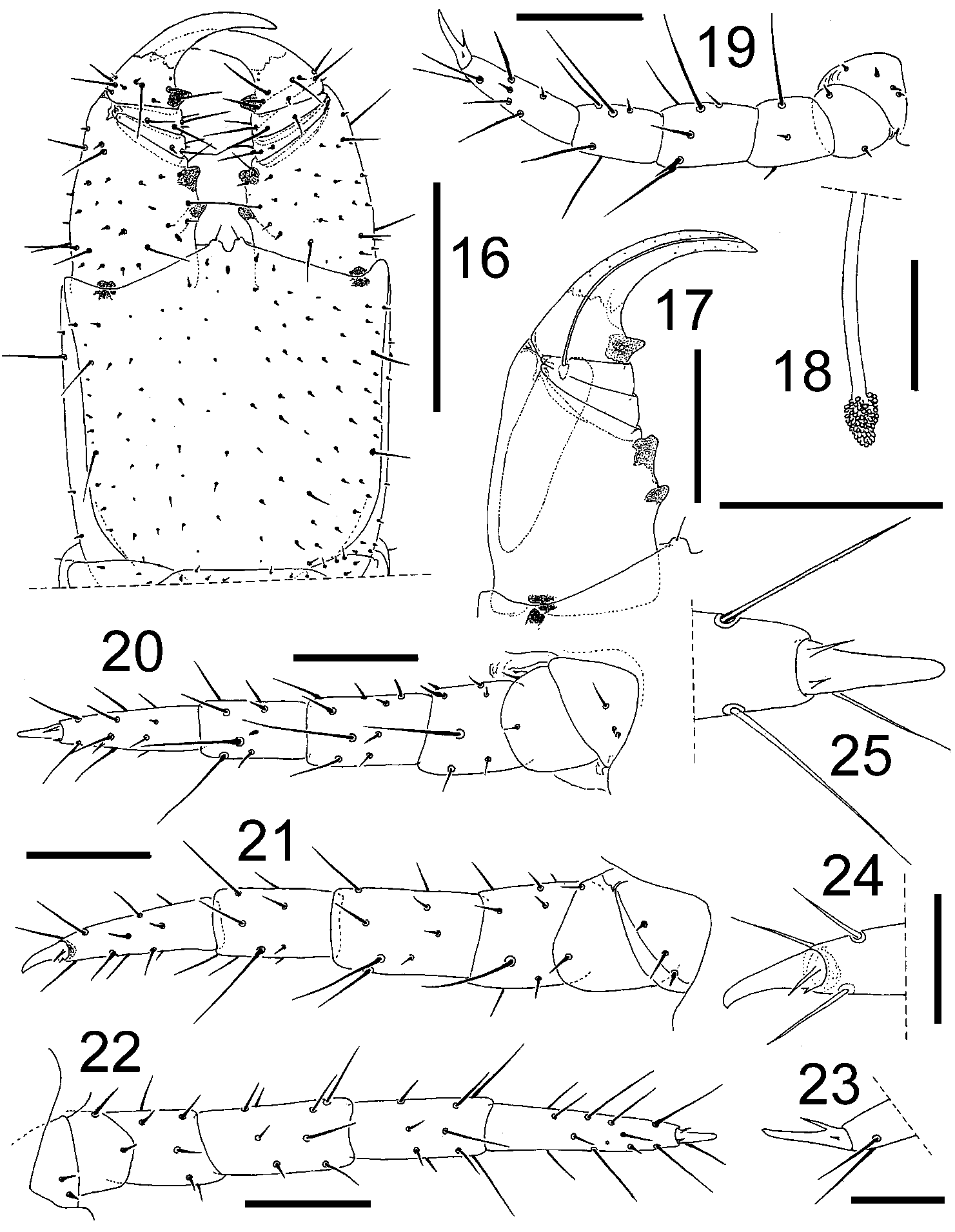
Forcipular segment: when closed, the telopodites reach the level of the anterior margin of the head or slightly project beyond. Forcipular tergum trapeziform with anterior and posterior margins, respectively, covered by the cephalic plate and the tergum of first legbearing segment; chaetotaxy represented by an irregular transverse row of ca. 4 setae on the middle and a very few additional smaller setae dispersed on the remaining surface. Coxosternum without chitinous lines, middle part of anterior border with two ochreous denticles. Telopodites: trochanteropraefemur with two denticles, both deeply pigmented, proximal denticle shorter than the distal one ( Figs. 16–17 View FIGURES 16 – 25 ). Femur and tibia without denticles. Tarsungulum basally with a well developed and deeply pigmented denticle; dorsal and ventral edge of the ungular blade not serrulate. Calyx of poison gland subtriangular ( Figs. 17–18 View FIGURES 16 – 25 ). Chaetotaxy of coxosternum and telopodites as in Fig. 16 View FIGURES 16 – 25 .
Walking legs: first pair shorter than the second one (ca. 0.85: 1). Praefemur, femur and tibia of legs I–XXV each ventrally with one specially long seta ( Figs. 19–21 View FIGURES 16 – 25 ). Remaining legs with shorter setae and similar chaetotaxy ( Fig. 22 View FIGURES 16 – 25 ). Claws ventrally with two basal parungues, the anterior larger than the posterior ( Figs. 23–25 View FIGURES 16 – 25 ).
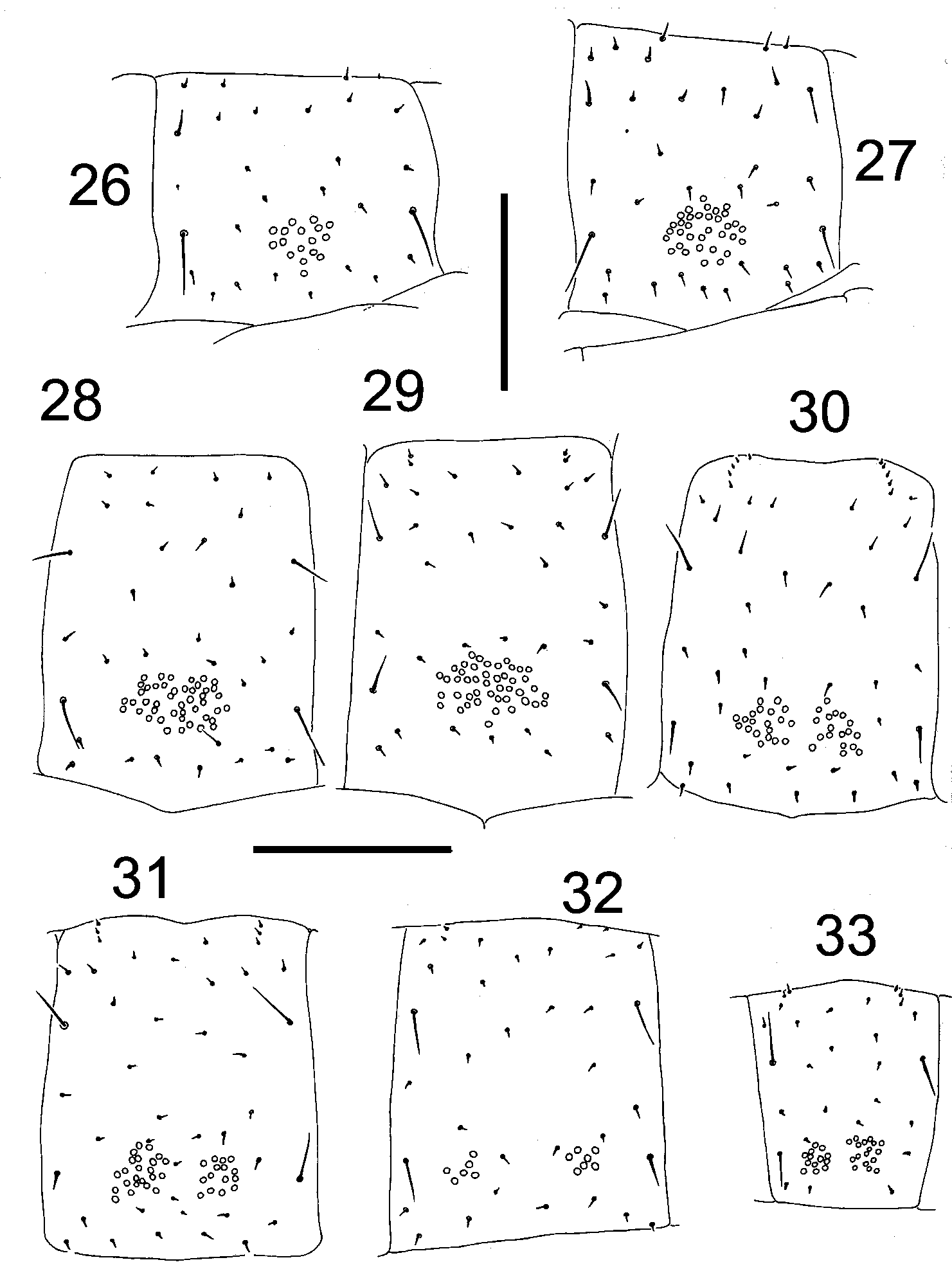
Sterna: pore fields present from the second to the penultimate sternum. Fields undivided on sterna II–XIII and divided in two subsymetrical areas on all remaining ones (XIVXLVIII). Form of fields changing along the trunk as in Figs. 26–33 View FIGURES 26 – 33 . Number of pores on selected sterna: on sternum II, 17 pores; on VI, 34; on XII, 45; on XIII, 44; on XIV, 20+18; on XV, 23+15; on XXXVII, 6+7; on XLVIII, 13+18.
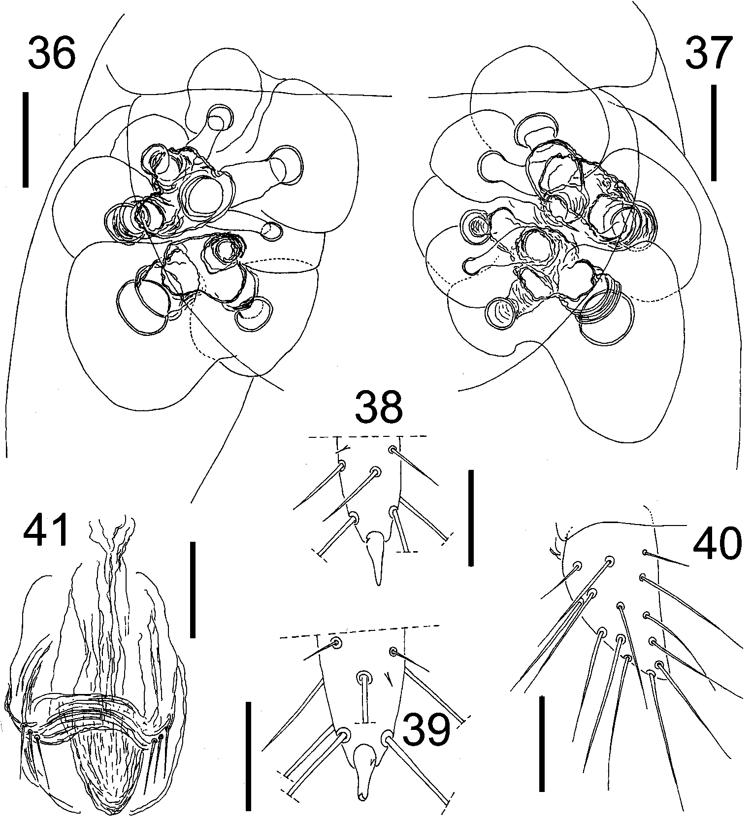
Last legbearing segment: without pleurites at the sides of praetergum. Praesternum divided along the sagittal plane; shape and chaetotaxy of tergum and sternum as in Figs. 34–35 View FIGURES 34 – 35 . Coxopleura slightly protruding at their distal ventral ends, setae numerous on the distal internal area, the remaining surface with less numerous larger setae. Coxal organs arranged in 2+2 clusters. Anterior clusters with ca. 4–5 organs, posterior with ca. 3–4 organs ( Figs. 35–37 View FIGURES 34 – 35 View FIGURES 36 – 41 ). Pores open on the membrane between coxopleuron and sternum, covered by the latter ( Fig. 35–37 View FIGURES 34 – 35 View FIGURES 36 – 41 ). Last legs with seven podomeres, form and chaetotaxy as in Figs. 34–35 View FIGURES 34 – 35 . Praetarsus unguiform, relatively smaller than those of the remaining legs, basally with a single parunguis ( Figs. 38–39 View FIGURES 36 – 41 ).
Terminal segments: intermediate tergum with posterior margin convex, intermediate sternum with posterior margin nearly straight. First genital sternum with posterior border approximately straight in the middle, concave along the sides. Gonopods uniarticulate with ca. 14 setae ( Figs. 35 View FIGURES 34 – 35 , 40 View FIGURES 36 – 41 ); penis dorsally with 3+3 apical setae ( Fig. 41 View FIGURES 36 – 41 ). Anal organs absent.
Remarks: The adult condition of this specimen is indicated by the tubula seminifera full of mature spermatozoa.
Female. Unknown.


| MLP |
Museo de La Plata |
No known copyright restrictions apply. See Agosti, D., Egloff, W., 2009. Taxonomic information exchange and copyright: the Plazi approach. BMC Research Notes 2009, 2:53 for further explanation.