
Clubiona qianlei J. Zhang, F. Zhang & H. Yu, 2022
|
publication ID |
https://doi.org/10.11646/zootaxa.5129.3.5 |
|
publication LSID |
lsid:zoobank.org:pub:02C5ACBF-29EA-45FF-B29C-867F2F810A7A |
|
DOI |
https://doi.org/10.5281/zenodo.6502255 |
|
persistent identifier |
https://treatment.plazi.org/id/03C1C17C-E769-367B-FF6D-F9ADFDE8FE51 |
|
treatment provided by |
Plazi |
|
scientific name |
Clubiona qianlei J. Zhang, F. Zhang & H. Yu |
| status |
sp. nov. |
Clubiona qianlei J. Zhang, F. Zhang & H. Yu View in CoL , sp. nov.
Figs 1–4 View FIGURE 1 View FIGURE 2 View FIGURE 3 View FIGURE 4 , 6 View FIGURE 6
Type material. Holotype: ♂, CHINA: Hubei Province, Xianning City, Tongshan County, Jiugongshan Nature Reserve , Shilonggou ( N29.41044598º, E114.64803038º, 480 m), 29.IV.2020, Q.L. Lu and Y. Zhong leg. GoogleMaps Paratype: 1♀ ( YHCLU0276 ), Jiugongshan Nature Reserve , Yunzhonghu ( N29.40443038º, E114.67254442º, 1240 m), 1.VII.2020, Q.L. Lu et al. leg GoogleMaps .; 1♂ ( YHCLU0318 ), same data as holotype GoogleMaps .
Etymology. This species is a patronymic named after Mr. Qianle Lu (Shenzhen City, China), collector of the types, who has greatly helped us in our research.
Diagnosis. The male of C. qianlei sp. nov. differs from those of all other group members by the absence of subapical barb on the RTA’s ventral process, and by the tegular apophysis width ca. 2/3 of bulb width ( Figs 2B, C View FIGURE 2 , 3B View FIGURE 3 ). In contrast, all other group members have subapical barb on the RTA’s ventral process, tegular apophysis narrower than half of bulb width, such as C. reclusa ( Almquist 2006: 374, figs 323a, b) and C. interjecta ( Figs 5A, C View FIGURE 5 ; Tang et al. 2005: 79, figs 3D, E). The female is similar to those of C. interjecta in the general appearance of the epigyne, but differs by: (1) copulatory openings slit-like, indistinct ( Figs 4A–C View FIGURE 4 ) (vs. nearly circular, relatively conspicuous; Fig. 5D View FIGURE 5 ; Tang et al. 2005: 79, fig. 3B; Zhu & Zhang 2011: 358, fig. 257B); (2) dorsal part of spermatheca (SPd) with hyaline outermost surface ( Figs 4D, E View FIGURE 4 ) (vs. surface sclerotized; Fig. 5E View FIGURE 5 ; Tang et al., 2005: 79, fig. 3C; Zhu & Zhang, 2011: 358, fig. 257B); (3) the two SPds separated by about one diameter ( Figs 4D, E View FIGURE 4 ) (vs. closely spaced or separated by less than 0.5 diameter; Fig. 5E View FIGURE 5 ; Tang et al. 2005: 79, fig. 3C; Zhu & Zhang 2011: 358, fig. 257B)
Description. Male ( holotype): Total length 5.90. carapace 2.95 long, 2.05 wide; abdomen 2.95 long, 1.62 wide.
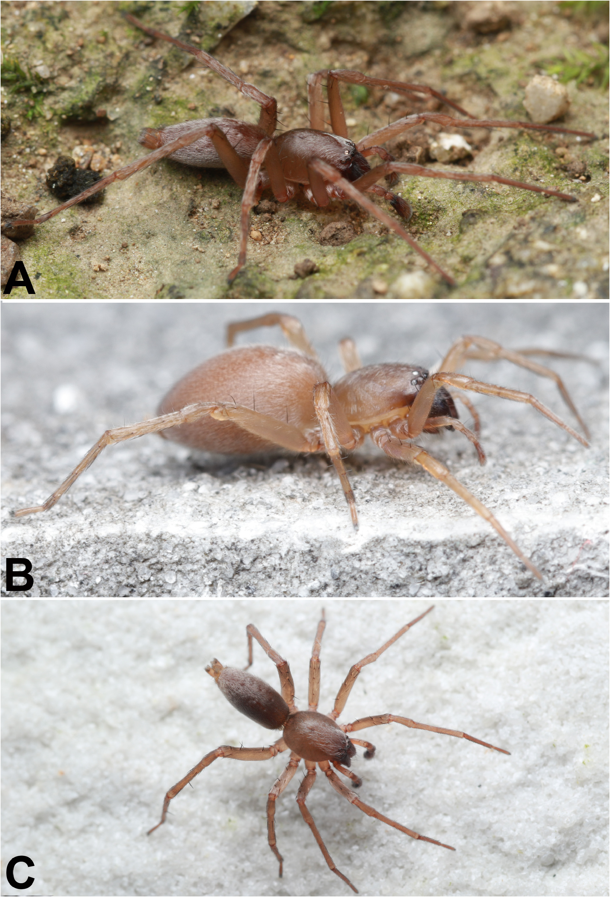
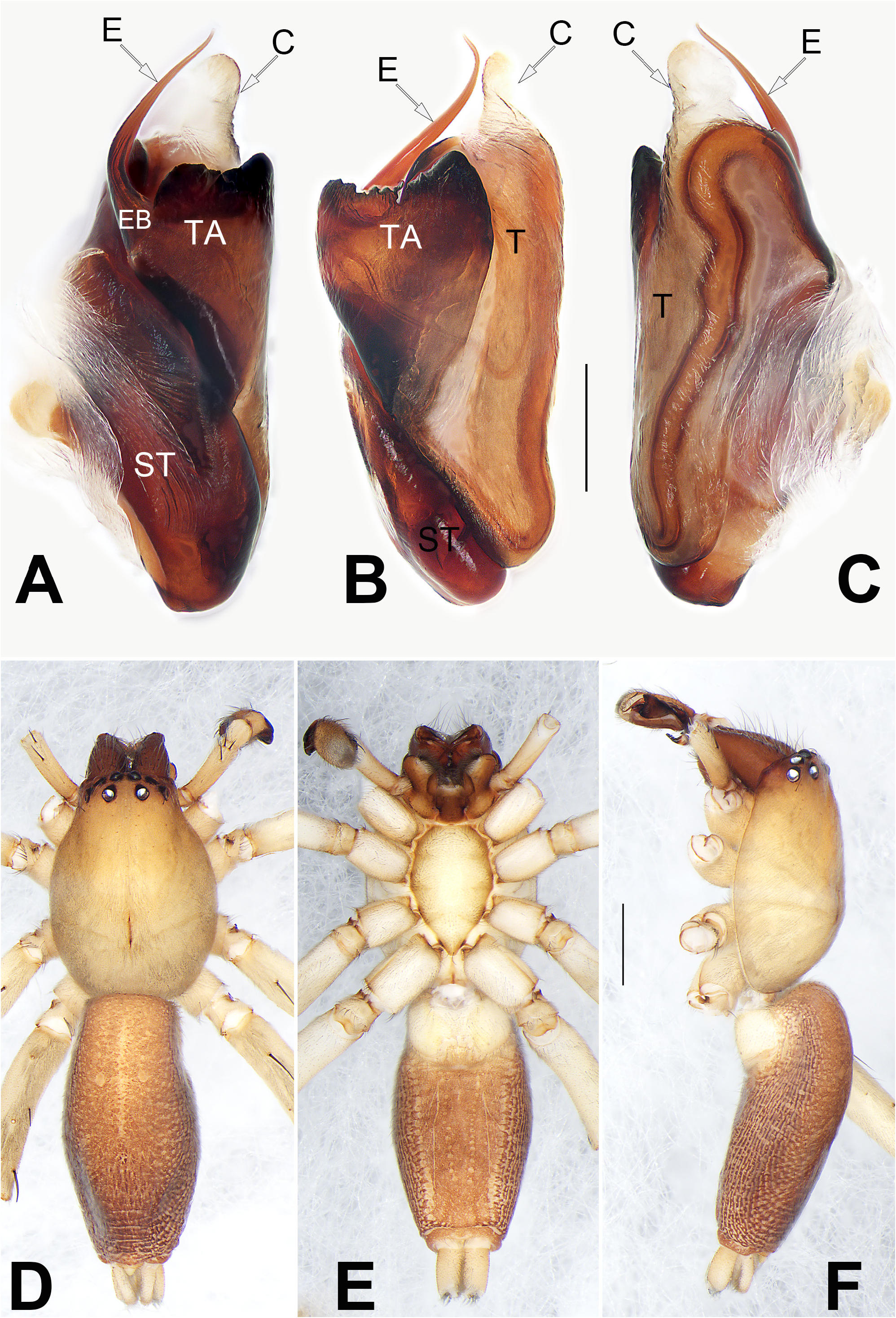
Living holotype dark brown ( Fig. 1A View FIGURE 1 ). Carapace yellowish-brown in ethanol ( Figs 3D, F View FIGURE 3 ), slightly darker in front, without a distinct pattern; cephalic region distinctly narrowed, thoracic groove and radial grooves indistinct; tegument smooth, clothed with fine hairs. Eyes: AER slightly recurved, PER slightly wider than AER, almost straight in dorsal view. Eye sizes and interdistances: AME 0.15, ALE 0.14, PME 0.18, PLE 0.16, AME–AME 0.12, AME–ALE 0.07, PME–PME 0.33, PME–PLE 0.21, MOQL 0.34, MOQA 0.37, MOQP 0.60. Chelicerae protruding, robust, uniformly brownish red, with four teeth on promargin and three on retromargin. Sternum ( Fig. 3E View FIGURE 3 ) yellowish white, 1.57 long, 1.02 wide. Labium coloured as chelicerae, endites brown. Legs light yellow, without distinct markings. Leg measurements: I 8.41 (2.37, 3.41, 1.66, 0.97), II 9.99 (2.55, 3.39, 3.11, 0.94), III 7.31 (2.21, 2.57, 1.90, 0.63), IV 10.36 (2.95, 3.44, 3.09, 0.88). Abdomen ( Figs 3D–F View FIGURE 3 ) elongate, oval, dorsum centrally with a lengthwise heart mark, reaching half of abdomen length, with a pair of muscular depressions located on middle level of heart mark; laterally with lengthwise reticular pattern, posteriorly furnished with a fuzzy pattern; venter medially with two longitudinal dotted lines.
Palp ( Figs 2 View FIGURE 2 A−D, 3A−C): Tibia short, ca. 1/3 length of cymbium. Retrolateral tibial apophysis (RTA) heavily sclerotized and bifurcated, slightly longer than tibia, broad at base; ventral process (VP) with sharp tip, shaped like a leaf in retrolateral view; dorsal process (DP) triangular, ca. 1/2 ventral process length. Genital bulb elongated and with a relatively flat tegulum, ca. 1.8 × longer than wide, with distinctive, sinuate sperm duct. Tegular apophysis (TA) represented by a large and flat sclerite, inverted triangular, anterior margin slightly concaved and rough, situated prolaterally on the tegulum, about 2/3 width of bulb. Embolar base (EB) inserted at approximately ten o’clock on tegulum, arising on the dorsal, hidden side of the tegular apophysis; free part of embolus (E) filamentous, as long as tegulum width, tip directed antero-mesally. Conductor (C) thick, finger-like, ca. 1/5 of tegulum length, originating from retrolateral, apical area of tegulum.
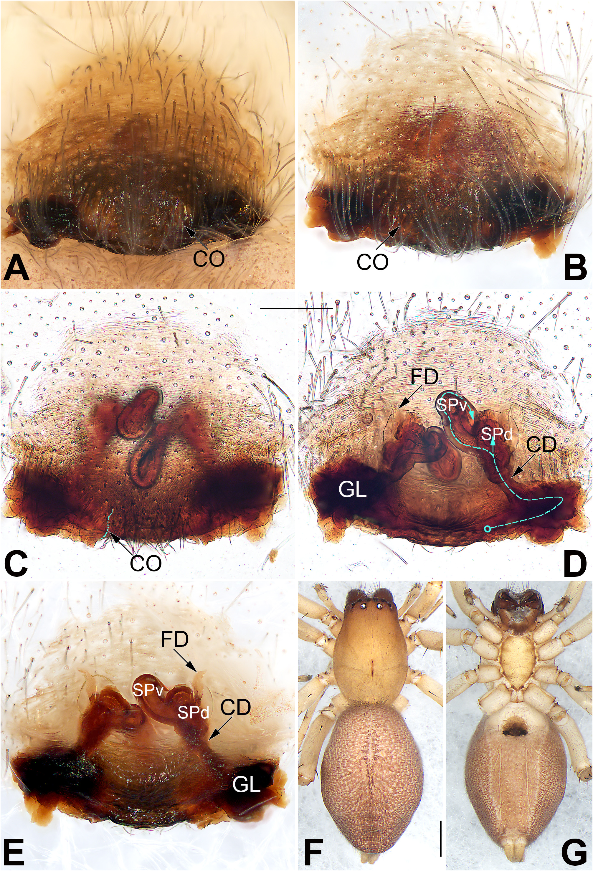
Female. As in male, but slightly larger in size and lighter in colour. Living paratype light brown ( Fig. 1B View FIGURE 1 ). Habitus in ethanol as Figs 4F, G View FIGURE 4 . Total length 6.80; carapace 2.92 long, 2.09 wide; abdomen 3.88 long, 2.86 wide. Eye sizes and interdistances: AME 0.16, ALE 0.18, PME 0.19, PLE 0.11, AME–AME 0.05, AME–ALE 0.06, PME– PME 0.25, PME–PLE 0.20. MOQL 0.45, MOQA 0.38, MOQP 0.58. Sternum 1.56 long, 0.94 wide. Measurements of legs: I 6.49 (1.89, 2.68, 1.16, 0.76), II 6.68 (1.93, 2.72, 1.31, 0.72), III 5.97 (1.76, 2.13, 1.57, 0.51), IV 9.17 (2.63, 3.09, 2.76, 0.69).
Epigyne ( Figs 4 View FIGURE 4 A−E): Epigyne plate a darkened disc, nearly triangular, anterior and lateral margins indistinct, posterior margin heavily sclerotized, middle part of posterior margin protruding; arrangement of the various parts of the vulva indistinctly visible through the tegument. Copulatory openings (CO) small, indistinct, represented by a short slit, situated at concave bends of epigynal plate posterior margin. Copulatory ducts (CD) firstly running along the posterior margin of epigyne, leading to large glands (GL), then ascending obliquely, finally connecting to anteriorly located spermathecae. Dorsal part of spermathecae (SPd) globular, innermost surface sclerotized, outermost surface hyaline, the two SPd separated by about one diameter. The ventral part of spermathecae (SPv) tubular or finger-like, curved medially, the two SPvs closely spaced. Fertilization ducts (FD) acicular, membranous, slightly shorter than diameter of SPv, located on dorsal surface of SPd.
Natural History. Leaf-dwelling spiders. Photos of live specimens were taken after the spiders dropped on the ground due to beating.
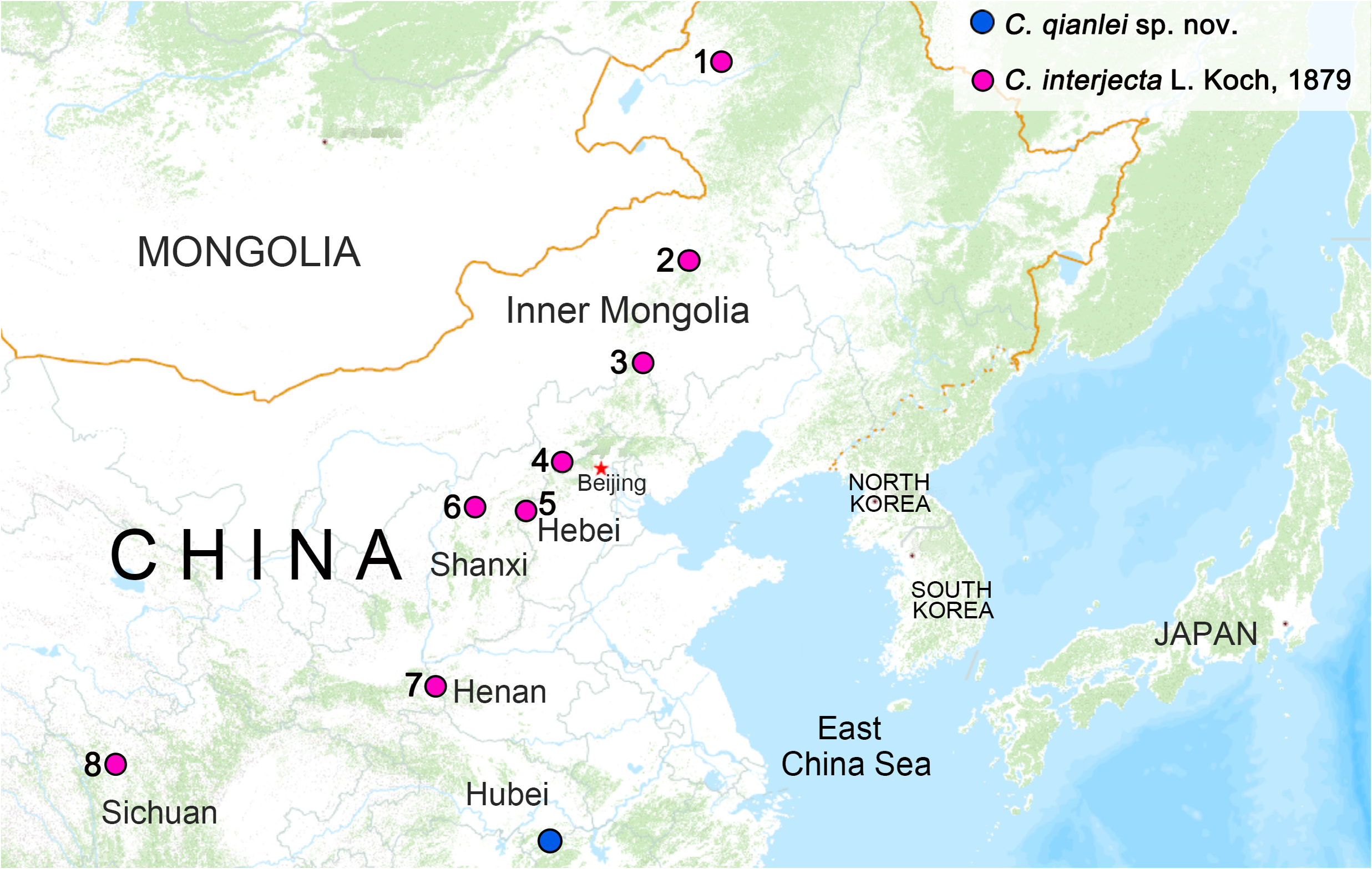
Distribution. Know only from the type locality, Mt. Jiugong, Hubei, China ( Fig. 6 View FIGURE 6 ).
Clubiona interjecta L. Koch, 1879
Figs 5−6 View FIGURE 5 View FIGURE 6
Clubiona interjecta L. Koch 1879: 89 View in CoL , pl. 3, fig. 7 ( ♂ ♀); Zhang & Hu 1989: 57, figs 3, 18 ( ♂ ♀); Song et al. 1999: 416, figs 246F−G, 249E−F ( ♂ ♀); Tang et al. 2005: 79, figs 3A−E ( ♂ ♀); Zhu & Zhang 2011: 358, f. 257A-E ( ♂ ♀), World Spider Catalog 2022 (full list of taxonomic references).
Material examined. CHINA: Hebei Province: Chengde City, Weichang County, 1♂ 5♀, Saihanba National Forest Park ( N42.584537º, E117.837489º, 1500 m), 3.VIII.2018, X.B. Guo, H. Wang, J.X. Lai and Y.F. Li leg GoogleMaps ; Shijiazhuang City, Pingshan County, 1♂, Tuoliang Nature Reserve ( N38.731813º, E113.824548º, 2000 m), 12. V.2018, X.B. Guo and Z.Y. Li leg GoogleMaps ; Zhangjiakou City, Zhuolu County, 27♀, Xiao Wutai Mountain Nature Reserve , Shanjiankou Village ( N40.029531º, E115.063527º, 1300 m), 6.VII.2018, X.B. Guo, S. Qiao, J.X. Lai and Y.F. Li leg GoogleMaps ; Shanxi Province: Xinzhou City, Ningwu County, 1♂, Luya mountain ( N38.837989º, E112.083789º, 1900 m), 6.VII.2011, C. Jin leg GoogleMaps ; Sichuan Province: Ganzi County, 1♂, Renguo village ( N31.667885º, E99.803766º, 3420 m), 20. VI.2018, X.B. Guo Leg. GoogleMaps
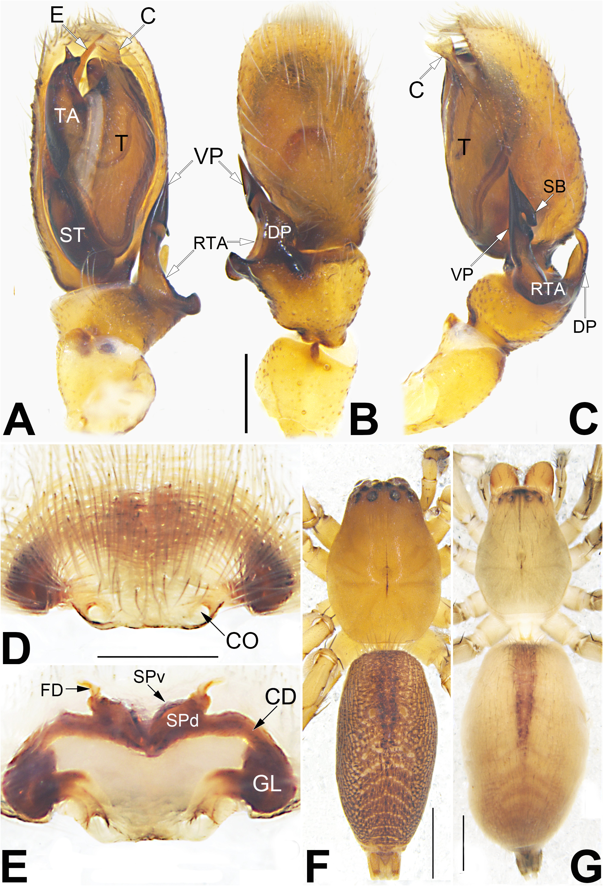
Emended diagnosis. As the only two members of the reclusa -group in China, Clubiona interjecta and C. qianlei sp. nov. share almost all of the group characters, but they can be separated by the shape of the copulatory organs, and by some somatic characters. C. interjecta can be distinguished from C. qianlei sp. nov. by the following characters: for the males, RTA’s ventral process with a subapical barb, dorsal process finger-like in C. interjecta ( Fig. 5C View FIGURE 5 ; Tang et al. 2005: 79, fig. 3E) (vs. subapical barb absent, dorsal process triangular; Fig. 2B View FIGURE 2 ); tegular apophysis about 1/3 width of bulb, distally narrowed and bearing a dentiform process ( Fig. 5A View FIGURE 5 ; Tang et al. 2005: 79, fig. 3D) (vs. tegular apophysis about 2/3 width of bulb, distal margin wide and rough; Figs 2C View FIGURE 2 , 3B View FIGURE 3 ); for the females, copulatory openings distinct, nearly circular in C. interjecta ( Fig. 5D View FIGURE 5 ; Tang et al. 2005: 79, fig. 3B) (vs. copulatory openings indistinct, slit-like; Figs 4 View FIGURE 4 A−C); SPd’ surface sclerotized, the two SPds nearly closely spaced ( Fig.5E View FIGURE 5 ; Tang et al. 2005: 79 fig. 3C) (vs. SPd’ outermost surface hyaline, the two SPds separated by about one diameter; Figs 4D, E View FIGURE 4 ). In addition, the two species can be separated by the abdominal pattern: dorsum of abdomen anteriorly with distinct, longitudinal heart mark, reaching 1/2 of abdomen length, posteriorly with 4~6 transverse chevrons in C. interjecta ( Figs 5F, G View FIGURE 5 ; Tang et al. 2005: 79, fig. 3A), but only with indistinct heart mark and muscular depressions in C. qianlei sp. nov. ( Figs 3D View FIGURE 3 , 4F View FIGURE 4 ).
Description. See Tang et al. (2005). Male palp as in Figs 5 View FIGURE 5 A−C, epigyne as in Figs 5D, E View FIGURE 5 , habitus as in Figs 5F, G View FIGURE 5 .
Distribution. Russia (West Siberia to Far East), Mongolia, China ( Inner Mongolia, Hebei, Shanxi, Henan, Sichuan, Jilin, Heiongjiang, as in Fig. 6 View FIGURE 6 ).

| V |
Royal British Columbia Museum - Herbarium |
| VI |
Mykotektet, National Veterinary Institute |
No known copyright restrictions apply. See Agosti, D., Egloff, W., 2009. Taxonomic information exchange and copyright: the Plazi approach. BMC Research Notes 2009, 2:53 for further explanation.
|
Kingdom |
|
|
Phylum |
|
|
Class |
|
|
Order |
|
|
Family |
|
|
Genus |
Clubiona qianlei J. Zhang, F. Zhang & H. Yu
| Zhang, Jianshuang, Chen, Lulu, Ding, Yanmei, Zhang, Feng & Yu, Hao 2022 |
Clubiona interjecta L. Koch 1879: 89
| Zhu, M. S. & Zhang, B. S. 2011: 358 |
| Tang, G. M. & Song, D. X. & Zhu, M. S. 2005: 79 |
| Song, D. X. & Zhu, M. S. & Chen, J. 1999: 416 |
| Zhang, G. R. & Hu, Y. J. 1989: 57 |
| Koch, L. 1879: 89 |