
Tridactylophagus sufflatus Hui, Mukherjee et Hazra., 2023
|
publication ID |
https://doi.org/10.11646/zootaxa.5230.3.2 |
|
publication LSID |
lsid:zoobank.org:pub:57670F00-A582-4F4F-8EDE-A3338E79FCC6 |
|
DOI |
https://doi.org/10.5281/zenodo.10555702 |
|
persistent identifier |
https://treatment.plazi.org/id/E8FE1E4F-2191-465C-B849-05EFB7258869 |
|
taxon LSID |
lsid:zoobank.org:act:E8FE1E4F-2191-465C-B849-05EFB7258869 |
|
treatment provided by |
Plazi |
|
scientific name |
Tridactylophagus sufflatus Hui, Mukherjee et Hazra. |
| status |
sp. nov. |
Tridactylophagus sufflatus Hui, Mukherjee et Hazra. sp. n.
GenBank Accession No. ON934632 View Materials
LSIDurn:lsid:zoobank.org:act: E8FE1E4F-2191-465C-B849-05EFB7258869
( Figs. 1A–K View FIGURE 1 )
Type material. Holotype male, India, West Bengal, Jalpaiguri, Lataguri (26.72 N, 88.76 E), 7th April, 2022, open light trap, Coll. P. Hui. GoogleMaps
Diagnosis. The adult male is distinguished from other species of the genus Tridactylophagus by the following characteristics: flattened, fist-shaped tarsomere I of the forelegs and distal end of the antennomere IV slightly bulged out on one side with a sensory spot.
Etymology.The name“ sufflatus ” refers to the Latinised version of the bulged out antennomere IV at the distal end.
Adult Male (n=1)
Total length 2.3 mm; length of metathorax 0.93 mm; width of metathorax 0.32 mm, length of antenna 0.64 mm.
Colour. Head light brown, thorax dark brown, abdomen light brown, eyes dark red, antenna light brown, mesothoracic wing dark brown, hind wing translucent with brown veins, and legs brown in colour.
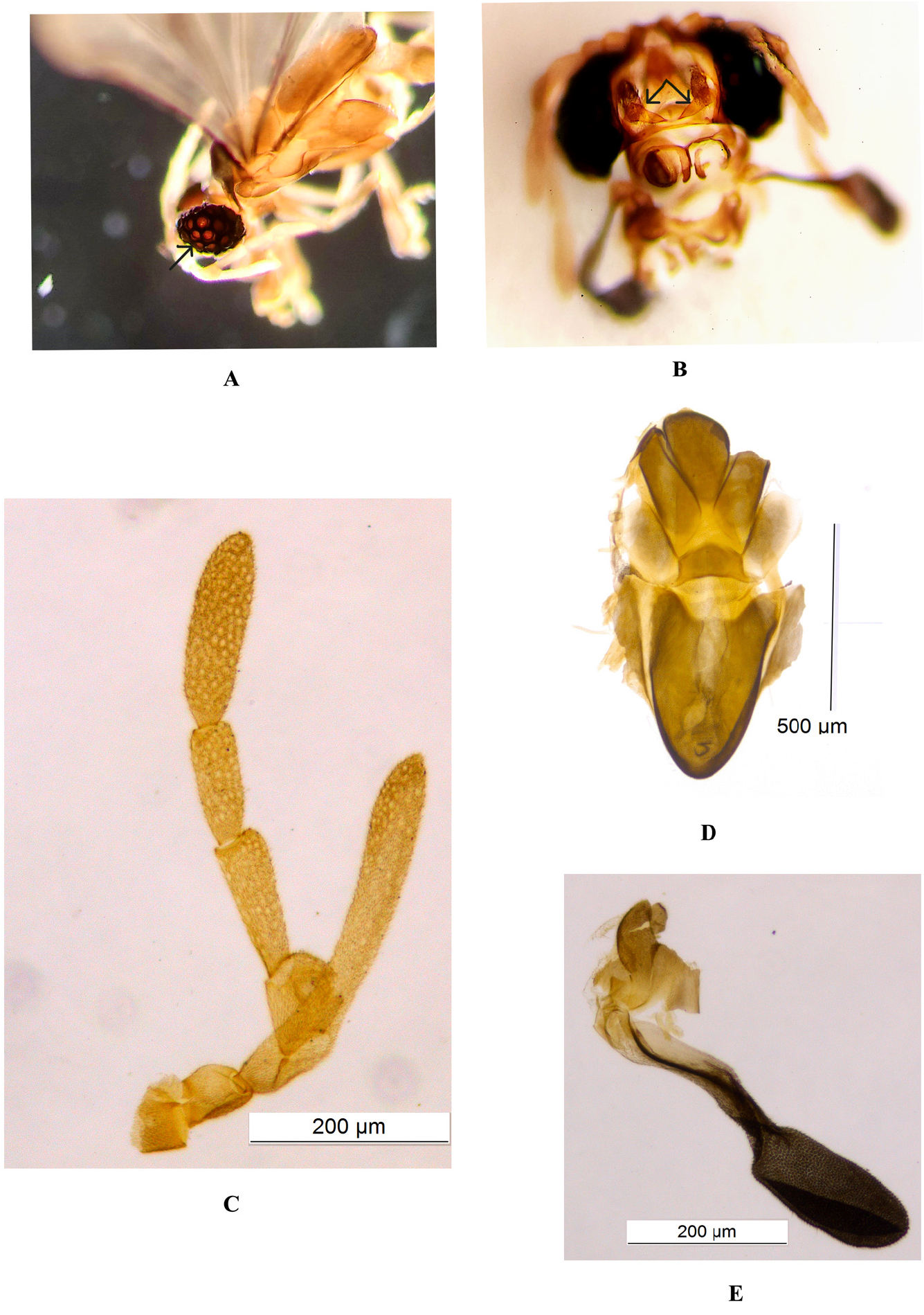
Head. Head extended laterally, width 0.5 mm, length 0.3 mm. Compound eyes prominent, having 19 hemispherical corneal lenses of ommatidia when viewed dorsally ( Fig. 1A View FIGURE 1 ) and ten of same when viewed ventrally ( Fig. 1B View FIGURE 1 ). Interommatidial space markedly pubescent.
Mandibles ( Fig. 1B View FIGURE 1 ). Strongly sclerotised, triangular with a broad base, not cross each other.
Maxillae. Very small, not discernible.
Antenna ( Fig. 1C View FIGURE 1 ). Seven segmented. Scapus (segment I) cup-shaped, pedicellus (segment II) cylindrical, segment III small with flabellum reaching almost at end of segment VI, segment IV round but slightly bulged out on one side with a sensory spot, segment V slightly longer than segments VI and VII. Lengths of antennal segments as follows: Antennomere I 0.05 mm, antennomere II 0.05 mm, antennomere III with flabellum 0.33 mm, antennomere IV 0.09 mm, antennomere V 0.14 mm, antennomere VI 0.11 mm, and antennomere VII 0.2 mm.
Thorax ( Fig. 1D View FIGURE 1 ). Metathorax. Prescutum 0.34 mm in length, and 0.15 mm in width; roughly triangular in shape with rounded edges. Scutum 0.33 mm in length, 0.09 mm in breadth, trapezoidal in shape with curved borders. Scutellum pentagonal, 0.08 mm in length and 0.16 mm in breadth. Postlumbium 0.11 mm in length and 0.21 mm in breadth. Postnotum 0.4 mm long with rounded tip.
Reduced mesothoracic wings ( Fig. 1E View FIGURE 1 ). 0.46 mm long. Apical part globular with 0.1 mm width; basal part narrow, stalk-like, 0.05 mm wide.
Wings ( Fig. 1F View FIGURE 1 ). Hind wings span 1.5 mm from base to middle of radial sector and 1.12 mm from base to edge of subcostal margin. C+Sc united and give impression of a single deeply pigmented vein. R 1 0.75 mm in length. Area between Sc and R 1 profoundly pigmented. C, Sc and R 1 extending about 2/3 rd length of anterior wing margin. Two detached veins, R 2 and R 3, between R 1 and R 4. R 2 0.52 mm long, broad, slightly curved, and located adjacent to anterolateral angle of wing. R 3 0.67 mm long, R 4 0.92 mm long, apical part well pigmented. R 5 0.72 mm in length, arising near apex of R 4. MA and CuA 1 1.22 mm and 0.97 mm in length respectively. CuP 0.67 mm in length and located close to anal margin.
Legs. Forelegs ( Fig. 1G View FIGURE 1 ) stout; coxa 0.21 mm long, trochantofemur 0.23 mm long, tibia 0.21 mm long, tarsomere I 0.06 mm long, tarsomere II 0.05 mm long, and tarsomere III 0.06 mm long. Tibia conical in shape, tarsomere I flattened and fist-shaped. Midlegs ( Fig. 1H View FIGURE 1 ). Coxa 0.28 mm long, trochantofemur 0.34 mm long, tibia 0.39 mm long; length of three tarsal segments as follows: tarsomere I 0.08 mm, tarsomere II 0.05 mm, tarsomere III 0.07 mm. Hindlegs ( Fig. 1I View FIGURE 1 ). Trochanter 0.11 mm long, femur 0.37 mm long, tibia 0.4 mm long; length of tarsal segments as follows: tarsomere I 0.07 mm, tarsomere II 0.07 mm, and tarsomere III 0.08 mm. Unlike fore legs, first tarsomere of mid and hind legs convex. All other tarsomeres triangular in shape.
Abdomen. Abdominal segments uniformly sclerotised. Segment IX 0.3 mm long, elongated for accommodation of aedeagus; proctiger 0.14 mm long, basal 2/3rd wide (0.05 mm), distal 1/3rd tapered, 0.05 mm long ( Fig. 1J View FIGURE 1 ).
Aedeagus ( Fig. 1K View FIGURE 1 ). Aedeagus short, 0.087 mm long, 0.07 mm wide at base, basally curved, apically hooked, abruptly narrowed towards apex; dorsal hook 0.01 mm long.
Remarks. The newly described species is included in the family Halictophagidae owing to seven segmented antennae, mandibles not crossing each other, three segmented tarsi without claw, and hook or anchor-shaped aedeagus. It is included in the subfamily Tridactylophaginae and the genus Tridactylophagus , owing to flabellum only on antennomere III. It shows similarity in wing venation with T. aduncus Maxumdar & Chaudhuri, 1999 but differs in the structure of antenna and metathorax. Parts of the thorax of the newly described species show similarities with several earlier described species; the prescutum shows similarity with T. orientalis ( Chaudhuri & Das Gupta, 1979) ; the scutellum shows similarity with T. buttonensis Kathirithamby, 1992 ; and the postnotum shows similarity with T. coniferus Yang, 1964 . Tridactylophagus sufflatus sp. n. draws affinity to T. similis Kinzelbach, 1971 in the shape of the aedeagus, though the apical portion of the aedeagus of T. similis seems to be narrower than that of the newly described species. The shape of the apical hook of the aedeagus of T. sufflatus sp. n. is also somewhat similar to T. aduncus , but the base of the newly described species is round and blunt, while that of T. aduncus is more or less angular.
There is only one COI sequence (JN082813.1) of the genus Tridactylophagus Subramaniam at NCBI, apart from the COI sequence ( ON934632 View Materials ) of the newly described species. So a molecular phylogenetic tree based on only two COI sequences could not be made.
Female. Unknown.
Host. Unknown.
No known copyright restrictions apply. See Agosti, D., Egloff, W., 2009. Taxonomic information exchange and copyright: the Plazi approach. BMC Research Notes 2009, 2:53 for further explanation.
|
Kingdom |
|
|
Phylum |
|
|
Class |
|
|
Order |
|
|
Family |
|
|
Genus |