
Eigenmannia loretana, Waltz & Albert, 2018
|
publication ID |
https://doi.org/10.11646/zootaxa.4399.3.9 |
|
publication LSID |
lsid:zoobank.org:pub:038FDEA5-16D4-492A-903E-87AB5C6662FF |
|
DOI |
https://doi.org/10.5281/zenodo.5980556 |
|
persistent identifier |
https://treatment.plazi.org/id/03BF8796-1716-9304-FF1E-FD96FAC53882 |
|
treatment provided by |
Plazi |
|
scientific name |
Eigenmannia loretana |
| status |
sp. nov. |
Eigenmannia loretana , new species
Figs. 1–6 View FIGURE 1 View FIGURE 2 View FIGURE 3 View FIGURE 4 View FIGURE5 View FIGURE 6 , Table 1
Holotype. MUSM 61210, 135 mm, 120 mm LEA, Peru, Loreto, Río Pacaya, Lago Tomana, southwest of Iquitos, 5°20’40”S, 74°30’00”W, J. S. Albert & W. G. R. Crampton, 21 Sep. 2002.
Paratypes. All from Peru. FMNH 134534 About FMNH , 1 About FMNH , 107 About FMNH mm, 91 mm LEA, Peru, Loreto, Río Pacaya, Lago Tomana , 5°17'6.13"S, 74°25'25.80"W, J. S. Albert & W. G. R. Crampton, 21 Sep. 2002 GoogleMaps ; MUSM 61212 , 2 , 60–98 mm (one specimen missing part of tail), 60–77 mm LEA (one specimen missing part of posterior anal fin), Peru, Loreto, Río Pacaya, Cocha Yanayacu , 5°16’43”S, 74°55’57”W, J. S. Albert & W. G. R. Crampton, 4 Aug 2000 GoogleMaps ; UF 126165, 1, 115 mm, 84 mm LEA, Peru, Loreto, Río Pacaya , J. S. Albert & W. Crampton, 19 Sep 2004 ; FMNH 134535 About FMNH , 2 About FMNH , 1 About FMNH C&S, 101–102 mm, 71–72 mm LEA, Peru, Loreto, Río Pacaya, Cocha Yarina , 5°24’37.3”S, 74°30’15.18”W, J. S. Albert & W. G. R. Crampton, 14 May 2003 GoogleMaps ; UF 129250, 1, 101 mm, 70 mm LEA, Peru, Loreto, Río Pacaya, Cocha Yarina , 5°24’37.3”S, 74°30’15.18”W, J. S. Albert & W. G. R. Crampton, 14 May 2003 GoogleMaps ; UF 114721, 1, 96 mm, 74 mm LEA, Peru, Loreto, Río Pacaya, Cocha Yanayacu , 5°16’43”S, 74°55’57”W, J. S. Albert & W. G. R. Crampton, 4 Aug 2000 GoogleMaps .
Non-types. All from Peru. UF 114494, 26, 82–113 mm, 61–82 mm LEA , Peru, Loreto, Quedabra 33 km southwest of Iquitos , J. S. Albert & W. G. R. Crampton, 6 Aug. 2000 ; UF 116557, 2, 93–141 mm, 1 C&S 141 mm, (one specimen missing part of tail), 93–103 mm LEA (one specimen missing part of posterior anal fin), Peru, Maynas, Loreto, Quedabra at km 23 on Iquitos-Nauta Road, J. Albert & W. G. R. Crampton, 27 Mar 2001 ; ANSP 202364 About ANSP , 12 About ANSP , 103– 130 About ANSP mm, 75–94.0 LEA, Peru, Loreto, Cocha Santa Thomas, affluent to Río Nanay , Iquitos, J. Craig & L. Kim, 21 Aug 2016 ; ANSP 177759 About ANSP , 132 About ANSP mm, 99 mm LEA, Mixana Reserve, near Iquitos , Loreto, Peru, 03°52'46"S, 73°29'33"W, J. Albert & W. G. R. Crampton, 0 8 April 2004 GoogleMaps ; AUM 38657, 16 View Materials , 89–111 View Materials mm, 56–80 mm LEA, Peru, Loreto, Cocha Santa Thomas, affluent to Río Nanay, Iquitos, J. Craig & L. Kim, 21 Aug 2016 ; MUSM 60215 , 39 , 56–121 mm (one specimen missing part of tail), 56–88 mm LEA (one specimen missing part of posterior anal fin), Peru, Loreto, Cocha Santa Thomas, affluent to Río Nanay, Iquitos , 3°47'56.9"S, 73°20'31.02"W, J. Craig & L. Kim, 21 Aug 2016 GoogleMaps ; MUSM 60216 , 2 C&S, 117–120 mm Peru, Loreto, Cocha Santa Thomas, affluent to Río Nanay, Iquitos , 3°47'56.9"S, 73°20'31.02"W, J. Craig & L. Kim, 21 Aug 2016 GoogleMaps ; MUSM 60217 , 3 C&S, 118– 128 mm, Peru, Loreto, Cocha Santa Thomas, affluent to Río Nanay, Iquitos , 3°47'56.9"S, 73°20'31.02"W, J. Craig & L. Kim, 21 Aug 2016 GoogleMaps .
Diagnosis. Table 2 provides a summary of diagnostic traits among species of the E. trilineata species group. Eigenmannia loretana can be distinguished from all congeners except members of the E. trilineata species group by the presence of a superior medial stripe ( vs. absent); from members of the E. humboldtii species group by a relatively small adult body size (< 150 mm vs. large adult body size,> 300 mm); from E. macrops by a relatively small eye (eye diameter less than snout length vs. greater than or equal to snout length); and from E. virescens by a terminal ( vs. subterminal) mouth and a smaller adult body size (vs. > 230 mm). Eigenmannia loretana can be distinguished from all other species in the E. trilineata species group by the length of the posterodorsal process of infraorbitals 1+2 (60–75% length of infraorbitals 1+2 vs. 40% in E. besouro and E. correntes ; 50% in E. matintapereira , E. meeki , E. trilineata , and E. waiwai ; and 100% in E. antonioi , E. desantanai , E. guairaca , E. microstoma , E. muirapinima , E. pavulagem , E. sayona , and E. vicentespelaea ). Eigenmannia loretana can be further differentiated from species in the E. trilineata group except E. antonioi and E. pavulagem by its premaxillary dentition, with 11–15 teeth distributed in three rows ( vs. eight–10 teeth in two rows in E. muirapinima ; nine–10 teeth in two rows in E. guairaca ; 16 teeth in three rows in E. microstoma ; 17 teeth in three rows in E. sayona ; 17–20 teeth in three rows in E. correntes ; 18–29 teeth in three to four rows in E. besouro ; 22–24 teeth in four rows in E. matintapereira ; 24–25 teeth in four rows in E. desantanai ; 25–26 teeth in four rows in E. vicentespelaea ; 31–33 teeth in four rows in E. trilineata ; 35–40 teeth in five rows in E. waiwai ; and 30–35 teeth in five to six rows in E. meeki ). Eigenmannia loretana can be further distinguished from E. antonioi and E. pavulagem by its endopterygoid dentition, with six to seven teeth distributed in a single row ( vs. eight–12 teeth in a single row in E. antonioi and eight to 11 teeth in a single row in E. pavulagem ), suborbital depth 28–36% HL ( vs. 18–27& HL in E. antonioi and 19–27% HL in E. pavulagem ), and by coloration with a uniformly dark brown head and light brown pectoral fins on recently-preserved specimens ( vs. darkened dorsal region of head gradually becoming lighter ventrally, and hyaline pectoral fins). Endopterygoid dentition also distinguishes E. loretana from all species in the E. trilineata group except E. correntes and E. guairaca (six to seven teeth distributed in a single row vs. eight to nine teeth in a single row in E. antonioi and E. sayona ; eight to 11 teeth in one or two rows in E. pavulagem ; nine to 12 teeth in one or two rows in E. matintapereira ; 11–16 teeth in one or two rows in E. microstoma ; eight to nine teeth in two rows in E. muirapinima ; 10–11 teeth in two rows in E. besouro ; 10–15 teeth in two rows in E. vicentespelaea ; 13–15 teeth in two rows in E. meeki ; 14–15 teeth in two rows in E. desantanai ; 14–17 teeth in two rows in E. waiwai ; and 16–17 teeth in two rows in E. trilineata .). Eigenmannia loretana can be further distinguished from E. correntes and E. guairaca by a total anal-fin rays count of 183–219 ( vs. 143–164 in E. correntes and 151–170 in E. guairaca ), and the size of dentary teeth increasing after the sixth to seventh tooth ( vs. all teeth similar in size). Eigenmannia loretana can be further distinguished from E. guairaca by pectoral-fin rays ii, 13–14 ( vs. ii, 11–12), suborbital depth, 28–36% HL ( vs. 22–28% HL), orbital diameter, 15.7–23.9% HL ( vs. 11.4–15.0% HL), head depth at supraoccipital, 86.8–96.7% HL ( vs. 75–86.4% HL). Eigenmannia loretana can be further distinguished from E. correntes by head length, 11.2–12.8% LEA ( vs. 13.1–14.9% LEA), snout length, 23.9–30.3% HL ( vs. 32.2–35.2% HL), interorbital distance, 33.5–40.7% HL ( vs. 25.2–30.1% HL), head depth at supraoccipital, 86.8–96.7% HL ( vs. 63.7–70.0% HL), and terminal mouth position ( vs. subterminal).
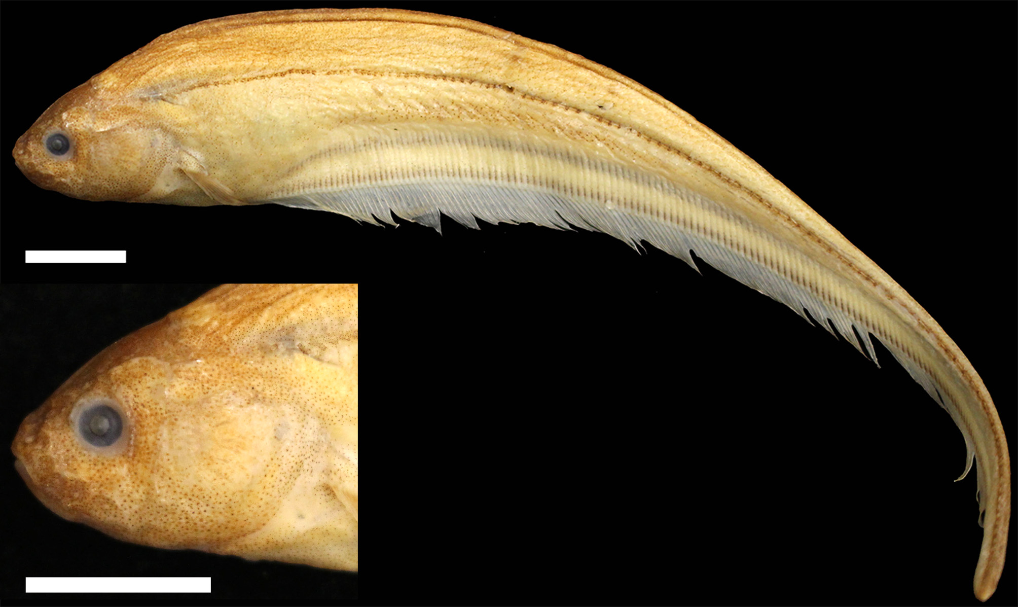
Description. Gymnotiform with relatively small adult body size, largest recorded specimen 135 mm. Morphometric data and meristic counts summarized in Table 1. No sexual dimorphism observed. Body elongate and laterally compressed, dorsal profile of body convex from posterior margin of head to vertical through middle of anal fin, then slightly angled downwards until caudal filament at insertion of last anal-fin ray. Ventral surface of body straight from base of posterior margin of head to first anal-fin ray, then slightly concave to end of caudal filament. Greatest body depth at posterior margin of pectoral-fin base. Head short and relatively round. Head mildly compressed laterally; greatest width at opercular region and greatest depth at vertical through supraoccipital. Dorsal profile of head slightly convex, from tip of snout to posterior supraoccipital margin. Ventral profile of head slightly convex from lower lip to branchial opening. Mouth terminal, at or slightly below horizontal line through eye. Lower jaw extending anterior to upper jaw (tip of snout) in some specimens. Premaxillary teeth 11–15 (n=3), distributed in three rows. Rictus at or slightly posterior to vertical line through anterior nostril. Posterior nares closer to eye than tip of snout. Eye circular, covered by skin, located laterally on head, closer to dorsal margin of head than ventral margin. Antorbital and infraorbitals 1–4 partial enlarged cylinders, infraorbitals 5–6 as narrow ossified tubes. Posterodorsal expansion of infraorbitals 1+2 equal to approximately 60–75% of the length on infraorbitals 1+2 ( Fig. 2 View FIGURE 2 ).
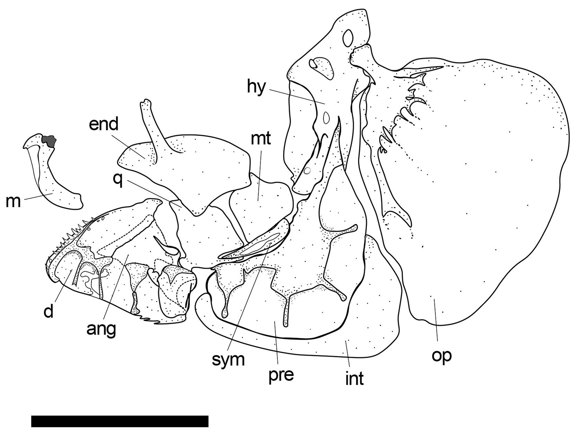
Suspensorium including opercular series and maxilla illustrated in Fig. 3 View FIGURE 3 . Maxilla sickle-shaped with smaller sickle-shaped anterodorsal process; descending blade of maxilla gradually increasing in width posteroventrally. Mandible illustrated in Figs. 3 View FIGURE 3 and 4 View FIGURE 4 . Dentary V -shaped, with 17–19 (n=3) teeth, distributed in one to two rows; teeth increasing in size towards rictus after sixth to seventh tooth; robust laterosensory canals present along lateroventral surface. Anguloarticular with narrow process on lateral surface, extending dorsally; small rectangular process on medial surface connecting to Meckel’s cartilage. Coronomeckelian bone roughly 20% length of Meckel’s cartilage. Retroarticular small, roughly rectangular, located at posteroventral margin of anguloarticular. Endopterygoid broadly overlapping quadrate and metapterygoid, roughly triangular, with well-developed ascending process and six to seven conical teeth distributed in a single row on ventral margin. Base of quadrate roughly square in shape extending into pointed triangular shape anterodorsally; quadrate articulating with preopercle and symplectic at base through posteroventral process and notch; circular process extending anteroventrally from base. Metapterygoid similar in shape to quadrate, without posterodorsal process and notch; triangular region pointed posterodorsally. Symplectic elongate and triangular. Preopercle crescent-shaped, with robust laterosensory canal tubes along lateral surface. Interopercle teardrop-shaped, with sharp, pointed posterodorsal expansion. Opercle roughly triangular, dorsal margin convex; opercular ridges present along lateral surface of anterior region; center weakly ossified, becoming increasingly laminar towards distal margins. Subopercle sickle-shaped, tapering posterodorsally, forming concave dorsal profile. Hyomandibula at roughly 100° to horizontal line through long axis of head; dorsal articulating head roughly one and a half times wider than ventral margin; laminar anterior shelf extending from widest of hyomandibula point to anteroventral margin.
Neurocranial bones shown in a full-head CT scan in Fig. 5 View FIGURE5 . Mesethmoid oriented at about 45° angle from ventral ethmoid, until reaching anterior margin of frontals. Vomer gradually tapering to points laterally near anterior margin of parasphenoid. Paired frontals convex in dorsal profile, roughly half length of total skull length. Supraorbital canal robust, forming a highly ossified shelf-like structure. Entire ventral surface of orbitosphenoid contacting dorsal margin of parasphenoid in adult specimens. Ventral surface of pterosphenoid largely not contacting dorsal margin of parasphenoid, forming lateral fenestrae, varying in size throughout ontogeny. Anterior fontanelle roughly 85% length of posterior fontanelle. Anterior half of posterior fontanelle surrounded by frontals, posterior half by parietals. Prootic and exoccipital with prominent foramenae. Supraoccipital extending dorsally to dorsal margin of parietals. Posttemporal bones fused with supracleithrae.
Scales cycloid, present from posterior margin of head to posterior point of caudal filament. Nine to 13 (n=20) longitudinal rows of scales above lateral line. Anal-fin pterygiophore scales small, roughly half the size of other scales. Pectoral-fin rays ii, 13–14 (n=20). Anal-fin origin at or slightly anterior to vertical line through pectoral fin base. Total anal-fin rays 183–219 (n=17). Caudal filament long and cylindrical, occasionally laterally compressed. Precaudal vertebrae 13 (n=3) or 14 (n=2). Displaced haemal spines two (n=1) or three (n=3).
Coloration in alcohol. Background color yellowish brown. Head dark brown in recently preserved specimens. Dorsal region of some specimens dark brown, gradually grading into lighter brown/yellow ventrally. Four longitudinal stripes present. Thin, dark lateral line stripe, one scale wide. Superior medial stripe thicker than lateral line stripe, gradually fading out near margin of body cavity. Dark inferior medial stripe and anal-fin base stripes present. Anal fins hyaline, pectoral fins occasionally pigmented in a light brown color.
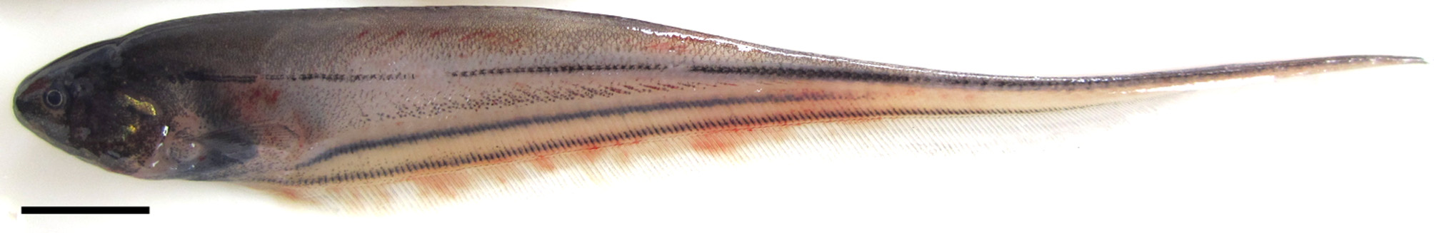
Live Coloration. See Fig. 6 View FIGURE 6 . Background color grey-opaque to transparent along body posterior to head. Head covered with chromatophores and colored dark brown. Pectoral fins light brown in some specimens. Four longitudinal stripes present; more apparent than in preserved specimens.
Distribution. Collection localities are shown in Figure 7 View FIGURE 7 . Known from the Western Amazon in Peru, from the Río Pacaya and affluents of the Río Nanay ( Fig. 1 View FIGURE 1 ). Specimens collected over sandy beaches and in floating vegetation along river and stream margins. The type locality was restricted to the Río Pacaya because of the cryptic nature of species in the E. trilineata complex.
Etymology. The new species is named in honor of the residents/inhabitants of Loreto, Peru. Feminine.


No known copyright restrictions apply. See Agosti, D., Egloff, W., 2009. Taxonomic information exchange and copyright: the Plazi approach. BMC Research Notes 2009, 2:53 for further explanation.
|
Kingdom |
|
|
Phylum |
|
|
Class |
|
|
Order |
|
|
Family |
|
|
Genus |