
Mesochra huysi, Suárez-Morales & Fuentes-Reinés, 2015
|
publication ID |
https://doi.org/ 10.1080/00222933.2015.1085604 |
|
DOI |
https://doi.org/10.5281/zenodo.4323824 |
|
persistent identifier |
https://treatment.plazi.org/id/03BF2E60-C576-FFE2-FE27-BEF7FBF4FE6D |
|
treatment provided by |
Carolina |
|
scientific name |
Mesochra huysi |
| status |
sp. nov. |
Mesochra huysi sp. nov.
( Figures 1–4 View Figure 1 View Figure 2 View Figure 3 View Figure 4 )
Material examined
Female holotype ( UARC390 ), male allotype ( UARC391 ), from shallow plankton samples, Laguna Navío Quebrado , Colombia (11°25 ʹ N, 73°05 ʹ W), partially dissected GoogleMaps ,
semi-permanent slides. Paratypes: one female, one male, plus two undissected females and two males, ethanol-preserved, vial ( UARC392 ). Three adult females and two adult males, dissected, mounted in slides, same locality, and collector (ECOCHZ- 09325). Additional material: 5 adult females, 5 adult males in author’ s ( JF-R) personal collection; these specimens are available for consultation upon request .
Type locality
Laguna Navío Quebrado , La Guajira, Colombia (11°25 ʹ N, 73°05 ʹ W) GoogleMaps .
Description of female
Body fusiform, slightly tapering posteriorly ( Figure 1A View Figure 1 ). Habitus in dorsal view as in Figure 1A View Figure 1 , in lateral view as in Figure 1B View Figure 1 . Total body length, measured from tip of rostrum
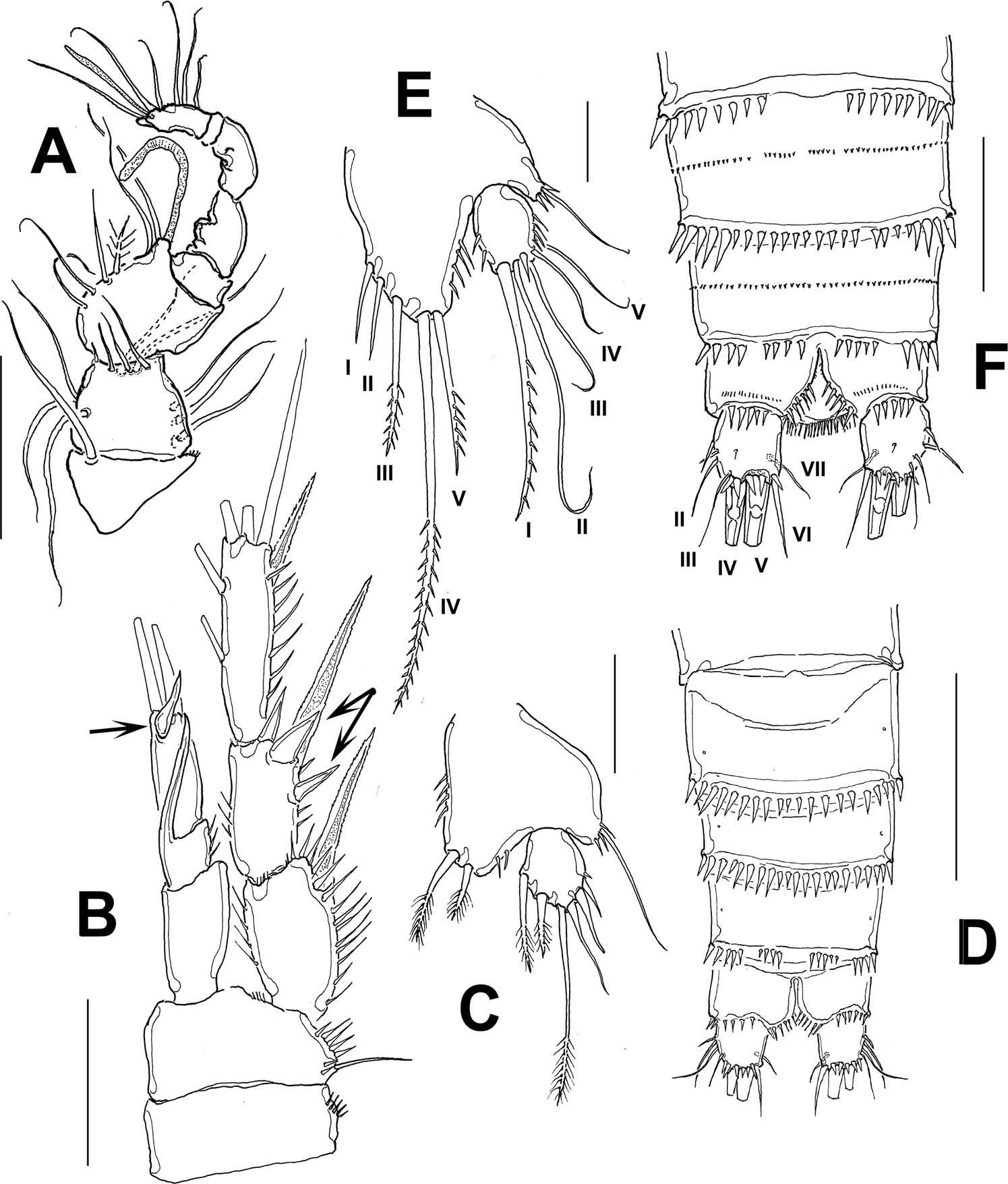
to posterior margin of caudal rami ranging from 336 to 403 μm (average 374 μm, n = 10; holotype: 336 μm). Rostrum distinct, flat in dorsal view, subtriangular, with rounded tip in lateral view. Dorsal surface of cephalosome and free prosomites with incised hyaline frill. Transverse row of microspinules on dorsal surface of first three pedigerous free somites, fourth free somite without dorsal spinulation. Genital double-somite with vestigial suture visible in ventral view, first genital somite with row of small spinules on mid-dorsal surface and posterior frill; second genital somite with transverse row of spinules and weak frill along posterior margin on dorsal surface ( Figure 1A View Figure 1 ); lateral and ventral margins with larger set of spinules ( Figures 1B View Figure 1 , 4D View Figure 4 ). First and second free urosomites with mid-dorsal row of minute spinules and row of spinules along posterior margin, spinules increasing in size laterally and ventrally ( Figure 4D View Figure 4 ). Anal somite with separate transverse rows of minute spinules on mid-dorsal surface, distal margin with short row of spinules along insertion of caudal rami. Anal operculum rounded, ornamented with slender spinules ( Figure 4F View Figure 4 ).
Caudal rami ( Figure 4F View Figure 4 ) subquadrate, with six setae, with spinules along ventral surface. Seta II with accompanying spinules, and about as long as caudal rami, seta III 1.2 times as long as seta II, seta V about 4.2 times longer than seta IV and seta VII as long as seta II.
Antennule. 6-segmented, with distal row of 8–9 spinules on first segment. Aesthetasc on third segment reaching well beyond last segment, this segment with small outer spine ( Figure 1C View Figure 1 ). Armature formula as follows: 1(0), 2(8), 3(7 + 1ae), 4(1), 5(1), 6(9 + ae).
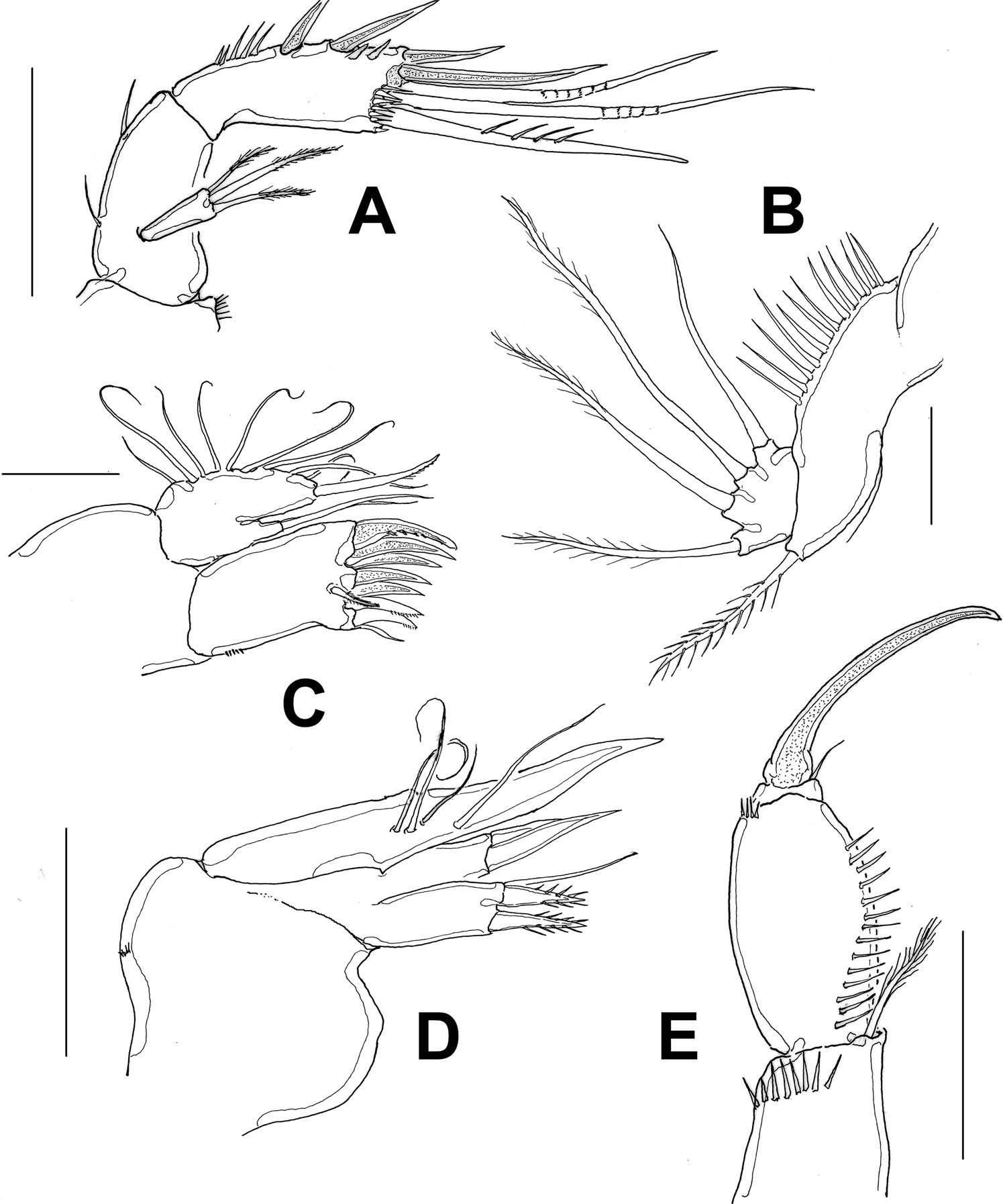
Antenna. Allobasis with two short setae, one proximal and one on subdistal position. Exopod short, cylindrical, 1-segmented, with three unequally long terminal setae, middle setae being longest. Endopod 1-segmented, inner margin with row of spinules on proximal 1/3 of segment, lateral armature represented by two subequal spines plus two outer spinules distally. Distal margin with five setal elements, two spines, two geniculate setae and one stout seta furnished with spinules ( Figure 2A View Figure 2 ).
Mandible. Gnathobase with strong, wide teeth and one plumose dorsal seta, basis elongate, armed with one basipodal seta and row of long spinules. Endopodal lobe armed with four setae ( Figure 2B View Figure 2 ).
Maxillule. Praecoxal arthrite with two setae. Distal margin with five strong curved claws and four setal elements. Coxa and basis partially fused; coxal endite with two slender setae. Basis with one seta. Endopodal and exopodal rami fused to basis, each represented by four and four setae, respectively ( Figure 2C View Figure 2 ).
Maxilla. Syncoxa ornamented with row of minute spinules, with two endites, each with three setal elements, two of them wide-based, spinulated, the other slender. Basis forming strong claw with one slender seta. Endopod represented by three slender setae ( Figure 2D View Figure 2 ).
Maxilliped. Syncoxa furnished with subdistal row of spinules, with inner distal plumose seta reaching half-length of basis. Basal segment robust, ornamented with longitudinal row of long spinules from proximal margin to distal 1/3 of segment; distal outer margin with row of small spinules. Endopod represented by weakly curved claw with an accompanying proximal seta ( Figure 2E View Figure 2 ).
P1 ( Figure 3A View Figure 3 ). Protopod ornamented with row of spinules along posterior margin, coxa with proximal rows of minute spinules, additional rows also along outer and posterior margins. Basis with curved row of spinules on middle surface; segment with strong spinules along distal margin, particularly near insertion of endopod; with outer and inner spine, latter stout, straight, reaching about 1/3 of ENP1. ENP 2-segmented, longer than EXP, first segment reaching well beyond length of EXP, about 7 times as long as wide, inner seta on first segment inserted almost at the half of segment; second segment ornamented with outer row of spinules, armed with three elements, inner subdistal one shorter than segment. EXP 3-segmented, reaching beyond insertion of inner seta on ENP1, EXP1 and EXP2 without inner setae, third segment with four elements, two of them geniculate.
P2 ( Figure 3B View Figure 3 ). Coxa and basis with weaker ornamentation than in P1. Basis with slender outer basipodal seta. ENP 2-segmented, shorter than EXP, not reaching midlength of EXP3. ENP1 with outer seta, without inner seta, ENP2 with five setal elements. EXP 3- segmented, EXP1 without inner seta, EXP2 with short inner seta, EXP3 with five elements.
P3 ( Figure 3C View Figure 3 ). Coxa and basis with weaker ornamentation than in P1, as in P2. Basis with slender outer basipodal seta. ENP 2-segmented, barely reaching distal end of EXP2; ENP1 with outer seta, without inner seta; ENP2 with five elements. EXP 3-segmented, EXP1 and EXP2 without inner setae, third segment with six elements.
P4 ( Figure 3D View Figure 3 ). Coxa and basis with weaker ornamentation than in P1, as in P2 and P3. Basis with slender outer basipodal seta. ENP 2-segmented, short, reaching only proximal 1/3 of EXP1; ENP1 with inner seta, without outer seta; ENP2 with five setal elements. EXP 3-segmented, EXP1 with inner seta, third segment with six elements, one of them a thickened pectinate seta (arrowed in Figure 3D View Figure 3 ).
P5 ( Figure 4C View Figure 4 ). Baseoendopod with single seta. EXP rounded, shorter than endopodal lobe, with five elements, inner middle distal seta longer than inner distal spine. Endopodal lobe armed with five elements ( Figure 4E View Figure 4 ). Setal formula (Arabic numerals = setae, Roman numerals = spines) of P1–P4 as in Table 1.
P6 ( Figure 1D View Figure 1 ) vestigial, represented by small crescent-shaped lobe ( Figure 1D View Figure 1 ), with one short seta.
Description of male
Habitus resembling that of female except for narrower prosome and urosome ( Figure 4D View Figure 4 ). Smaller than female, total body length, measured from tip of rostrum to posterior margin of caudal rami ranging from 263 to 315 μm (average 289 μm, n = 5; holotype: 263 μm). Somitic ornamentation as in female. Urosomites 1–3 with ventral row of large spinule along posterior margin ( Figure 4D View Figure 4 ) which are coarser than those along the dorsal surface. Row of spinules along ventral margin of preanal somite with discontinuous row of spinules, as opposed to female, with continuous such row. Anal somite as in female, with smaller spinules on ventral surface and along insertion of caudal rami. Armature of caudal rami as in female.
Antennule. Geniculate, 8-segmented ( Figure 4A View Figure 4 ).
Mouthparts, P1, P2, and P4 as in female.
P3 ( Figure 4B View Figure 4 ). Coxa and basis with weaker ornamentation than in female P1, as in female P2. Basis with slender outer basipodal seta. ENP 3-segmented, shorter than EXP, reaching proximal 1/4 of EXP3; ENP1 unarmed, ENP2 without inner seta but with inner sinuous apophysis reaching apical margin of ENP3; ENP3 with two distal setae and a single claw-like or flame-shaped ( Gómez and Fiers 1997) element (arrowed in Figure 4B View Figure 4 ). EXP 3-segmented, EXP1 and EXP2 with strong, long outer spine. EXP2 ornamented with noticeably strong, wide-based spinules (double arrow in Figure 4B View Figure 4 ). EXP3 with 6 setal elements, two of them on inner margin ( Figure 4B View Figure 4 ).
P5 ( Figure 4C View Figure 4 ). Baseoendopod with cluster of spinules at point of insertion of seta. EXP rounded, reaching slightly beyond distal margin of endopodal lobe, with six setal elements, middle seta being longest. Endopodal lobe ornamented with few small spinules along inner and outer margins, armed with two spines of unequal length.
Etymology
The new species is named after Dr Rony Huys (Natural History Museum, London, UK), for his outstanding contributions to the taxonomic knowledge of the Harpacticoida .
Remarks
Mesochra huysi sp. nov. most closely resembles M. parva Thomson, 1946 , M. pacifica Gómez and Fiers, 1997 , and M. pseudoparva Gómez and Fiers, 1997 ; they share an identical armature formula of P1–P4. The female fifth leg armature, with five setal elements on the fifth leg EXP also resembles that of M. parva ( Hamond 1971, fig. 13), M. pacifica ( Gómez and Fiers, 1997, fig. 5b) and M. pseudoparva ( Gómez and Fiers 1997, fig. 11b). However, M. huysi sp. nov. can be separated from these species when comparing the insertion of the inner seta P1 ENP1. It is at the same level of the distal margin of P1 EXP 3 in both M. parva ( Hamond 1971, fig. 14) and M. pseudoparva ( Gómez and Fiers 1997, fig. 10a), it is inserted beyond the distal end of P1EXP3, in the distal 1/3 of the ENP 1 in M. pacifica ( Gómez and Fiers 1997, fig. 4a), while in M. huysi this seta is at the level of half the length of P1EXP3 ( Figure 3A View Figure 3 ). Also, the length/width ratio of the P1ENP1 is different in these species: is similar in both M. parva (from Hamond 1971, fig. 14) and M. pseudoparva (from Gómez and Fiers 1997, fig. 10a), 6.0 in M. pacifica (from Gómez and Fiers 1997, fig. 4a) and 5.0 in the new species, M. huysi . The length ratio of the inner seta of P1ENP2/ length of P1ENP2 reveals some additional differences among these species. The seta is short, only about half as long as the second endopodal segment in both M. parva ( Hamond 1971, fig. 14) and M. huysi ( Figure 3A View Figure 3 ) but it is as long as the second endopodal segment in M. pacifica ( Gómez and Fiers 1997, fig. 4a) and it is longest, 1.3 longer than the P1ENP2, in M. pseudoparva ( Gómez and Fiers 1997, fig. 10a). In addition, the distal end of P3 ENP reaches the proximal 1/3 of P3 EXP 3 in M. parva ( Hamond 1971, fig. 16) while in M. pseudoparva ( Gómez and Fiers 1997, fig. 10d), M. huysi ( Figure 3C View Figure 3 ), and M. pacifica ( Gómez and Fiers 1997, fig. 4c), the ENP reaches the distal margin of P3EXP2. The P4 ENP reaches the distal margin of P4 EXP 2 in M. parva ( Hamond 1971, fig. 17), half of EXP2, almost reaching the insertion of the inner seta of EXP 2 in M. pseudoparva ( Gómez and Fiers 1997, fig. 11a), and the proximal 1/3 of P4 EXP 2 in both M. huysi ( Figure 3D View Figure 3 ) and M. pacifica ( Gómez and Fiers 1997, fig. 5a). The anal operculum is naked in M. parva ( Hamond 1971, fig. 6) and M. pseudoparva ( Gómez and Fiers 1997, fig. 8a) whereas it is furnished with fine spinules in both M. huysi ( Figures 1A View Figure 1 , 4F View Figure 4 ) and also in M. pacifica ( Gómez and Fiers 1997, fig. 6a, b).
The male of M. huysi sp. nov. differs from the male of its congeners M. pseudoparva , M. pacifica and M. parva by the features of the outer spinules on the first and second segments of P3 EXP; such spinules are remarkably strong in M. huysi while in the other three species, M. parva ( Hamond 1971, fig. 20), M. pacifica ( Gómez and Fiers 1997, fig. 7b) and M. pseudoparva ( Gómez and Fiers 1997, fig. 13a) these elements are slender, regular spinules. The apophysis of P3 ENP2 differs among these species. This structure does not reach the distal end of P3ENP 3 in M. pseudoparva ( Gómez and Fiers 1997, fig. 13a), M. pacifica ( Gómez and Fiers 1997, fig. 7b) and M. huysi ( Figure 4B View Figure 4 ) but in M. parva it is clearly shorter, not reaching the midlength of P3ENP3 ( Hamond 1971, fig. 20). The claw-like or flame-shaped elements in P3 ENP3 are unreported or absent in both M. parva ( Hamond 1971, fig. 20) and M. pacifica ( Gómez and Fiers 1997, fig. 7b), but one is present in M. huysi (arrowed in Figure 4B View Figure 4 ) and two in M. pseudoparva ( Gómez and Fiers 1997, fig. 13a, b). As mentioned by Gómez and Fiers (1997), these structures are likely to be overlooked by its size and position in some cases or even confused with other setal elements; their homologies or function cannot be established yet. The male P5 EXP has six setal elements in M. huysi , M. pseudoparva ( Gómez and Fiers 1997, fig. 13c) and M. pacifica ( Gómez and Fiers 1997, fig. 7c), while only five are present in M. parva ( Hamond 1971, fig. 21).
Following Wells’ (2007) key, our specimens are grouped among several species of Mesochra (key KG 33) ( M. sewelli Lang, 1948 , M. meridionalis Sars, 1905 , M. pseudoparva , M. parva , M. wolskii Jakubisiak, 1933 , M. lindbergi Petkovski, 1964 , M. rostrata Gurney, 1927 and M. aestuarii Gurney, 1921 ) by its possession of a combination of characters including the segmentation of P1 rami, the number of segments on P2ENP and P4ENP, and the number of setae on the EXP3 of P2–P4. The new species is then compared (KG 33/2) with M. pacifica and M. suifunensis Borutsky, 1952 , both sharing the same ornamentation of the anal operculum, armature of ENP2 of P2–P4 and armature of male and female fifth legs. Mesochra suifunensis clearly differs from the new species, M. huysi , in the lack of inner seta on ENP1 of P2 and P3; these setae are present in the new species. Also, the male of M. suifunensis lacks an apophysis in the P3, which is present in M. huysi . Therefore, the unique combination of characters of these specimens from Colombia appears to be enough evidence to consider them as belonging to a new species of Mesochra .
Distribution and ecology
Hamond (1971) recognized 31 valid species of Mesochra and presented a key based on Lang’ s (1948) work. Fiers and Rutledge (1990) recognized, apart from Mielke’ s (1974) Mesochra sp., 34 valid species. Gómez and Fiers (1997) added M. pacifica and M. pseudoparva . The number of species increased with the addition of three species from Iceland ( Gómez and Steinarsdóttir 2007), and one from South Korea ( Lee and Chang 2008). Gaviria-Melo et al. (2013) recognized up to 45 species of Mesochra ; hence, with the addition of this new species from Colombia, the number of species in the genus rises to 46. Overall, from the valid species of Mesochra , only about 25% of them have been recorded in the Americas, Brazil harbouring the highest number of species of this genus ( Reid 1998). According to Fiers and Rutledge’ s (1990) and to our comparative analysis, only 11 species ( M. lindbergi Petkovski, 1964 , M. dulcicula Jakobi, 1956 , M. suifunensis Borutsky, 1952 , M. meridionalis Sars, 1905 , M. aestuari Gurney, 1921 , M. wolskii Jakubisiak, 1933 , M. rostrata Gurney, 1927 , M. sewelli Lang, 1948 , M. parva Thomson, 1946 , M. pacifica Gómez & Fiers, 1997 , and M. pseudoparva Gómez & Fiers, 1997 have a 2-segmented P1 ENP combined with a 222 spine formula (number of spines on the ultimate exopodal segment of P2–P4). The new species M. huysi can be assigned to this group. Only four species of this group ( M. dulcicula , M. pacifica , M. pseudoparva and the new species, M. huysi ) are known to be distributed in the Americas. There are only 10 species of the genus known from the continent ( Gómez and Fiers 1997; Suárez-Morales et al. 2006; Sarmento and Parrera Santos 2012; Gómez and Morales-Serna 2014), a figure that reaches 11 with the addition of the new species. Only two of these species of Mesochra have been known from the Caribbean, the nominal M. pygmaea (Claus, 1863) from Barbados ( Coull 1970), which is probably a species complex ( Wells 2007), and a Mesochra sp. from the Mexican Caribbean coast ( Suárez-Morales et al. 2006). This is the third record of a species of this genus from the Caribbean.
Mesochra huysi is currently known from a single locality, Laguna Navío Quebrado ( Colombia). It was found in the limnetic region associated to a salinity of 28 psu. This large (surface area = 10.7 km 2) and shallow (depth range = 30– 110 cm) lagoonal system has a temperature ranging between 28 and 31°C, and pH values were 7.8–8.3. This is the same kind of lagoonal environment from which its closest congeners, C. pacifica , C. parva and C. pseudoparva ( Hamond 1971; Gómez and Fiers 1997) have been collected. Currently, up to 14 species of Harpacticoida have been recorded in this water body ( Fuentes-Reinés and Suárez-Morales 2014).
Key to the American species of Mesochra (females)
1 P1ENP 2-segmented ............................................................................................. 2 P1ENP 3-segmented ............................................................................................. 6
2 P2-P4EXP3 with outer spine formula 333 ..................... M. lilljeborgi Boeck, 1865 P2-P4EXP3 with outer spine formula 222 ........................................................... 3
3 Antennules with 6 segments, female and male P5EXP with five or six elements.... 4 Antennules with 7 segments, female P5EXP with 4 elements. ............................. .................................................................................... M. dulcicula Jakobi, 1956
4 Anal operculum naked, female and male P5EXP with five elements, male P3ENP3 with two claw-like elements. ..... M. pseudoparva Gómez & Fiers, 1997 Anal operculum with fine cilium, female and male P5EXP with five or six elements. .......................................................................................................... 5
5 Spine on P1ENP1 inserted beyond distal margin of P1EXP3, female and male P5EXP with five elements. Male P3ENP3 with no claw-like elements .................. ........................................................................... M. pacifica Gómez & Fiers, 1997 Spine on P1ENP1 inserted at about half-length of P1EXP3, female P5EXP with five elements, male with six. Male P3ENP3 with one claw-like element ............................................................................................ M. huysi sp. nov.
6 P1ENP1 long, going beyond P1EXP3. ................................................................ 7 P1ENP1 short, reaching the end of P1EXP2 ................ M. mexicana Wilson, 1971
7 P1ENP1 less than 5 times as long as wide ........................................................... 8 P1ENP1 more than 5 times as long as wide ........................................................ 9
8 Length of inner seta of P1ENP2/length P1ENP2 ratio about 1, anal operculum with teeth or spines ........................................................ M. alaskana Wilson, 1958 Length of inner seta of P1ENP2/length P1ENP2 ratio about 1.5, anal operculum without teeth, P2EXP with six setae. ............................ M. rapiens ( Schmeil, 1894)
9 Inner seta of P1 ENP inserted at the level of distal margin of P1EXP2, P2EXP3 with six setal elements ................................................... M. stellfeldi Jakobi, 1954 Inner seta of P1ENP1 inserted at a different level, P2EXP3 with seven setal elements ............................................................................................................. 10
10 Inner seta of P1ENP1 inserted at proximal 1/3 of P1EXP3 ............................... .............................................................................. M. paranaensis Jakobi, 1954 Inner seta of P1ENP1 inserted at distal 1/3 of P1EXP3..... ............................ ............................................................................. M. pygmaea (Claus, 1863) *
*As mentioned by Wells (2007) this nominal species is probably a widely distributed species complex; American records should be treated and compared in detail.


No known copyright restrictions apply. See Agosti, D., Egloff, W., 2009. Taxonomic information exchange and copyright: the Plazi approach. BMC Research Notes 2009, 2:53 for further explanation.
|
Kingdom |
|
|
Phylum |
|
|
Class |
|
|
Order |
|
|
Family |
|
|
Genus |