
Aristogethes Audisio & Cline, 2009
|
publication ID |
https://doi.org/10.5281/zenodo.5319334 |
|
persistent identifier |
https://treatment.plazi.org/id/03BE87CC-F657-FFC5-BA62-FCDFFD7EFD95 |
|
treatment provided by |
Felipe (2021-08-28 07:26:47, last updated by Plazi 2023-11-05 05:53:56) |
|
scientific name |
Aristogethes Audisio & Cline |
| status |
gen. nov. |
17. Aristogethes Audisio & Cline , gen. nov.
( Figs. 17 a–m View Fig )
Type species. Meligethes translatus Grouvelle, 1913: 393 (by present designation) [= Meligethes (Acanthogethes) atratus Reitter, 1872: 244 , 259, nec Nitidula atrata A. G. Olivier, 1790: 18 ); = Aristogethes translatus (Grouvelle, 1913) comb. nov.].
Generic description and diagnosis. Inclusive species vary greatly in size (1.8–3.7 mm length), and share the following combination of characters.
Body color and pubescence: pubescence silvery-whitish to golden, in most species strongly developed, long and dense, recumbent, frequently partially obscuring the variably colored dorsal body surface (black to reddish-brown); pronotal and elytral sides narrowly flattened, typically same color as disc. Lateral margin of pronotum and elytra fimbriate, each seta usually 0.5–0.6× as long as those on elytral disc; posterior margin of pronotum with moderately long, usually distally multifid microsetae, microsetae uniformly distributed on middle region anterior to scutellum ( Fig. 17g View Fig ).
Dorsal habitus: body moderately convex, variably shaped ( Figs. 17a, b View Fig ); dorsal punctures on discal portion of pronotum larger than eye facets, usually deeply impressed and densely distributed; anterior margin of clypeus truncate or moderately emarginate, narrowly bordered ( Figs. 17c, d View Fig ), without small, faintly distinct, medial bulge; circum-ocular furrows (occipital sulci) on dorsal side of head in most species complete, narrow, and deeply impressed ( Fig. 17c View Fig ), absent in a few species, (i.e. AUDISIO et al. 1998; Fig. 17d View Fig ); eyes large and usually moderately projecting laterally ( Figs. 17a–d View Fig ); pronotum with distinct obtuse posterior angles, never directed posteriorly ( Figs. 17a, b View Fig ); scutellum regularly punctured on most of exposed portion ( Fig. 17g View Fig ); elytra with punctures simple or completely transversely strigose; elytral humeral angle moderately distinct, not protruding laterally ( Figs. 17a, b View Fig ); elytral humeral striae not distinct; elytral pre-sutural striae faintly distinct, originating at scutellar vertex, terminating close to elytral apex, and delimiting on each elytron a flat, poorly defined narrow sutural border, usually narrower than proximal portion of 3 rd antennomere; elytral apices truncately rounded in both sexes ( Fig. 17a View Fig ); pygidium partially exposed, moderately convex, apically rounded in both sexes ( Figs. 17a, b View Fig ).
Ventral habitus: antennal furrows markedly delimited, nearly parallel-sided, slightly convergent posteriorly; mentum subpentagonal, submental impression short and transverse ( Fig. 17e View Fig ); antennal furrows on anterior margin of prosternum moderately to strongly raised but relatively short ( Fig. 17e View Fig ); prosternal process relatively wide, subapical dilated portion 2.3–2.6× as wide as maximum width of 1 st antennomere, apex usually blunt ( Fig. 17f View Fig ); lateral borders of prosternal process delimiting moderately deep and narrowly impressed furrows, distally terminating over predistal lateral expansions, nearly reaching the smooth posterior margin ( Fig. 17f View Fig ); posterior margin of mesoventrite simple, never medially incised ( Fig. 17f View Fig ); male impressions on metaventrite more or less strongly developed; first two visible abdominal ventrites simple in both sexes, without tufts of setae; caudal marginal lines of metacoxal cavities nearly simple, slightly and regularly deviating posterio-medially from posterior margin of metacoxal cavities ( Fig. 17k View Fig ), with deep arched impression of outer ‘axillary’ line ( Fig. 17k View Fig ); ‘axillary’ space on first abdominal ventrite reduced, ‘axillary’ angle usually subacute ( Fig. 17k View Fig ); relatively wide and short, deeply impressed arched impressions on basal portion of last visible abdominal ventrite, frequently partially covered by distal portion of penultimate visible abdominal ventrite ( Fig. 17h View Fig ).
Appendages: male 1 st antennomere 0.7–0.8× as long as width of protibiae excluding distal teeth ( Figs. 17a, b View Fig ); 3 rd antennomere in both sexes usually 2.5–2.6× longer than wide, 0.9–1.0× as long as but distinctly thinner than 2 nd antennomere ( Fig. 17e View Fig ); 4 th and 5 th antennomeres in both sexes subequal, short, slightly longer than wide; antennal club compact, small, simple, comprising last 3 antennomeres in both sexes (8 th antennomere scarcely widened, 0.5–0.6× as wide as 9 th antennomere) ( Fig. 17e View Fig ), distinctly narrower than width of protibiae, sexual dimorphism absent; labial palpi moderately long in both sexes ( Fig. 17e View Fig ), terminal segment nearly1.6–1.8× as long as wide; maxillary palpi long in both sexes ( Fig. 17e View Fig ), terminal segment 2.6–3.0× as long as wide; mandible mid-sized ( Figs. 17a, b View Fig ), apex acuminate, no sexual dimorphism present; tarsal claws simple, bluntly toothed, or strongly and acutely toothed at base ( Fig. 17m View Fig ; Figs. 55–57 in AUDISIO et al. 1998); tarsi of normal size and shape, 0.5–0.7× as long as corresponding tibiae ( Figs. 17a, b View Fig ); protibiae with a series of variably shaped, frequently uneven, and more or less sharply acuminate, perpendicular or oblique teeth on lateral margin ( Figs. 17a, b View Fig ; Figs. 9–18 View Fig View Fig View Fig View Fig View Fig View Fig View Fig View Fig View Fig View Fig in AUDISIO et al. 1998), with or without a group of larger distal teeth; meso- and metatibiae on lateral margin bearing a single and usually even row of relatively long and robust spurs, without U-shaped sinuosity at distal third; meso- and metatibiae of variable width, usually slender and narrow ( Figs. 17a, b View Fig ), never subtrapezoidal or axe-shaped; tibial sexual dimorphism present in a few species ( Fig. 19 View Fig in AUDISIO et al. 1998); tarsal plates of prolegs usually distinctly wider in males; posterior margin of metafemora in both sexes without tubercles, spines, or gibbosities.
Male genitalia: processes along inner side of parameres absent ( Figs. 22 View Fig –44 in AUDISIO et al. 1998), with more or less deeply incised or subtruncate distal margin, and always without deep median longitudinal desclerotization from proximal portion of tegmen extending to medial distal excision; median lobe of aedeagus variable, without lateral emargination, narrowed and variably shaped distally, most species with acuminate or truncate distal projections.
Female genitalia (ovipositor): variably shaped, usually large; styli moderately long or short, simple, cylindrical, usually unpigmented, inserted close to apex of contiguous gonostyloids (Figs. 45–52 in AUDISIO et al. 1998); each gonostyloid lightly sclerotized and occasionally moderately to darkly pigmented distally, with a simple, never indentate outer portion of basicoxites, and a single, narrow, lightly pigmented and sclerotized arcuate area along outer subdistal portion of gonostyloids. ‘Central point’ of ovipositor usually nearly centrally located, with or without proximad directed spicule.
Etymology. The generic name is derived from a combination of the Greek ‘ αριστος’ (= excellent, the best), to emphasize the usually long and elegant dorsal pubescence of most inclusive species, and from ‘- gethes ’, to emphasize its phylogenetic relationship with Meligethes . Gender masculine.
Biology. The biology of inclusive species is only partially known, but appears to be similar throughout the genus. Members of Aristogethes gen. nov. are likely all associated as larvae with flowers of Sterculiaceae . This specialization likely occurred in a single ecological shift followed by a marked radiation and morphological diversification. Members of the speciose genus Hermannia L. are the recognized larval host-plants of most described Southern African species ( AUDISIO et al. 1998, and unpublished data; AUDISIO & DE BIASE 2007).
Phylogenetic position. Available morphological and molecular ( TRIZZINO et al. 2009) datasets provide weak support for tentative placement of Aristogethes gen. nov. within Meligethinae , with potential relationships with Afrogethes gen. nov. and allied taxa, or with ‘ Meligethes ’ perfectus Easton, 1960 and allied species in the group of African-Indian species with uncertain generic affinities (see Afrogethes gen. nov.).
Taxonomy and geographic distribution. Aristogethes gen. nov. includes 19 described African species, which were attributed to three formerly recognized species-groups, i.e. the ‘ Meligethes pubescens / plumbeus ’, ‘ M. incognitus ’, and ‘ M. eremita ’ species-groups ( AUDISIO et al. 1998, and unpublished data; AUDISIO & DE BIASE 2007).
Aristogethes argentarius (Audisio, Kirk-Spriggs South Africa: E Cape, KwaZulu-Natal
& Kirejtshuk, 1998) comb. nov.
Aristogethes aurivestis (Audisio, Kirk-Spriggs South Africa: W Cape
& Kirejtshuk, 1998) comb. nov.
Aristogethes bisignifer (Kirejtshuk, 1996) comb. nov. N Namibia
Aristogethes colonnellii ( Audisio & De Biase, 2007) South Africa: W Cape comb. nov.
Aristogethes confertus (Reitter, 1872) comb. nov. South Africa: W and E Cape
Aristogethes eremita (Audisio, Kirk-Spriggs South Africa: W Cape, NW Province, S Namibia & Kirejtshuk, 1998) comb. nov.
Aristogethes fuerschi ( Spornraft & Audisio, 1995) South Africa: Mpumalanga, Free State, KwaZulucomb. nov. Natal
Aristogethes hermanniae (Audisio, Kirk-Spriggs South Africa: E Cape
& Kirejtshuk, 1998) comb. nov.
Aristogethes incognitus ( Grouvelle, 1910) comb. nov. Tanzania
Aristogethes marshalli ( Grouvelle, 1914) comb. nov. South Africa: E Cape, KwaZulu-Natal
Aristogethes massivus (Audisio, Kirk-Spriggs South Africa: Limpopo
& Kirejtshuk, 1998) comb. nov.
Aristogethes namaqwaensis (Audisio, Kirk-Spriggs South Africa: W Cape
& Kirejtshuk, 1998) comb. nov.
Aristogethes pecten (Audisio, Kirk-Spriggs South Africa: W Cape, Free State
& Kirejtshuk, 1998) comb. nov.
Aristogethes pilosus ( Easton, 1960) comb. nov. Kenya, Cameroon
Aristogethes plumbeus (Reitter, 1872) comb. nov. South Africa: W Cape
Aristogethes pubescens (Reitter, 1872) comb. nov. South Africa: W Cape
Aristogethes rufofuscus (Audisio, Kirk-Spriggs South Africa: Limpopo; Namibia
& Kirejtshuk, 1998) comb. nov.
Aristogethes translatus (Grouvelle, 1913) comb. nov. South Africa: W Cape
Aristogethes verniceus ( Kirejtshuk, 1990) comb. nov. Namibia
AUDISIO P., KIRK-SPRIGGS A. H. & KIREJTSHUK A. G. 1998: The Meligethes of the M. pubescens species-group from Southern Africa (Coleoptera: Nitidulidae, Meligethinae). Entomologica Scandinavica 29: 169 - 198.
AUDISIO P. & DE BIASE A. 2007: A new Meligethes of the M. pubescens species-group from South Africa (Coleoptera, Nitidulidae, Meligethinae). Bonner Zoologische Beitrage 55: 151 - 157.
EASTON A. M. 1960: The Meligethes of East Africa (Coleoptera: Nitidulidae). Transactions of the Royal Entomological Society of London 112: 263 - 318.
GROUVELLE A. 1910: Coleoptera. 15. Clavicornes. Pp. 309 - 334. In: SJOSTEDT Y. (ed.): Wissenschaftliche Ergebnisse der Schwedischen zoologischen Expedition nach dem Kilimandjaro, dem Meru und den umgebenden Massaisteppen Deutsch-Ostafrikas 1905 - 1906, unter leitung von prof. dr. Yngve Sjostedt. Vol. 1. Herausgegeben mit Unterstutzung von der Koniglichen Schwedischen Akademie der Wissenschaften, Stockholm.
GROUVELLE A. 1914: Descriptions de deux Meligethes de l'Afrique occidentale (Col. Nitidulidae). Bulletin de la Societe Entomologique de France 1914: 295 - 298.
KIREJTSHUK A. G. 1990: Novye taksony zhukov-blestyanok vostochnogo polushariya. Chast' 4. [New taxa of the Nitidulidae (Coleoptera) of the Eastern Hemisphere. Part 4]. Trudy Zoologicheskogo Instituta, Akademiya Nauk SSSR 211: 84 - 103 (in Russian).
OLIVIER A. G. 1790: Entomologie, ou Histoire Naturelle des Insectes, avec leur caracteres generiques et specifiques, leur description, leur synonymie, et leur figure enlumiee. Coleopteres, Tome 2. Baudouin, Paris, 9 - 34 + xxii pp.
SPORNRAFT K. & AUDISIO P. 1995: Ein weiterer neuer Meligethes aus Sudafrika (Coleoptera, Nitidulidae). Nachrichtenblatt der Bayerischen Entomologen 44: 69 - 73.
TRIZZINO M., AUDISIO P., ANTONINI G., DE BIASE A. & MANCINI E. 2009: Comparative analysis of sequences and secondary structures of the rRNA internal transcribed spacer 2 (ITS 2) in pollen-beetles of the subfamily Meligethinae (Coleoptera, Nitidulidae): potential use of slippage-derived sequences in molecular systematics. Molecular Phylogenetics and Evolution 51: 215 - 226.
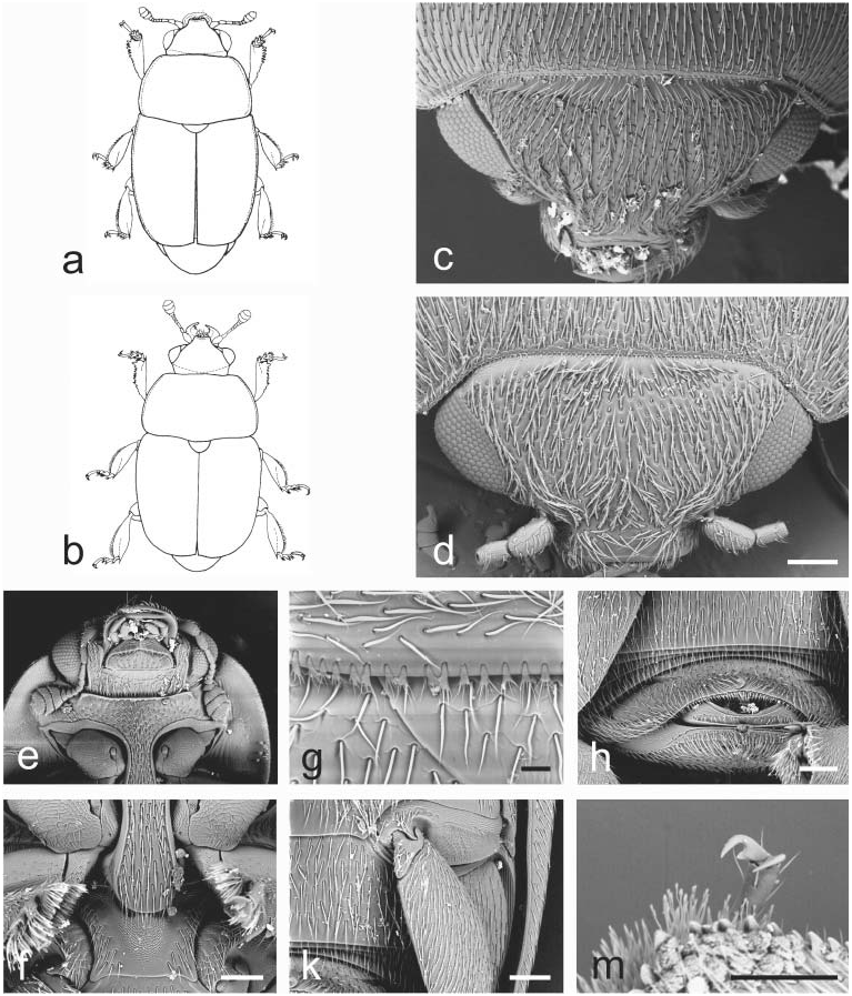
Fig. 17. Aristogethes Audisio & Cline, gen. nov.: a – A. translatus (Grouvelle, 1913); b – A. pecten (Audisio, Kirk-Spriggs & Kirejtshuk, 1998); c, e–k – A. pubescens (Reitter, 1872); d, m – A. marshalli (Grouvelle, 1914). a, b – male habitus (a – length 2.6 mm; b – length 2.4 mm); c, d – dorsal view of head; e – ventral view of head and anterior portion of prosternum; f – prosternal process and mesoventrite; g – anterior portion of scutellum and microsetae on middle of posterior margin of pronotum; h – exposed portion of last visible abdominal ventrite; k – caudal marginal line of metacoxal cavity; m – last tarsomere of middle leg. Scale bars: Figs. d, f, h, k, m = 100 μm; Fig. g = 20 μm.

Fig. 9. Boragogethes Audisio & Cline, gen. nov.: a, d, k, m – B. symphyti (Heer, 1841); b–c, e–h – B. rosenhaueri (Reitter, 1871). a, b – male habitus (a – length 3.0 mm; b – length 2.5 mm); c, d – dorsal view of head; e – microsetae on posterior margin of pronotum; f – ventral view of head and anterior portion of prosternum; g – prosternal process and mesoventrite; h – exposed portion of last visible abdominal ventrite; k – caudal marginal lines of metacoxal cavities; m – outer margin of mesotibia. Scale bars: Figs. c, d, f, g, h, m = 100 μm; Fig. e = 20 μm; Fig. k = 200 μm.
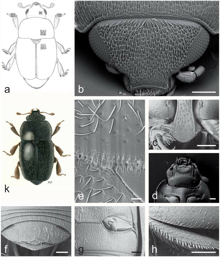
Fig. 10. Afrogethes Audisio & Cline, gen. nov.: a – A. tristis (Sturm, 1845); b–d, h – A. reticulatus (Reitter, 1872); e – A. alani (Kirejtshuk, 1988); f–g – A. planiusculus (Heer, 1841); k – A. isoplexidis (Wollaston, 1854). a, k – male habitus (a – length 2.6 mm; k – length 2.5 mm); b – dorsal view of head; c – prosternal process; d – ventral view of head and anterior portion of prosternum; e – microsetae on middle posterior margin of pronotum; f – exposed portion of last visible abdominal ventrite; g – caudal marginal lines of metacoxal cavities; h – outer margin of mesotibia. Scale bars: Figs. b, c, d, f, g, h = 100 μm; Fig. e = 20 μm.
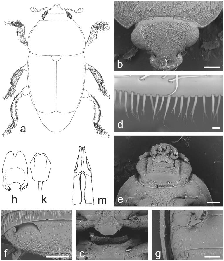
Fig. 11. Indogethes Audisio & Cline, gen. nov.: a–m – I. curvipes (Grouvelle, 1908). a – male habitus (pubescence and mandibles not illustrated; length 3.5 mm); b – dorsal view of head; c – prosternal process and mesoventrite; d – microsetae on middle of posterior margin of pronotum; e – ventral view of head and anterior portion of prosternum; f – exposed portion of last visible abdominal ventrite; g – caudal marginal line of metacoxal cavity; h–k – male genitalia (h – length 0.5 mm; k – 0.4 mm); m – ovipositor (length 0.7 mm). Scale bars: Figs. b, c, e, f, g = 200 μm; Fig. d = 10 μm.

Fig. 12. Bolbocerogethes Audisio & Cline, gen. nov.: a–e – B. pallipes (Boheman, 1851). a – male habitus (length 2.6 mm); b – ovipositor (modified from SPORNRAFT & KIREJTSHUK (1993); length 0.6 mm); c–d – male genitalia (c, d – length 0.3 mm); e – prosternal process (width 0.3 mm).

Fig. 13. Genistogethes Audisio & Cline, gen. nov.: a – G. immundus (Kraatz, 1858); b–h – G. punctatus (C. N. F. Brisout de Barneville, 1863). a – male habitus (length 2.7 mm); b – dorso-lateral view of head; c – ventral view of head and anterior portion of prosternum; d – caudal marginal line of metacoxal cavity; e – anterior portion of scutellum and microsetae on middle of posterior margin of pronotum; f – exposed portion of last visible abdominal ventrite; g – prosternal process and mesoventrite; h – middle leg with outer margin of mesotibia. Scale bars: Figs. b, c, d, f, g, h = 100 μm; Fig. e = 30 μm.
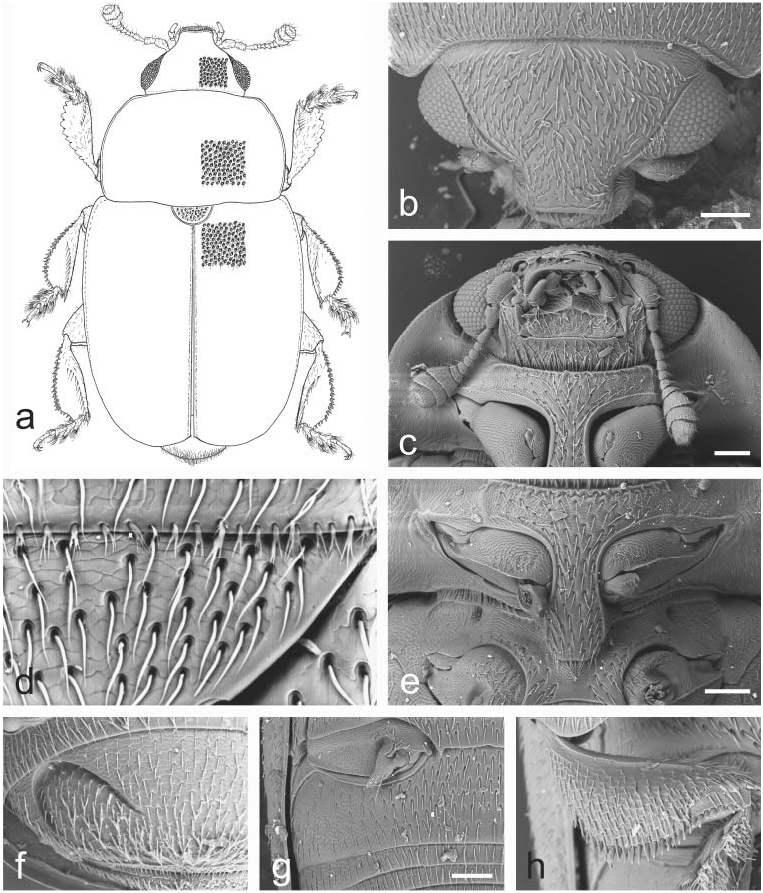
Fig. 14. Fabogethes Audisio & Cline, gen. nov.: a – F. opacus (Rosenhauer, 1856); b–h – F. nigrescens (Stephens, 1830). a – male habitus (length 2.7 mm); b – dorso-lateral view of head; c – ventral view of head and anterior portion of prosternum; d – anterior portion of scutellum and microsetae on middle of posterior margin of pronotum; e – prosternal process and mesoventrite; f – exposed portion of last visible abdominal ventrite; g – caudal marginal line of metacoxal cavity; h – middle leg with outer margin of mesotibia. Scale bars: Figs. b, c, e, g = 100 μm.

Fig. 15. Thymogethes Audisio & Cline, gen. nov.: a, h – T. egenus (Erichson, 1845); b – T. subfumatus (Ganglbauer, 1899); c–g, k – T. nigritus (Lucas, 1849). a, b – male habitus (a – length 2.5 mm; b – length 2.7 mm); c – ventral view of head and anterior portion of prosternum; d – anterior portion of scutellum and microsetae on middle of posterior margin of pronotum; e – dorsal view of head; f – prosternal process and mesoventrite; g – caudal marginal line of metacoxal cavity; h – exposed portion of last visible abdominal ventrite; k – middle leg with outer margin of mesotibia. Scale bars: Figs. c, e, f, g, h = 100 μm; Fig. d = 20 μm.
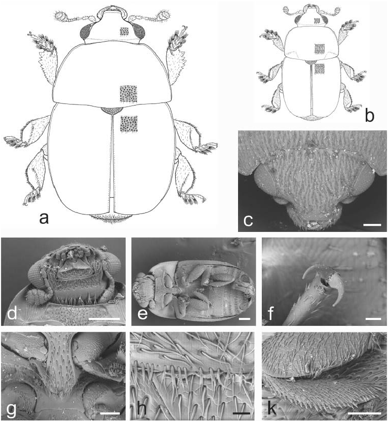
Fig. 16. Sagittogethes Audisio & Cline, gen. nov.: a – S. ater (C. N. F. Brisout de Barneville, 1863); b – S. lindbergi (Rebmann, 1940); c, f–g – S. maurus (Sturm, 1845); d–e, h–k – S. obscurus (Erichson, 1845). a, b – male habitus (a – length 2.9 mm; b – length 2.5 mm); c – dorsal view of head; d – ventral view of head and anterior portion of prosternum; e – ventral view of body; f – last tarsomere of a middle leg; g – prosternal process and mesoventrite; h – anterior portion of scutellum and microsetae on middle posterior margin of pronotum; k – middle leg with outer margin of mesotibia. Scale bars: Figs. c, g, k = 100 μm; Figs. d, e = 200 μm; Fig. f = 20 μm; Fig. h = 30 μm.

Fig. 18. JelinekigethesAudisio & Cline, gen. nov.: a–e – J. danielssoni (Audisio, 1995).a – male habitus; b–c – male genitalia; d – ovipositor; e – male protibia. Figs. a–e – refer to AUDISIO (1995) for scale.
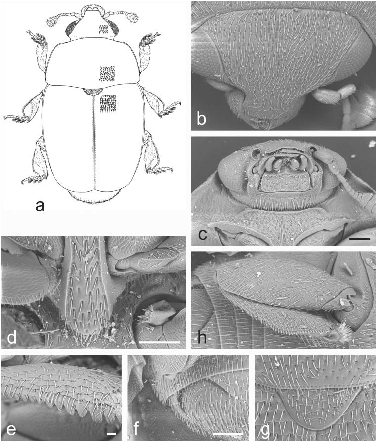
Fig. 19. Astylogethes Kirejtshuk, 1992: a–h – A. subrugosus (Gyllenhal, 1808). a – male habitus (length 2.4 mm); b – dorsal view of head; c – ventral view of head and anterior portion of prosternum; d – prosternal process and mesoventrite; e – outer margin of protibia; f – exposed portion of last visible abdominal ventrite; g – scutellum and microsetae on middle of posterior margin of pronotum; h – caudal marginal line of metacoxal cavity. Scale bars: Figs. c, d, f = 100 μm; Fig. e = 20 μm.

Fig. 22. Rubiogethes Audisio & Cline, gen. nov.: a–k – R. newtoni (Kirejtshuk, 1990). a – female habitus; b – dorsal view of head; c – ventral view of head and anterior portion of prosternum; d – scutellum and microsetae on middle of posterior margin of pronotum; e – prosternal process and mesoventrite; f – exposed portion of last visible abdominal ventrite; g – caudal marginal line of metacoxal cavity; h – middle leg with outer margin of mesotibia; k – protibia. Scale bars: Figs. a, b = 200 μm; Figs. c, d, e, f, g = 100 μm; Figs. h, k = 20 μm.
No known copyright restrictions apply. See Agosti, D., Egloff, W., 2009. Taxonomic information exchange and copyright: the Plazi approach. BMC Research Notes 2009, 2:53 for further explanation.
|
Kingdom |
|
|
Phylum |
|
|
Class |
|
|
Order |
|
|
Family |