
Lamiogethes Audisio & Cline, 2009
|
publication ID |
https://doi.org/10.5281/zenodo.5319334 |
|
persistent identifier |
https://treatment.plazi.org/id/03BE87CC-F63B-FFD2-BA6C-FDD6FBA5F9B2 |
|
treatment provided by |
Felipe (2021-08-28 07:26:47, last updated by Plazi 2023-11-05 05:53:56) |
|
scientific name |
Lamiogethes Audisio & Cline |
| status |
gen. nov. |
23. Lamiogethes Audisio & Cline , gen. nov.
( Figs. 23 a–n View Fig )
Type species. Meligethes ruficollis Reitter, 1872: 244 , 258 (by present designation) [= Lamiogethes ruficollis (Reitter, 1872) comb. nov.].
Generic description and diagnosis. Inclusive species vary greatly in size (1.4–3.3 mm length), and share the following combination of characters.
Body color and pubescence: pubescence silvery-whitish or golden, fine, usually not welldeveloped, recumbent, never obscuring the usually brown, blackish-brown, or reddish-brown dorsal body surface; pronotal and elytral sides narrowly flattened, frequently paler than disc; lateral margin of pronotum and elytra with a series of faintly distinct, small and short setae, each seta 0.3–0.5× as long as those on elytral disc; posterior margin of pronotum typically with long, usually distally bifid microsetae (frequently reduced in members of Lamiogethes convexus species-group), frequently absent at least along narrow middle portion anterior to scutellum ( Figs. 23e, f View Fig ) in Afrotropical species-groups.
Dorsal habitus: body highly convex to relatively flat, even within members of the same species-group ( AUDISIO 1996), variably shaped, shortly oval to long and parallel-sided ( Figs. 23a, b View Fig ); dorsal punctures on discal portion of pronotum usually larger than eye facet, and typically moderately to deeply impressed and densely distributed ( Fig. 23d View Fig ), but depth and distribution highly variable; anterior margin of clypeus truncate, slightly or distinctly sinuate medially, usually without small, faintly distinct, medial bulge, faintly distinctly bordered, lateral angles rounded or obtuse ( Fig. 23d View Fig ); circum-ocular furrows (occipital sulci) on dorsal side of head narrow, moderately to deeply impressed, usually obliterated posteriorly, incomplete ( Fig. 23d View Fig ); eyes large and usually moderately projecting laterally ( Figs. 23a, b, d View Fig ); pronotum with obtusely distinct to rounded posterior angles, never directed posteriorly ( Figs. 23a, b View Fig ); areas adjacent to posterior outer portions of pronotum usually impunctate and glabrous; scutellum minutely punctured on exposed portion ( Figs. 23e, f View Fig ); elytra with simple to more or less distinctly transversely strigose punctures ( Figs. 23a, e, f View Fig ); elytral humeral striae usually indistinct; elytral pre-sutural striae distinct, originating at scutellar vertex, terminating close to elytral apex, and delimiting on each elytron a flatly raised and narrow sutural border, border largest at posterior third but narrower than proximal portion of 3 rd antennomere; elytral apices truncately rounded in both sexes ( Fig. 23a View Fig ); pygidium partially exposed, moderately convex, apically rounded in both sexes ( Figs. 23a, b View Fig ).
Ventral side: antennal furrows markedly delimited, nearly parallel-sided, slightly sinuate, slightly divergent posteriorly; mentum subpentagonal ( Fig. 23c View Fig ), transverse, trapezoidal; prosternal antennal furrows on anterior margin of prosternum strongly raised and relatively long ( Fig. 23c View Fig ); prosternal process variably shaped, subapical dilated portion 2.0–3.6× as wide as maximum width of 1 st antennomere, usually bluntly convex and microscopically indistinctly crenulate distally ( Figs. 23h, k View Fig ); lateral borders of prosternal process delimiting shallowly impressed but wide and distinct furrows, distally terminating over predistal lateral expansions ( Figs. 23h, k View Fig ); posterior margin of mesoventrite never medially incised, frequently slightly to markedly arcuately convex posteriorly ( Figs. 23h, k View Fig ); male impressions on metaventrite and tubercles variably developed; first two visible abdominal ventrites simple in both sexes, without tufts of setae; caudal marginal lines of metacoxal cavities nearly simple, usually subparallel and more or less narrowly contiguous to posterior margin of metacoxal cavities, with shallow arched impression of outer ‘axillary’ line ( Fig. 23g View Fig ); ‘axillary’ space on first abdominal ventrite usually large, ‘axillary’ angle usually widely obtuse ( Fig. 23g View Fig ); large, long, and deeply impressed arched impressions present on basal portion of last visible abdominal ventrite, frequently partially covered by distal portion of penultimate visible abdominal ventrite ( Fig. 23g View Fig ); apex of last abdominal ventrite frequently more or less distinctly emarginate in males with shining tubercles or arcuate ridges.
Appendages: male 1 st antennomere 0.8–0.9× as long as width of protibiae excluding distal teeth ( Figs. 23 a, b View Fig ); 3 rd antennomere usually moderately short in both sexes, 2.1–2.2× as long as wide, 0.8–1.0× as long as but distinctly thinner than 2 nd antennomere ( Fig. 23c View Fig ); 4 th and 5 th antennomeres subequal in both sexes, short, nearly as long as wide; antennal club compact, mid-sized, simple, comprising last 3 antennomeres in both sexes (8 th antennomere scarcely widened, 0.4–0.5× as wide as 9 th antennomere) ( Fig. 23c View Fig ), narrower than width of protibiae, sexual dimorphism absent; labial palpi relatively short in both sexes ( Fig. 23c View Fig ), terminal segment ~1.6–1.8× as long as wide; maxillary palpi moderately long and slender in both sexes ( Fig. 23c View Fig ), terminal segment 2.2–2.3× as long as wide; mandible mid-sized ( Fig. 23a, b, c View Fig ), apex moderately acuminate, no sexual dimorphism; tarsal claws variable, simple, not toothed at base ( Fig. 23n View Fig ), bluntly toothed, or strongly and sharpy toothed ( Fig. 23m View Fig ); tarsi of variable size and shape, 0.5–0.8× as long as corresponding tibiae ( Figs. 23a, b, n View Fig ); protibiae with a series of variable, uneven, small and blunt to large and sharp teeth on distal portion or on most of lateral margin ( Figs. 23a, b, g View Fig ); meso- and metatibiae on lateral margin bearing a single and usually even row of long and robust pegs ( Figs. 23a, b, n View Fig ), without U-shaped sinuosity at distal third; meso- and metatibiae moderately slender and relatively narrow ( Figs. 23a, b, n View Fig ), never subtrapezoidal or axe-shaped; sexual dimorphism only rarely expressed in metatibial shape; tarsal plates of prolegs distinctly wider in males; posterior margin of metafemora simple in both sexes, without tubercles or projections.
Male genitalia: processes along inner side of parameres absent ( Figs. 24–25 View Fig View Fig in EASTON 1959b; Figs. 54–57, 58– 69 in EASTON 1960; Figs. 149–152 in AUDISIO 1993b; Figs. 1–18 View Fig View Fig View Fig View Fig View Fig View Fig View Fig View Fig View Fig View Fig View Fig View Fig View Fig View Fig View Fig View Fig View Fig View Fig in AUDISIO 1996), distal margin variably incised, without deep median longitudinal desclerotization from proximal portion of tegmen extending to medial distal V-shaped excision; median lobe of aedeagus variably shaped, without lateral emargination, narrowed and obtuse, acuminate or spatulate distally, rarely with minute excisions or emarginations.
Female genitalia (ovipositor): small or large, variably shaped; styli short to long, cylindrical, inserted close to apex of contiguous gonostyloids (Fig. 58 in EASTON 1959b; Figs. 105–111 in EASTON 1960; Figs. 162–163 in AUDISIO 1993b; Figs. 21–22 View Fig View Fig and 24–29 View Fig View Fig View Fig View Fig View Fig View Fig in AUDISIO 1996); each gonostyloid lightly sclerotized and rarely darkly pigmented distally, with a simple, never indentate outer portion of narrow basicoxites, and a single, narrow, more or less pigmented and sclerotized arcuate area along outer subdistal portion of gonostyloids. ‘Central point’ of ovipositor usually centrally located, or placed slightly more distad than middle, usually without or rarely with proximad directed spicule.
Etymology. The generic name is derived from the host-plant family of inclusive species, i.e. Lamiaceae , and from ‘- gethes ’, to emphasize its phylogenetic relationship with Meligethes . Gender masculine.
Biology. The biology of inclusive species is incompletely known, but appears to be homogeneous. Members of Lamiogethes gen. nov. are likely all associated as larvae with flowers of Lamiaceae (= Labiatae ), in particular Lamium L., Salvia L., Stachys L., Becium Lindl , Leucas Burm. ex R. Br. , Leonotis R. Br. , and allied genera in Palaearctic and Afrotropical areas ( EASTON 1960; AUDISIO 1993b, 1996, and unpublished data).
Phylogenetic position. Available morphological datasets provide good evidence for a clade including Lamiogethes gen. nov., Rubiogethes gen. nov., Paleogethes gen. nov., Astylogethes , and Stachygethes gen. nov. However, phylogenetic relationships between these taxa are only partially supported with molecular data ( TRIZZINO et al. 2009).
Taxonomy and geographic distribution. Lamiogethes gen. nov. is numerically the second largest Meligethinae genus, including nearly one hundred described species as well as ~20 additional identified but undescribed species, mainly distributed in Tropical Africa, Madagascar, the Indian Subcontinent, Europe, Anatolia, and the Eastern Palaearctic ( KIREJTSHUK 1992b; AUDISIO 1993b, 1996; JELÍNEK & AUDISIO 2007). Inclusive species are attributed to four or five formerly recognized species-groups, i.e. the ‘ Meligethes difficilis ’, ‘ M. ruficollis / gloriosus ’, ‘ M. convexus ’, and ‘ M. politus / phalacroides ’ species-groups.
Lamiogethes abductus ( Audisio, Jelínek & Cooter, 2005) NE China comb. nov.
Lamiogethes accretus (Kirejtshuk, 1988) comb. nov. South Africa: Mpumalanga, NW Province, KwaZulu- Natal, Free State, E Cape ; Lesotho
Lamiogethes aeneoviridinitens (Audisio, 1993) comb. nov. NE Turkey, Caucasus
Lamiogethes amabilis (Kirejtshuk, 1988) comb. nov. India
Lamiogethes amei ( Audisio & Kirejtshuk, 1988) comb. nov. N Turkey, Caucasus
Lamiogethes andrewesi (Grouvelle, 1908) comb. nov. India
Lamiogethes angustatus (Küster, 1848) comb. nov. S Europe
Lamiogethes asignifer (Kirejtshuk, 1996) comb. nov. N Namibia
Lamiogethes assamensis (Kirejtshuk, 1980) comb. nov. N India, Assam
Lamiogethes atomus ( Grouvelle, 1904) comb. nov. Congo, Zanzibar
Lamiogethes atramentarius (Förster, 1849) comb. nov. Central Europe
Lamiogethes atrovirens (Jelínek, 1982) comb. nov. N Turkey, Caucasus
Lamiogethes becivorus ( Audisio, 1996) comb. nov. South Africa: NW Province
Lamiogethes besucheti (Kirejtshuk, 1988) comb. nov. Sri Lanka
Lamiogethes bidens (C. N. F. Brisout de Barneville, 1863) Europe, Caucasus comb. nov.
Lamiogethes bolognai (Audisio, 1977) comb. nov. N Turkey, Caucasus
Lamiogethes brunnicornis ( Sturm, 1845) comb. nov. Europe, W Siberia, Middle Asia, N Africa Lamiogethes bucciarellii (Audisio, 1976) comb. nov. SE Europe, Turkey, Caucasus, Middle East Lamiogethes buyssoni (C. N. F. Brisout de Barneville, 1882) Europe, Anatolia, Caucasus comb. nov.
Lamiogethes byrrhoides ( Audisio, 1996) comb. nov. South Africa: NW Province; Namibia ; Botswana Lamiogethes candidus (Easton, 1964) comb. nov. Congo
Lamiogethes chlorocupreus (Audisio, Jelínek & S China
Cooter, 2005) comb. nov.
Lamiogethes conjungens ( Grouvelle, 1910) comb. nov. S China
Lamiogethes convexus ( Boheman, 1851) comb. nov. South Africa: KwaZulu-Natal, Free State Lamiogethes cooteri (Audisio, 1989) comb. nov. Montenegro
Lamiogethes cribrosus (Grouvelle, 1908) comb. nov. India
Lamiogethes descarpentriesi (Kirejtshuk, 1980) comb. nov. India
Lamiogethes desolatus (Easton, 1964) comb. nov. Congo
Lamiogethes dieckmanni (Audisio & Jelínek, 1984) N Turkey, N Iran, Caucasus comb. nov.
Lamiogethes difficilis ( Heer, 1841) comb. nov. Palaearctic Region
Lamiogethes difficiloides ( Audisio, Jelínek & Cooter, 2005) S China comb. nov.
Lamiogethes equus ( Easton, 1960) comb. nov. Kenya
Lamiogethes fairmairei (Grouvelle, 1913) comb. nov. Madagascar
Lamiogethes gibbulus ( Spornraft & Kirejtshuk, 1993) South Africa: KwaZulu-Natal, Free State, NW comb. nov. Province; Lesotho
Lamiogethes glebi (Kirejtshuk, 1988) comb. nov. Sri Lanka
Lamiogethes gloriosus ( Grouvelle, 1910) comb. nov. Tanzania
Lamiogethes grouvellei (Plavilstshikov, 1924) comb. nov. Madagascar
Lamiogethes haemorrhoidalis (Förster, 1849) comb. nov. Europe, Near East, W Middle Asia
Lamiogethes imitans (Kirejtshuk, 1988) comb. nov. Namibia
Lamiogethes instabilis (Grouvelle, 1906) comb. nov. Madagascar
Lamiogethes jelineki (Audisio, 1976) comb. nov. S Europe, Caucasus
Lamiogethes kasparyani ( Kirejtshuk, 1984) comb. nov. SE Siberia, NE China
Lamiogethes kaszabi (Audisio, 1979) comb. nov. Georgian Caucasus
Lamiogethes kirejtshuki (Audisio, 1979) comb. nov. N Turkey, Caucasus
Lamiogethes kunzei ( Erichson, 1845) comb. nov. Europe, Caucasus, N Turkey
Lamiogethes leati ( Easton, 1956) comb. nov. Sicily, N Africa, N Iran
Lamiogethes leileri ( Kirejtshuk, 2002) comb. nov. Kenya
Lamiogethes leucasi ( Easton, 1960) comb. nov. Kenya 1)
Lamiogethes levis (Kirejtshuk, 1995) comb. nov. Sri Lanka
Lamiogethes lornae ( Easton, 1960) comb. nov. Kenya
Lamiogethes luminosus (Reitter, 1873) comb. nov. India
1) It is likely that L. leucasi ( Easton, 1960) is a junior synonym of L. similis (Grouvelle, 1899) .
Lamiogethes luteolus (Reitter, 1873) comb. nov. India
Lamiogethes madagascariensis ( Grouvelle, 1896) Madagascar comb. nov.
Lamiogethes madurensis (Kirejtshuk, 1988) comb. nov. India
Lamiogethes medvedevi ( Kirejtshuk, 1978) comb. nov. NE Turkey, Caucasus
Lamiogethes merkli ( Kirejtshuk, 2001) comb. nov. Kenya
Lamiogethes mirator (Kirejtshuk, 1979) comb. nov. Russian Far East: Primorie
Lamiogethes mitis (Kirejtshuk, 1996) comb. nov. Namibia
Lamiogethes mixtus (Grouvelle, 1908) comb. nov. India
Lamiogethes montivagus ( Easton, 1960) comb. nov. Tanzania
Lamiogethes morosus ( Erichson, 1845) comb. nov. Palaearctic Region
Lamiogethes nakanei (Easton, 1957) comb. nov. E Siberia, Japan, NE China
Lamiogethes nigriceps (Easton, 1964) comb. nov. Congo
Lamiogethes nobilis (Easton, 1964) comb. nov. Congo
Lamiogethes oberprieleri ( Audisio, 1996) comb. nov. South Africa: W Cape
Lamiogethes ochropus ( Sturm, 1845) comb. nov. Europe, Siberia, N Korea
Lamiogethes opacidorsum (Kirejtshuk, 1996) comb. nov. Namibia
Lamiogethes paraconvexus ( Audisio, 1996) comb. nov. South Africa: Mpumalanga
Lamiogethes paschalis (Spornraft, 1975) comb. nov. Central and southern Italy
Lamiogethes pedicularius ( Gyllenhal, 1808) comb. nov. Europe, Siberia, N Middle Asia
Lamiogethes perparvulus (Grouvelle, 1913) comb. nov. Madagascar
Lamiogethes persicus (Faldermann, 1835) comb. nov. Europe, Near East, W Siberia, N Middle Asia, NW China
Lamiogethes phalacroides ( Grouvelle, 1896) comb. nov. Madagascar
Lamiogethes politus (Motschulsky, 1863) comb. nov. Sri Lanka
Lamiogethes potanini (Kirejtshuk, 1979) comb. nov. China: Sichuan
Lamiogethes pubiusculus (Kirejtshuk, 1988) comb. nov. South Africa: KwaZulu-Natal
Lamiogethes relativus ( Kirejtshuk & Viklund, 2002) Kenya comb. nov.
Lamiogethes rubiginosus (Grouvelle, 1909) comb. nov. Tanzania, Zanzibar
Lamiogethes rubricollis (Grouvelle, 1903) comb. nov. India
Lamiogethes ruficollis (Reitter, 1872) comb. nov. Tropical Africa, South Africa
Lamiogethes rufithorax Grouvelle, 1894 comb. nov. India
Lamiogethes rufulus ( Grouvelle, 1916) comb. nov. Congo
Lamiogethes sellaris ( Easton, 1960) comb. nov. Kenya
Lamiogethes serripes (Gyllenhal, 1827) comb. nov. Europe, Siberia, N Middle Asia
Lamiogethes shimoyamai (Sadan. Hisamatsu, 1964) Japan comb. nov.
Lamiogethes shrilankaensis (Kirejtshuk, 1988) comb. nov. Sri Lanka
Lamiogethes similis (Grouvelle, 1899) comb. nov. Ethiopia, Kenya 1)
Lamiogethes simillimus ( Kirejtshuk, 1984) comb. nov. Russian Far East, NE China
Lamiogethes simulans (Easton, 1959) comb. nov. Ethiopia
Lamiogethes spadix (Easton, 1954) comb. nov. S Arabian Peninsula
Lamiogethes subglobosus (Reitter, 1875) comb. nov. South Africa: W Cape
Lamiogethes sulcatus (C. N. F. Brisout de Barneville, 1863) Europe, Near East, Siberia comb. nov.
Lamiogethes tilmani ( Easton, 1968) comb. nov. Nepal, SW China: Tibet, N Pakistan, NW India Lamiogethes verdcourti ( Easton, 1960) comb. nov. Kenya
Lamiogethes vietnamensis (Kirejtshuk, 1979) comb. nov. Vietnam
Lamiogethes xyphosuroides ( Kirejtshuk, 1989) comb. nov. South Africa: NW Province, Mpumalanga
AUDISIO P. & KIREJTSHUK A. G. 1988: Novyy vid roda Meligethes Stephens gruppy difficilis s Kavkaza i obzor zapadnopalearkticheskikh vidov, blizkikh k M. viduatus (Heer) (Coleoptera; Nitidulidae). (A new species of the Meligethes difficilis group from the Caucasus, and review of the West-Palaearctic species related to M. viduatus (Heer) (Coleoptera, Nitidulidae )). Entomologicheskoe Obozrenie 67: 559 - 568 (in Russian, English title and summary).
AUDISIO P. 1993 b: Coleoptera Nitidulidae - Kateretidae. Fauna d'Italia. Vol. 32. Calderini Edizione, Bologna, xvi + 971 pp.
AUDISIO P. 1996: New and little-known South African Meligethes Stephens of the M. convexus group (Coleoptera: Nitidulidae: Meligethinae). African Entomology 4: 213 - 230.
AUDISIO P., JELINEK J. & COOTER J. 2005 c: New and little-known species of Meligethes Stephens, 1830 from China (Coleoptera: Nitidulidae). Acta Entomologica Musei Nationalis Pragae 45: 111 - 127.
BOHEMAN C. H. 1851: Insecta Caffraria annis 1838 - 1845 a J. A. Wahlberg collecta, amici auxilio suffultus, descripsit Carolus H. Boheman. Pars I., Fasciculus 2. Coleoptera (Buprestides, Elaterides, Cebrionites, Rhipicerides, Cyphonides, Lycides, Lampyrides, Telephorides, Melyrides, Clerii, Terediles, Ptiniores, Palpatores, Silphales, Histeres, Scaphidilia, Nitidulariae, Cryptophagidae, Byrrhii, Dermestini, Parnidae, Hydrophilidae). Officina Norstedtiana, Holmiae, pp. 299 - 626, pl. 2.
BRISOUT DE BARNEVILLE C. N. F. 1863: [new taxa]. In: GRENIER A. (ed.): Catalogue des Coleopteres de France, et materiaux pour servir a la faune des Coleopteres Francais. A. Grenier, Paris, iv + 9 - 79 + 1 - 135 pp.
EASTON A. M. 1956: The Meligethes of North Africa (Coleoptera, Nitidulidae). Memoires de la Societe des Sciences Naturelles et Physiques du Maroc, Serie Zoologie 2 (1955): 7 - 70.
EASTON A. M. 1959 b: The Meligethes of Abyssinia (Col.; Nitid.). Transactions of the Royal Entomological Society of London 111: 367 - 403.
EASTON A. M. 1960: The Meligethes of East Africa (Coleoptera: Nitidulidae). Transactions of the Royal Entomological Society of London 112: 263 - 318.
EASTON A. M. 1968: The Meligethes of High Nepal (Coleoptera, Nitidulidae). Pp. 40 - 48. In: Khumbu Himal, Vol. 3. Universitats-Verlag Wagner, Innsbruck - Munchen.
ERICHSON W. F. 1845 - 1848: [I., II. Lieferungen]. Pp. 1 - 320. In: Naturgeschichte der Insecten Deutschlands. Erste Abtheilung. Coleoptera. Dritter Band. Nikolaische Buchhandlung, Berlin, iv + [ii] + 968 pp. [issued in parts: pp. 1 - 320: 1845; pp. 321 - 480: 1846; pp. 481 - 800: 1847; pp. 801 - 968: 1848].
GROUVELLE A. 1896: Descriptions de Clavicornes d'Afrique et du Madagascar. Annales de la Societe Entomologique de France 65: 71 - 94.
GROUVELLE A. 1904: Descriptions de Clavicornes nouveaux du Musee de Bruxelles. Annales de la Societe Entomologique de Belgique 48: 181 - 185
GROUVELLE A. 1910: Coleoptera. 15. Clavicornes. Pp. 309 - 334. In: SJOSTEDT Y. (ed.): Wissenschaftliche Ergebnisse der Schwedischen zoologischen Expedition nach dem Kilimandjaro, dem Meru und den umgebenden Massaisteppen Deutsch-Ostafrikas 1905 - 1906, unter leitung von prof. dr. Yngve Sjostedt. Vol. 1. Herausgegeben mit Unterstutzung von der Koniglichen Schwedischen Akademie der Wissenschaften, Stockholm.
GROUVELLE A. 1916: Clavicornes du Musee du Congo. Deuxieme memoire. Revue Zoologique Africaine 4 (1914 - 1916): 129 - 141.
GYLLENHAL L. 1808: Insecta Suecica: Classis I, Coleoptera sive Eleuterata. Tomus 1. F. J. Leverentz, Scaris, xii + 572 pp.
HEER O. 1841: Fauna Coleopterorum Helvetica. Pars 1., fasciculus 3. Impensis Orelii, Fuesslini et Sociorum, Turici, pp. 361 - 652 pp.
JELINEK J. & AUDISIO P. 2007: Family Nitidulidae. Pp. 459 - 491. In: LOBL I. & SMETANA A. (eds): Catalogue of Palaearctic Coleoptera. Vol. 4: Elateroidea - Derodontoidea - Bostrichoidea - Lymexyloidea - Cleroidea - Cucujoidea. Apollo Books, Stenstrup, 935 pp.
KIREJTSHUK A. G. 1978: Novye vidy zhukov-blestyanok roda Meligethes Stephens (Coleoptera, Nitidulidae) iz SSSR i obzor gruppy vidov, blizkikh k M. umbrosus Sturm. (New species of the genus Meligethes Stephens (Coleoptera, Nitidulidae) from the USSR and review of the group of species related to Meligethes umbrosus Sturm). Entomologicheskoe Obozrenie 57: 578 - 595 (in Russian, English title).
KIREJTSHUK A. G. 1984: New taxa of Nitidulidae (Coleoptera) from the Indo-Malayan fauna. Annales Historico- Naturales Musei Nationalis Hungarici 76: 169 - 195.
KIREJTSHUK A. G. 1989: Novye taksony zhukov-blestyanok (Coleoptera, Nitidulidae) vostochnogo polushariya (Chast' III). [New taxa of the Nitidulidae (Coleoptera) of the East hemisphere (Part III)]. Trudy Zoologicheskogo Instituta, Akademiya Nauk SSSR 208: 64 - 89 (in Russian).
KIREJTSHUK A. G. 1992 b: 59, 61. Sem. Nitidulidae - Blestyanki. Pp. 114 - 209. In: LER P. A. (ed.): Opredelitel'nasekomykh Dal'nego Vostoka SSSR v shesti tomakh. Tom III. Zhestkokrylye, ili zhuki. [Key to the insects of the Far East of the USSR in six volumes. Vol. 3, Coleoptera]. Nauka, Sankt-Petersburg, 704 pp (in Russian).
KIREJTSHUK A. G. 2001: Notes on the systematics of the African Nitidulidae (Coleoptera). Annales Historico- Naturales Musei Nationalis Hungarici 93: 17 - 89.
KIREJTSHUK A. G. & VIKLUND B. 2002: Contribution to the knowledge on the subgenus Meligethes (Clypeogethes Scholtz, 1932) from Kenya (Coleptera, Nitidulidae). Annales Historico-Naturales Musei Nationalis Hungarici 94: 9 - 21.
SPORNRAFT K. & KIREJTSHUK A. G. 1993: Uber alte und neue sudafrikanische Meligethes-Arten (Coleoptera, Nitidulidae). Mitteilungen der Munchner Entomologischen Gesellschaft 83: 47 - 75.
STURM J. 1845: Deutschlands Fauna in Abbildungen nach der Natur mit Beschreibungen. V. Abtheilung. Die Insecten. Zweites Bandchen. Kafer. Nurnberg, 114 pp + pls. ccciv - cccix.
TRIZZINO M., AUDISIO P., ANTONINI G., DE BIASE A. & MANCINI E. 2009: Comparative analysis of sequences and secondary structures of the rRNA internal transcribed spacer 2 (ITS 2) in pollen-beetles of the subfamily Meligethinae (Coleoptera, Nitidulidae): potential use of slippage-derived sequences in molecular systematics. Molecular Phylogenetics and Evolution 51: 215 - 226.

Fig. 23. Lamiogethes Audisio & Cline, gen. nov.: a – L. paschalis (Spornraft, 1975); b – L. convexus (Boheman, 1851); c–e, g–h, m – L. ruficollis (Reitter, 1872); k, f, n – L. difficilis (Heer, 1841). a, b – male habitus (a – length 3.0 mm; b – length 2.7 mm); c – ventral view of head and of anterior portion of prosternum; d – dorsal view of head; e–f – scutellum and microsetae on middle of posterior margin of pronotum; g – ventral view of body; h–k – prosternal process and mesoventrite; m – last tarsomere of middle leg; n – outer margin of mesotibia. Scale bars: Figs. c, d, e, f, h, n = 100 μm; Fig. g = 1 mm; Fig. m = 20 μm.

Fig. 24. Chromogethes Kirejtshuk, 1989: a – C. formosus (Kirejtshuk, 1989); b–h – C. mastax (Audisio & De Biase, 2004); k – C. splendidulus (Reitter, 1873). a – male habitus (length 2.0 mm); b, k – dorsal view of head; c – scutellum and microsetae on middle of posterior margin of pronotum; d – ventral view of head and anterior portion of prosternum; e – exposed portion of last visible abdominal ventrite; f – tarsal claws of middle leg; g – outer margin of mesotibia; h – prosternal process and mesoventrite. Scale bars: Figs. b, d, e, g, k = 100 μm; Fig. c, f = 30 μm; Fig. h = 200 μm.
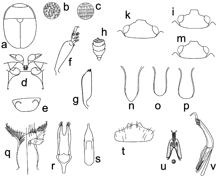
Fig. 25. Cyclogethes Kirejtshuk, 1979: a–h, k, p–v – C. orientalis Kirejtshuk, 1979; i, o – C. abnormis Kirejtshuk, 1979; m – C. fuscipennis Jelínek, 2000; n – C. aldridgei Kirejtshuk, 1980. a – male habitus; b–c – punctation of pronotum and elytra; d – prosternal process, mesoventrite, and metaventrite; e – exposed portion of last visible abdominal ventrite; f – protibia; g – mesotibia; h – antennal club; k, i, m – dorsal view of head; n, o, p – prosternal process; q – labium and left palp; r–s – dorsal view of male genitalia; t – labrum; u – ovipositor; v – lateral view of male genitalia. Drawings a–h, k, q–v from KIREJTSHUK (1979a); drawings i, m–p from JELÍNEK (2000b); refer to KIREJTSHUK (1979a) and to JELÍNEK (2000b) for scale.
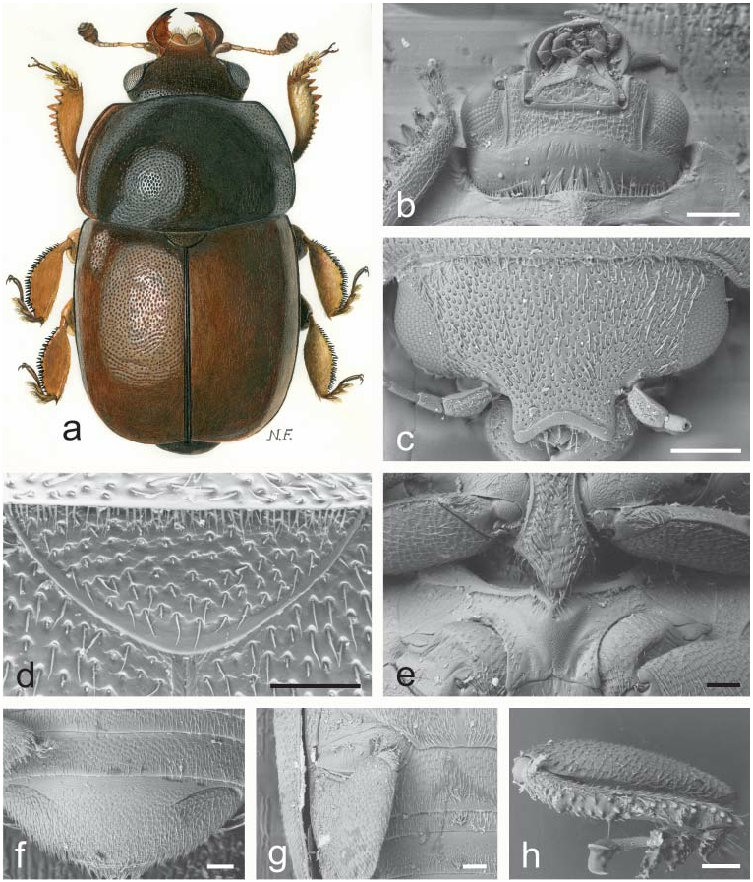
Fig. 1. Acanthogethes Reitter, 1871: a–h – A. fuscus (Olivier, 1790). a – male habitus (length 3.5 mm); b – ventral view of head and anterior portion of prosternum; c – dorsal view of head; d – scutellum and microsetae on posterior margin of pronotum; e – prosternal process and mesoventrite; f – exposed portion of last visible abdominal ventrite; g – caudal marginal lines of metacoxal cavities; h – middle leg illustrating outer margin of mesotibia. Scale bars: Figs. b, c = 200 μm; Figs. d, e, f, g, h = 100 μm.
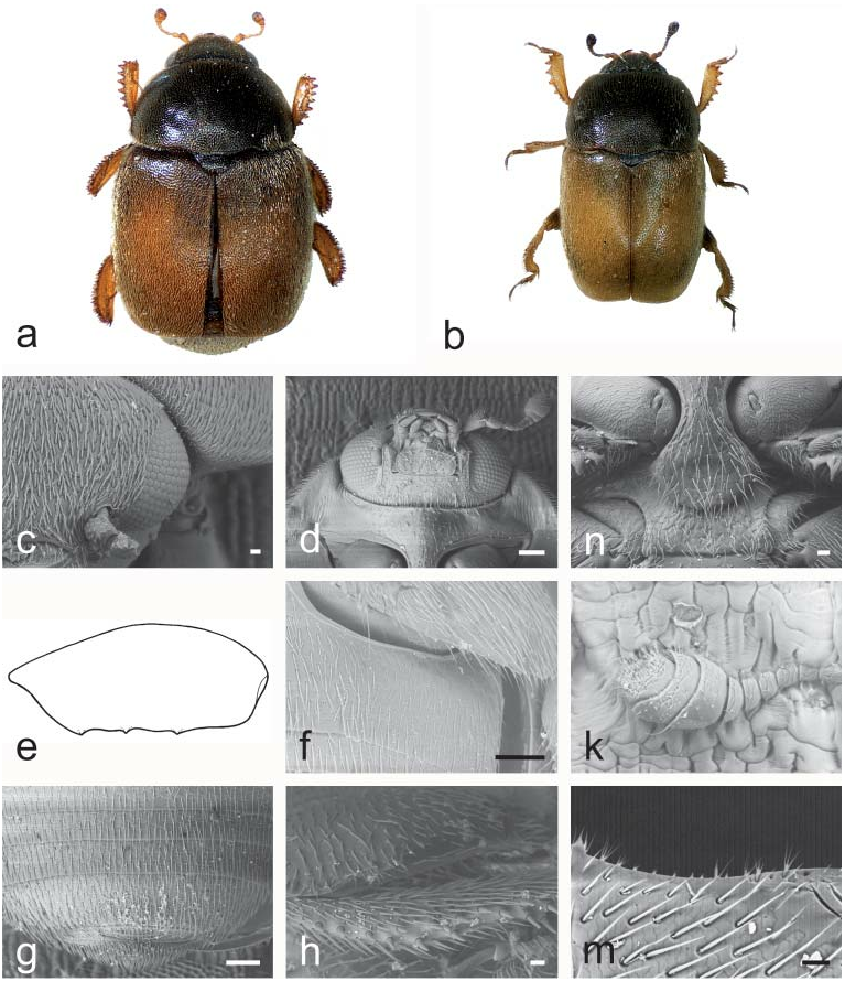
Fig. 2. Asterogethes Audisio & Cline, gen. nov.: a – A. endroedyi (Kirejtshuk & Audisio, 1995); b–d, f–n – A. arcuatus (Reitter, 1872); e – A. rufiventris (Reitter, 1872). a, b – male habitus (a – length 3.2 mm; b – length 2.4 mm); c – dorso-lateral view of head; d – ventral view of head and anterior portion of prosternum; e – outline of male metafemur (length 0.5 mm); f – caudal marginal lines of metacoxal cavities; g – exposed portion of last visible abdominal ventrite; h – middle leg with illustrating outer margin of mesotibia; k – antenna; m – pronotal setae and microsetae on posterior margin of pronotum; n – prosternal process and mesoventrite. Scale bars: Figs. c, h, m, n = 20μm; Figs. d, f, g = 100 μm.

Fig. 3. Odontholariopsis Audisio & Cline, gen. nov.: a, g – O. haagii (Reitter, 1872); b–f, h – O. nebulosus (Reitter, 1872). a – male habitus (length 2.6 mm); b – dorsal view of head; c – ventral view of head and anterior portion of prosternum; d – middle leg illustrating outer margin of mesotibia; e – scutellum and microsetae on posterior margin of pronotum; f – prosternal process and mesoventrite; g – outline of male metafemur (length 0.5 mm); h – exposed portion of last visible abdominal ventrite. Scale bars: Figs. b, c, f, h = 100 μm; Fig. d = 30 μm; Fig. e = 20 μm.

Fig. 4. Lariopsis Kirejtshuk, 1989: a – L.vultuosus (Kirejtshuk & Audisio, 1995); b–k – L. variabilis (Reitter, 1872). a – male habitus (length 3.3 mm); b, c – dorso-lateral view of head; d – ventral view of head and anterior portion of prosternum; e – prosternal process and mesoventrite; f – middle leg with outer margin of mesotibia; g – microsetae on middle posterior margin of pronotum; h – exposed portion of last visible abdominal ventrite; k – caudal marginal lines of metacoxal cavities. Scale bars: Figs. b, c, d, e, h, k = 100 μm; Fig. f = 30 μm; Fig. g = 10 μm.
Fig. 5. Neolariopsis Audisio & Cline, gen. nov.: a, c–h – N. cercoides (Reitter, 1872); b – N. thalycroides (Kirejtshuk & Audisio, 1995). a, b – male habitus (a – length 2.1 mm, b – length 2.1 mm); c – dorsal view of head; d – ventral view of head and anterior portion of prosternum; e – middle leg with outer margin of mesotibia; f – prosternal process and mesoventrite; g – exposed portion of last visible abdominal ventrite; h – antenna. Scale bars: Figs. c, d, f = 100 μm; Figs. e, h = 20 μm; Fig. g = 30 μm.

Fig. 6. Clypeogethes Scholz, 1932 and Xerogethes Audisio & Cline, gen. nov.: a, c – C. chlorocyaneus (Jelínek & Audisio, 1977); b, d–e – C. elongatus (Rosenhauer, 1856); k–n – C. lepidii (Miller, 1851); f–g – X. osellai (Audisio & Jelínek, 2000); h – X. rotundicollis (C. N. F. Brisout de Barneville, 1863). a – male habitus (length 2.5 mm); b, c, h – ovipositors; d–e, f–g – male genitalia; k – exposed portion of last visible abdominal ventrite; m – dorsal view of head; n – ventral view of head and anterior portion of prosternum. Figs. b–h: refer to AUDISIO (1993b) and AUDISIO et al. (2000) for scale. Scale bars: Figs. k, m, n = 100 μm.

Fig. 7. Xerogethes Audisio & Cline, gen. nov.: a – X. osellai (Audisio & Jelínek, 2000); b–g – X. rotundicollis (C. N. F. Brisout de Barneville, 1863). a – male habitus (length 2.0 mm); b – dorso-lateral view of head; c – ventral view of head and anterior portion of prosternum; d – prosternal process and mesoventrite; e – exposed portion of last visible abdominal ventrite; f – caudal marginal lines of metacoxal cavities; g – microsetae on middle of posterior margin of pronotum. Scale bars: Figs. b, c, d, e, f = 100 μm; g = 30 μm.

Fig. 8. Idiogethes Kirejtshuk, 1977: a–e – I. angustitarsus Kirejtshuk, 1977. a – male habitus (length 2.2 mm); b – dorsal view of head; c – antenna; d – anterior leg; e – mesotibia. Figs. b–e: refer to KIREJTSHUK (1977a) for scale.

Fig. 9. Boragogethes Audisio & Cline, gen. nov.: a, d, k, m – B. symphyti (Heer, 1841); b–c, e–h – B. rosenhaueri (Reitter, 1871). a, b – male habitus (a – length 3.0 mm; b – length 2.5 mm); c, d – dorsal view of head; e – microsetae on posterior margin of pronotum; f – ventral view of head and anterior portion of prosternum; g – prosternal process and mesoventrite; h – exposed portion of last visible abdominal ventrite; k – caudal marginal lines of metacoxal cavities; m – outer margin of mesotibia. Scale bars: Figs. c, d, f, g, h, m = 100 μm; Fig. e = 20 μm; Fig. k = 200 μm.
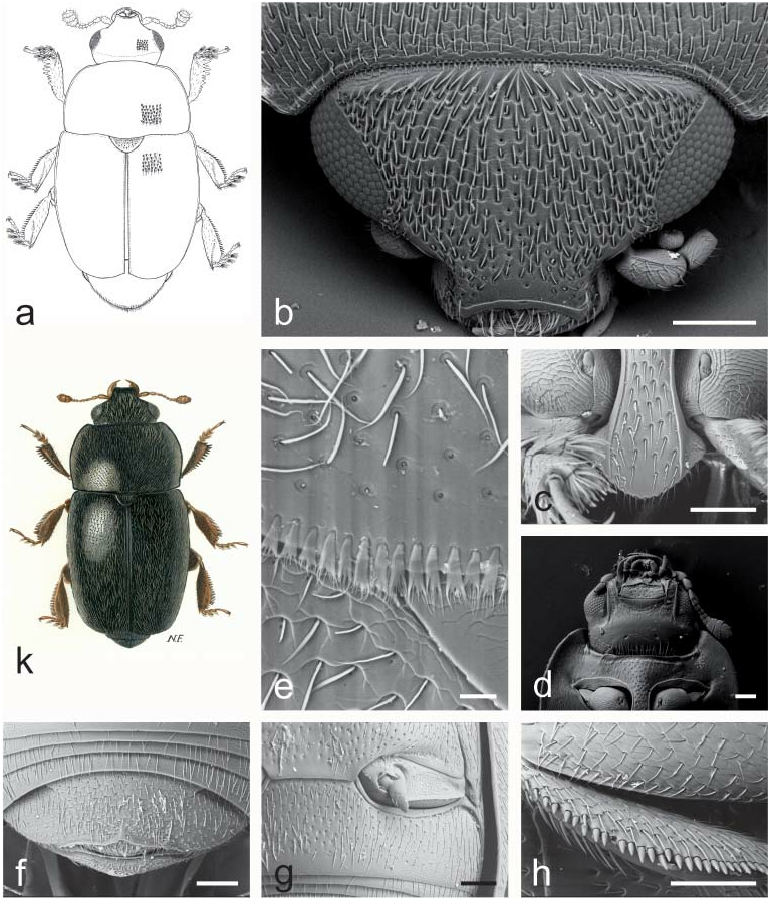
Fig. 10. Afrogethes Audisio & Cline, gen. nov.: a – A. tristis (Sturm, 1845); b–d, h – A. reticulatus (Reitter, 1872); e – A. alani (Kirejtshuk, 1988); f–g – A. planiusculus (Heer, 1841); k – A. isoplexidis (Wollaston, 1854). a, k – male habitus (a – length 2.6 mm; k – length 2.5 mm); b – dorsal view of head; c – prosternal process; d – ventral view of head and anterior portion of prosternum; e – microsetae on middle posterior margin of pronotum; f – exposed portion of last visible abdominal ventrite; g – caudal marginal lines of metacoxal cavities; h – outer margin of mesotibia. Scale bars: Figs. b, c, d, f, g, h = 100 μm; Fig. e = 20 μm.
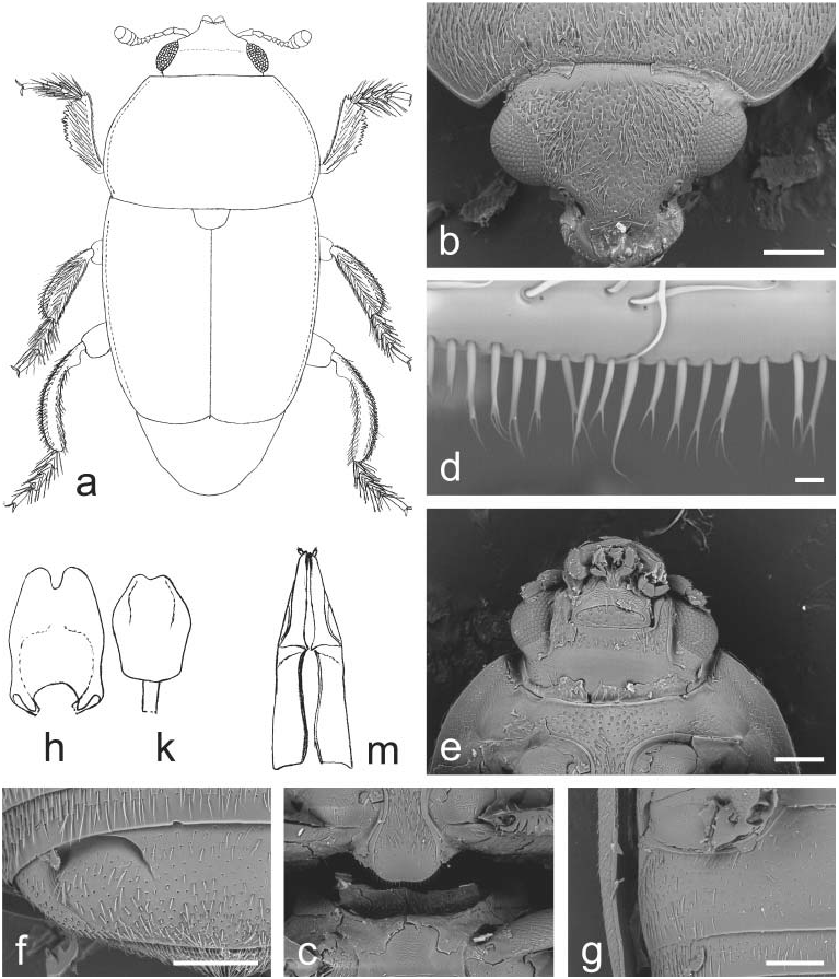
Fig. 11. Indogethes Audisio & Cline, gen. nov.: a–m – I. curvipes (Grouvelle, 1908). a – male habitus (pubescence and mandibles not illustrated; length 3.5 mm); b – dorsal view of head; c – prosternal process and mesoventrite; d – microsetae on middle of posterior margin of pronotum; e – ventral view of head and anterior portion of prosternum; f – exposed portion of last visible abdominal ventrite; g – caudal marginal line of metacoxal cavity; h–k – male genitalia (h – length 0.5 mm; k – 0.4 mm); m – ovipositor (length 0.7 mm). Scale bars: Figs. b, c, e, f, g = 200 μm; Fig. d = 10 μm.

Fig. 12. Bolbocerogethes Audisio & Cline, gen. nov.: a–e – B. pallipes (Boheman, 1851). a – male habitus (length 2.6 mm); b – ovipositor (modified from SPORNRAFT & KIREJTSHUK (1993); length 0.6 mm); c–d – male genitalia (c, d – length 0.3 mm); e – prosternal process (width 0.3 mm).

Fig. 13. Genistogethes Audisio & Cline, gen. nov.: a – G. immundus (Kraatz, 1858); b–h – G. punctatus (C. N. F. Brisout de Barneville, 1863). a – male habitus (length 2.7 mm); b – dorso-lateral view of head; c – ventral view of head and anterior portion of prosternum; d – caudal marginal line of metacoxal cavity; e – anterior portion of scutellum and microsetae on middle of posterior margin of pronotum; f – exposed portion of last visible abdominal ventrite; g – prosternal process and mesoventrite; h – middle leg with outer margin of mesotibia. Scale bars: Figs. b, c, d, f, g, h = 100 μm; Fig. e = 30 μm.
Fig. 14. Fabogethes Audisio & Cline, gen. nov.: a – F. opacus (Rosenhauer, 1856); b–h – F. nigrescens (Stephens, 1830). a – male habitus (length 2.7 mm); b – dorso-lateral view of head; c – ventral view of head and anterior portion of prosternum; d – anterior portion of scutellum and microsetae on middle of posterior margin of pronotum; e – prosternal process and mesoventrite; f – exposed portion of last visible abdominal ventrite; g – caudal marginal line of metacoxal cavity; h – middle leg with outer margin of mesotibia. Scale bars: Figs. b, c, e, g = 100 μm.

Fig. 15. Thymogethes Audisio & Cline, gen. nov.: a, h – T. egenus (Erichson, 1845); b – T. subfumatus (Ganglbauer, 1899); c–g, k – T. nigritus (Lucas, 1849). a, b – male habitus (a – length 2.5 mm; b – length 2.7 mm); c – ventral view of head and anterior portion of prosternum; d – anterior portion of scutellum and microsetae on middle of posterior margin of pronotum; e – dorsal view of head; f – prosternal process and mesoventrite; g – caudal marginal line of metacoxal cavity; h – exposed portion of last visible abdominal ventrite; k – middle leg with outer margin of mesotibia. Scale bars: Figs. c, e, f, g, h = 100 μm; Fig. d = 20 μm.
Fig. 16. Sagittogethes Audisio & Cline, gen. nov.: a – S. ater (C. N. F. Brisout de Barneville, 1863); b – S. lindbergi (Rebmann, 1940); c, f–g – S. maurus (Sturm, 1845); d–e, h–k – S. obscurus (Erichson, 1845). a, b – male habitus (a – length 2.9 mm; b – length 2.5 mm); c – dorsal view of head; d – ventral view of head and anterior portion of prosternum; e – ventral view of body; f – last tarsomere of a middle leg; g – prosternal process and mesoventrite; h – anterior portion of scutellum and microsetae on middle posterior margin of pronotum; k – middle leg with outer margin of mesotibia. Scale bars: Figs. c, g, k = 100 μm; Figs. d, e = 200 μm; Fig. f = 20 μm; Fig. h = 30 μm.
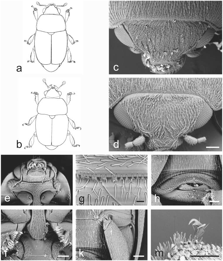
Fig. 17. Aristogethes Audisio & Cline, gen. nov.: a – A. translatus (Grouvelle, 1913); b – A. pecten (Audisio, Kirk-Spriggs & Kirejtshuk, 1998); c, e–k – A. pubescens (Reitter, 1872); d, m – A. marshalli (Grouvelle, 1914). a, b – male habitus (a – length 2.6 mm; b – length 2.4 mm); c, d – dorsal view of head; e – ventral view of head and anterior portion of prosternum; f – prosternal process and mesoventrite; g – anterior portion of scutellum and microsetae on middle of posterior margin of pronotum; h – exposed portion of last visible abdominal ventrite; k – caudal marginal line of metacoxal cavity; m – last tarsomere of middle leg. Scale bars: Figs. d, f, h, k, m = 100 μm; Fig. g = 20 μm.

Fig. 18. JelinekigethesAudisio & Cline, gen. nov.: a–e – J. danielssoni (Audisio, 1995).a – male habitus; b–c – male genitalia; d – ovipositor; e – male protibia. Figs. a–e – refer to AUDISIO (1995) for scale.

Fig. 21. Paleogethes Audisio & Cline, gen. nov.: a–k – P. wollastoni (Easton, 1950). a – male habitus (length 1.9 mm); b – dorsal view of head; c – protibia; d – ventral view of head and anterior portion of prosternum; e – middle leg with outer margin of mesotibia; f – prosternal process and mesoventrite; g – anterior portion of scutellum and microsetae on middle of posterior margin of pronotum; h – exposed portion of last visible abdominal ventrite; k – ventral view of body. Scale bars: Figs. b, d, e, f, h = 100 μm; Fig. c = 20 μm; Fig. g = 10 μm; Fig. k = 200 μm.

Fig. 22. Rubiogethes Audisio & Cline, gen. nov.: a–k – R. newtoni (Kirejtshuk, 1990). a – female habitus; b – dorsal view of head; c – ventral view of head and anterior portion of prosternum; d – scutellum and microsetae on middle of posterior margin of pronotum; e – prosternal process and mesoventrite; f – exposed portion of last visible abdominal ventrite; g – caudal marginal line of metacoxal cavity; h – middle leg with outer margin of mesotibia; k – protibia. Scale bars: Figs. a, b = 200 μm; Figs. c, d, e, f, g = 100 μm; Figs. h, k = 20 μm.
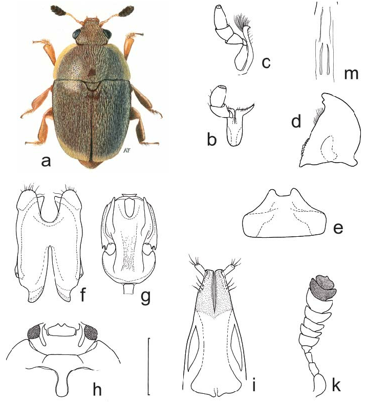
Fig. 26. Anthystrix Kirejtshuk, 1981: a, f–g, i – A.squamosa Kirejtshuk, 1981; b–e, h, k, m – A. longiclava Kirejtshuk & Easton, 1988. a – male habitus (length 2.5 mm); b – labium and right labial palpus; c – right maxilla and palpus; d – left mandible; e – labrum; f–g – male genitalia; h – ventral view of head and anterior portion of prosternum; i – distal portion of ovipositor; k – male antennal club; m – major sclerites of male endophallus. Drawings b–m: refer to AUDISIO et al. (2009a) for scale.

Fig. 27. Tarchonanthogethes Audisio & Cline, gen. nov.: a, c–i – T. rotundiclava (Kirejtshuk & Easton, 1988); b – T.sp.; k – T. martini (Grouvelle, 1899). a, b – male habitus (a – length 2.6 mm; b – length 2.0 mm); c–d – male genitalia (c – length 0. 43 mm; d – length 0.38 mm); e – major sclerites of male endophallus (length 0.20 mm); f – scutellum and microsetae on middle of posterior margin of pronotum; g – ventral view of body; h – male antenna (length 0.65 mm); i – dorsal view of head; k – distal portion of ovipositor (length 0.4 mm). Scale bars: Figs. f, i = 100 μm; Fig. g = 300 μm.

Fig. 28. Sebastiangethes Audisio, Kirk-Spriggs & Cline, 2008: a–i – S. anthystrixoides Audisio, Kirk-Spriggs & Cline, 2008. a – male habitus (length 2.7 mm); b – right maxilla and palpus; c – left mandible; d – labrum; e–f – male genitalia; g – major sclerites of male endophallus; h – distal portion of ovipositor; i – ventral view of head and anterior portion of prosternum. Drawings b–i: refer to AUDISIO et al. (2008) for scale.

Fig. 29. Xenostrongylogethes Audisio & Cline, gen. nov.: a–h – X. luculentus (Kirejtshuk & Easton, 1988). a – male habitus (length 2.5 mm); b – protibia (length 0.32 mm); c – male antenna (length 0.50 mm); d – ventral view of head and anterior portion of prosternum (pronotal width 1.22 mm); e–f – male genitalia (e – length 0.42 mm; f – length 0.47 mm); g – major sclerites of male endophallus (length 0.42 mm); h – distal portion of ovipositor (length 0.49 mm).
No known copyright restrictions apply. See Agosti, D., Egloff, W., 2009. Taxonomic information exchange and copyright: the Plazi approach. BMC Research Notes 2009, 2:53 for further explanation.
|
Kingdom |
|
|
Phylum |
|
|
Class |
|
|
Order |
|
|
Family |