
Cyclogethes Kirejtshuk, 1979
|
publication ID |
https://doi.org/10.5281/zenodo.5319334 |
|
DOI |
https://doi.org/10.5281/zenodo.10542371 |
|
persistent identifier |
https://treatment.plazi.org/id/03BE87CC-F631-FFDA-BAAB-FC00FD45FBFA |
|
treatment provided by |
Felipe (2021-08-28 07:26:47, last updated 2024-01-21 05:13:01) |
|
scientific name |
Cyclogethes Kirejtshuk, 1979 |
| status |
|
25. Cyclogethes Kirejtshuk, 1979
( Figs. 25 a–v View Fig )
Cyclogethes Kirejtshuk, 1979: 359 .
Type species. Cyclogethes orientalis Kirejtshuk, 1979: 362 (by original designation).
Generic redescription and diagnosis. Inclusive species vary moderately in size (1.7–2.4 mm length), and share the following combination of characters.
Body color and pubescence: pubescence variable, short, golden to silvery-whitish and moderately dense, recumbent, never obscuring the predominantly orange-brown to blackishbrown dorsal body surface, pronotal and elytral sides narrowly flattened and frequently paler than disc; lateral margin of pronotum and elytra with a series of faintly distinct, small and short setae, each seta 0.3–0.5× as long as those on elytral disc; posterior margin of pronotum with long, distally bifid or trifid microsetae, microsetae also present along middle portion anterior to scutellum.
Dorsal habitus: body moderately convex, oval, moderately wide ( Fig. 25a View Fig ); dorsal punctures on discal portion of pronotum as large as or larger than eye facet, moderately to deeply impressed ( Fig. 25b View Fig ); anterior margin of clypeus medially truncate, simple, i.e. without small distinct medial bulge ( Figs. 25 i, k, m View Fig ), not bordered; circum-ocular furrows (occipital sulci) on head absent; eyes large and usually markedly projecting laterally; posterior angles of pronotum distinct, blunt, obtuse, not directed posteriorly; scutellum uniformly punctured on most of exposed portion; elytra finely and completely transversely strigose ( Fig. 25c View Fig ); elytral humeral angle distinct, obtuse, never protruding laterally ( Fig. 25a View Fig ); elytral humeral striae indistinct; elytral pre-sutural striae faintly visible, originating at scutellar vertex or slightly posterior, terminating at elytral apex, and delimiting on each elytron a faintly distinct, flat, unraised sutural border, widest at posterior third, narrower than proximal width of 3 rd antennomere; elytral apices obtusely rounded in both sexes ( Fig. 25a View Fig ); pygidium partially exposed, moderately convex, apically rounded in both sexes ( Fig. 25e View Fig ).
Ventral habitus: antennal furrows markedly delimited, moderately convergent posteriorly; mentum subpentagonal ( Fig. 25t View Fig ); prosternal antennal furrows on anterior margin of prosternum distinctly delimited, faintly raised, slightly divergent, variable in length, never reaching posteriorly to the anterior margin of procoxal cavity; prosternal process variably shaped, moderately to markedly wide, subapical portion 2.0–2.5× as wide as maximum width of 1 st antennomere, apex bluntly acuminate to rounded ( Figs. 25n, o, p View Fig ); lateral borders of prosternal process delimiting shallowly impressed distinct furrows, distally terminating over predistal lateral expansions (as in Fig. 27g View Fig ); posterior margin of mesoventrite simple, never medially incised ( Fig. 25d View Fig ); scarcely evident sexual dimorphism in impressions on metaventrite; first two visible abdominal ventrites simple in both sexes, without tufts of setae, caudal marginal lines of metacoxal cavities always simple, subparallel and moderately contiguous to posterior margin of metacoxal cavities, ‘axillary’ impression nearly absent; ‘axillary’ space on first abdominal ventrite variably developed (e.g. larger in C. orientalis and allied species, more reduced in C. abnormis and allied species), ‘axillary’ angle widely obtuse in C. orientalis and allied species, nearly right angled in C. abnormis and allied species; moderately marked arched impressions on basal portion of last visible abdominal ventrite ( Fig. 25e View Fig ), occasionally partially covered by distal portion of penultimate visible abdominal ventrite.
Appendages: male 1 st antennomere 0.8–1.1× as long as width of protibiae excluding distal teeth; 3 rd antennomere 2.2–2.4× as long as wide in both sexes, distinctly longer and thinner than 2 nd; 4 th and 5 th antennomeres usually subequal in both sexes, relatively short, usually moderately longer than wide; antennal club moderately compact, nearly symmetric, comprising the last 3 antennomeres in both sexes ( Fig. 25h View Fig ; Fig. 8 View Fig in JELÍNEK 2000b), sexual dimorphism absent; labial palpi short in both sexes ( Fig. 25q View Fig ), terminal segment 1.2–1.4× as long as wide; maxillary palpi long and slender in both sexes, terminal segment ~2.0–2.2× as long as wide; mandible mid-sized, moderately short, apex bifid and moderately acuminate, sexual dimorphism absent; tarsi of normal size and shape, 0.5–0.7× as long as corresponding tibiae ( Fig. 25f View Fig ); tarsal claws variable, bluntly angulate to simple, not toothed at base; protibiae with simple and crenulate teeth on outer margins ( Fig. 25f View Fig ); meso- and metatibiae on lateral margin bearing a single and regular row of long and thin, yellowish pegs, without U-shaped sinuosity at distal third; meso- and metatibiae of variable width, abruptly dilated inwards in basal portion, subparallel-sided ( Fig. 25g View Fig ; Figs. 11 View Fig , 22 View Fig , 23 View Fig in JELÍNEK 2000b); sexual dimorphism nearly absent in tibial shape; tarsal plates of prolegs wider in males; posterior margin of metafemora simple in both sexes, without tubercles or projections.
Male genitalia: tegmen variably shaped, processes along inner side of parameres absent ( Fig. 25r View Fig ; Figs. 12 View Fig , 16 View Fig , 18 View Fig , 20 View Fig in JELÍNEK 2000b), without deep median longitudinal desclerotization from proximal portion of tegmen extending to medial distal V-shaped excision; median lobe of aedeagus variably shaped, without lateral emargination, acuminate, spatulate or emarginated distad ( Fig. 25s View Fig ; Figs. 14 View Fig , 17 View Fig , 19 View Fig , 21 View Fig in JELÍNEK 2000b).
Female genitalia (ovipositor): variably shaped, usually small; styli long and distinct, simple and usually pigmented, inserted not far from blunt apex of contiguous gonostyloids, each gonostyloid lightly sclerotized and distally pigmented, with a simple, never indentate outer portion of narrow basicoxites ( Fig. 25u View Fig ; Figs. 37 View Fig , 43 View Fig in KIREJTSHUK 1979a), single pigmented and more sclerotized arcuate area present along outer subdistal portion of gonostyloids. ‘Central point’ of ovipositor located more proximad than middle, without proximad directed spicule.
Etymology. The generic name was obviously derived from Greek ‘κύκλος’ (= circle), which is indicative of the short, wide, and markedly arcuated at sides body shape of the type species, and from ‘- gethes ’, to emphasize its phylogenetic relationship with Meligethes . Gender masculine.
Biology. Larval biology remains unknown. There is a morphological similarity of Cyclogethes (especially members of the M. abnormis species-group) with some members of southern and eastern African Tarchonanthogethes gen. nov., which is suggestive that larval development may be analogously (see below) associated with male inflorescences of arboreal Asteraceae , however this assumption is speculative and requires further fieldwork to substantiate. Adults have been collected in tropical and subtropical forest habitats, frequently on whitish flowers of Castanopsis (Fagaceae) , and on other forest trees that are attractive to beetles generally (S. Bílý, pers. comm. 2008).
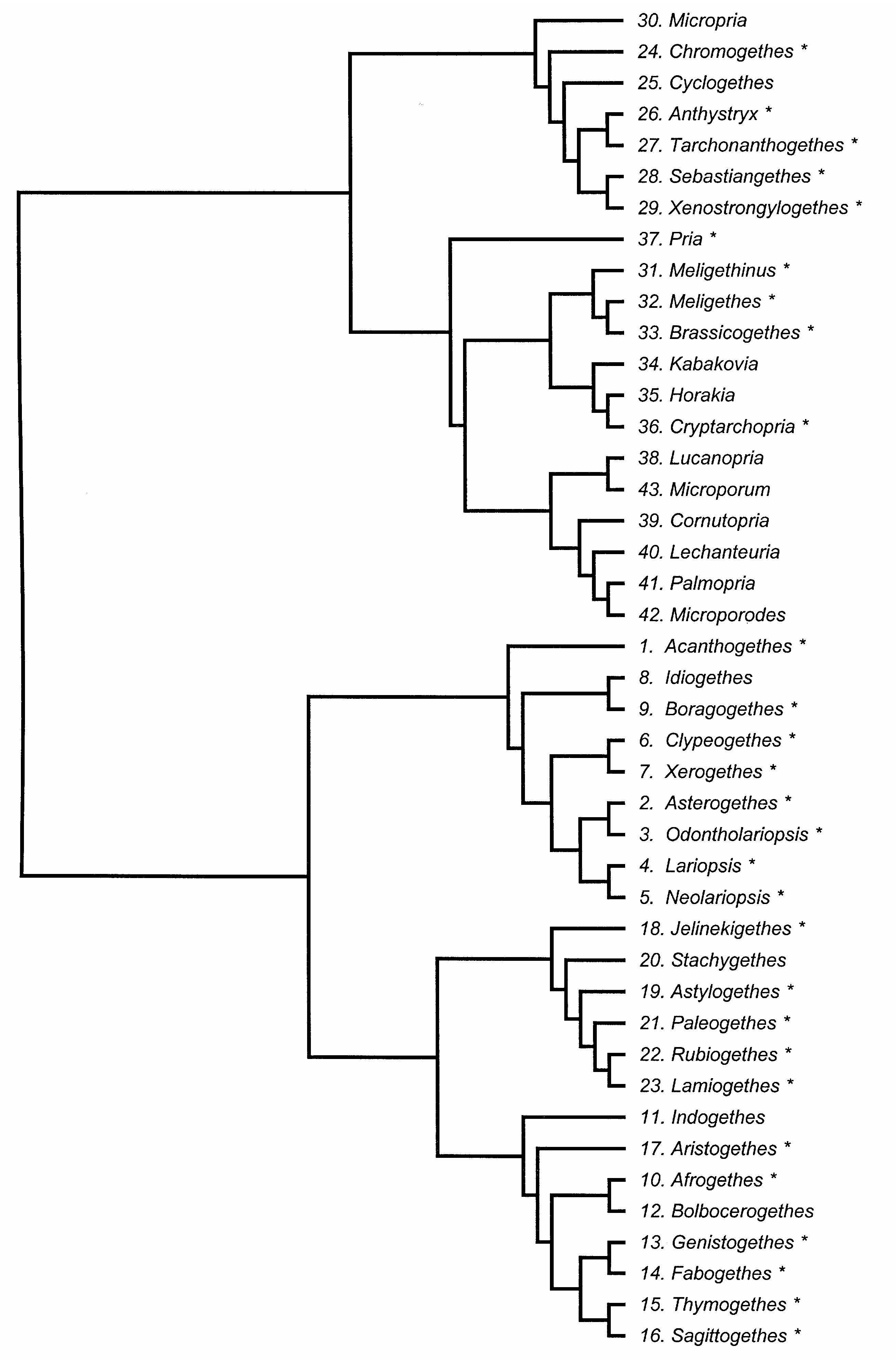
Phylogenetic position. Morphological data suggests a relatively close phylogenetic relationship of Cyclogethes with African members of the ‘ Anthystrix complex of genera’ (i.e. Anthystrix , Sebastiangethes , Tarchonanthogethes gen. nov., and Xenostrongylogethes gen. nov.; AUDISIO et al. 2008, and this paper; TRIZZINO et al. 2009), especially to some undescribed species of Tarchonanthogethes gen. nov. from southern and eastern Africa. However, discovery of larval host-plants and comparison of molecular data on members of this genus have not yet been accomplished and would undoubtedly allow for a more robustly supported phylogenetic placement of this taxon.
Taxonomy and geographic distribution. As reported by JELÍNEK (2000b), this genus is so far represented by five species manifesting a transformation series involving several characters, and are to be separated into two amply distinct groups, i.e. the ‘ orientalis ’, and ‘ abnormis ’
species-groups. Cyclogethes abnormis Kirejtshuk, 1979 Vietnam; Thailand; S China: Yunnan 3); N India: Darjeeling
Cyclogethes aldridgei Kirejtshuk, 1980 N India, Nepal, S China: Yunnan
Cyclogethes fuscipennis Jelínek, 2000 Thailand
Cyclogethes orientalis Kirejtshuk, 1979 Vietnam, Thailand
AUDISIO P., KIRK-SPRIGGS A. H., CLINE A. R., TRIZZINO M., ANTONINI G., MANCINI E. & DE BIASE A. 2008: A new genus of pollen-beetle from South Africa (Coleoptera: Nitidulidae), with discussion of the generic classification of the subfamily Meligethinae. Insect Systematics and Evolution 39: 419 - 430.
JELINEK J. 2000 b: New species of the genus Cyclogethes (Coleoptera: Nitidulidae: Meligethinae). Klapalekiana 36: 81 - 88.
KIREJTSHUK A. G. 1979 a: Dva novykh roda i novye vidy zhukov-blestyanok podsem. Meligethinae (Coleoptera, Nitidulidae) iz Vietnama. (Two new genera and new species of the subfam. Meligethinae (Coleoptera, Nitidulidae) from Vietnam). Entomologicheskoe Obozrenie 58: 355 - 368 (in Russian, English title).
TRIZZINO M., AUDISIO P., ANTONINI G., DE BIASE A. & MANCINI E. 2009: Comparative analysis of sequences and secondary structures of the rRNA internal transcribed spacer 2 (ITS 2) in pollen-beetles of the subfamily Meligethinae (Coleoptera, Nitidulidae): potential use of slippage-derived sequences in molecular systematics. Molecular Phylogenetics and Evolution 51: 215 - 226.
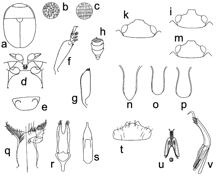
Fig. 25. Cyclogethes Kirejtshuk, 1979: a–h, k, p–v – C. orientalis Kirejtshuk, 1979; i, o – C. abnormis Kirejtshuk, 1979; m – C. fuscipennis Jelínek, 2000; n – C. aldridgei Kirejtshuk, 1980. a – male habitus; b–c – punctation of pronotum and elytra; d – prosternal process, mesoventrite, and metaventrite; e – exposed portion of last visible abdominal ventrite; f – protibia; g – mesotibia; h – antennal club; k, i, m – dorsal view of head; n, o, p – prosternal process; q – labium and left palp; r–s – dorsal view of male genitalia; t – labrum; u – ovipositor; v – lateral view of male genitalia. Drawings a–h, k, q–v from KIREJTSHUK (1979a); drawings i, m–p from JELÍNEK (2000b); refer to KIREJTSHUK (1979a) and to JELÍNEK (2000b) for scale.

Fig. 27. Tarchonanthogethes Audisio & Cline, gen. nov.: a, c–i – T. rotundiclava (Kirejtshuk & Easton, 1988); b – T.sp.; k – T. martini (Grouvelle, 1899). a, b – male habitus (a – length 2.6 mm; b – length 2.0 mm); c–d – male genitalia (c – length 0. 43 mm; d – length 0.38 mm); e – major sclerites of male endophallus (length 0.20 mm); f – scutellum and microsetae on middle of posterior margin of pronotum; g – ventral view of body; h – male antenna (length 0.65 mm); i – dorsal view of head; k – distal portion of ovipositor (length 0.4 mm). Scale bars: Figs. f, i = 100 μm; Fig. g = 300 μm.

Fig. 8. Idiogethes Kirejtshuk, 1977: a–e – I. angustitarsus Kirejtshuk, 1977. a – male habitus (length 2.2 mm); b – dorsal view of head; c – antenna; d – anterior leg; e – mesotibia. Figs. b–e: refer to KIREJTSHUK (1977a) for scale.
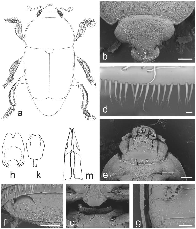
Fig. 11. Indogethes Audisio & Cline, gen. nov.: a–m – I. curvipes (Grouvelle, 1908). a – male habitus (pubescence and mandibles not illustrated; length 3.5 mm); b – dorsal view of head; c – prosternal process and mesoventrite; d – microsetae on middle of posterior margin of pronotum; e – ventral view of head and anterior portion of prosternum; f – exposed portion of last visible abdominal ventrite; g – caudal marginal line of metacoxal cavity; h–k – male genitalia (h – length 0.5 mm; k – 0.4 mm); m – ovipositor (length 0.7 mm). Scale bars: Figs. b, c, e, f, g = 200 μm; Fig. d = 10 μm.

Fig. 22. Rubiogethes Audisio & Cline, gen. nov.: a–k – R. newtoni (Kirejtshuk, 1990). a – female habitus; b – dorsal view of head; c – ventral view of head and anterior portion of prosternum; d – scutellum and microsetae on middle of posterior margin of pronotum; e – prosternal process and mesoventrite; f – exposed portion of last visible abdominal ventrite; g – caudal marginal line of metacoxal cavity; h – middle leg with outer margin of mesotibia; k – protibia. Scale bars: Figs. a, b = 200 μm; Figs. c, d, e, f, g = 100 μm; Figs. h, k = 20 μm.

Fig. 23. Lamiogethes Audisio & Cline, gen. nov.: a – L. paschalis (Spornraft, 1975); b – L. convexus (Boheman, 1851); c–e, g–h, m – L. ruficollis (Reitter, 1872); k, f, n – L. difficilis (Heer, 1841). a, b – male habitus (a – length 3.0 mm; b – length 2.7 mm); c – ventral view of head and of anterior portion of prosternum; d – dorsal view of head; e–f – scutellum and microsetae on middle of posterior margin of pronotum; g – ventral view of body; h–k – prosternal process and mesoventrite; m – last tarsomere of middle leg; n – outer margin of mesotibia. Scale bars: Figs. c, d, e, f, h, n = 100 μm; Fig. g = 1 mm; Fig. m = 20 μm.

Fig. 12. Bolbocerogethes Audisio & Cline, gen. nov.: a–e – B. pallipes (Boheman, 1851). a – male habitus (length 2.6 mm); b – ovipositor (modified from SPORNRAFT & KIREJTSHUK (1993); length 0.6 mm); c–d – male genitalia (c, d – length 0.3 mm); e – prosternal process (width 0.3 mm).
Fig. 16. Sagittogethes Audisio & Cline, gen. nov.: a – S. ater (C. N. F. Brisout de Barneville, 1863); b – S. lindbergi (Rebmann, 1940); c, f–g – S. maurus (Sturm, 1845); d–e, h–k – S. obscurus (Erichson, 1845). a, b – male habitus (a – length 2.9 mm; b – length 2.5 mm); c – dorsal view of head; d – ventral view of head and anterior portion of prosternum; e – ventral view of body; f – last tarsomere of a middle leg; g – prosternal process and mesoventrite; h – anterior portion of scutellum and microsetae on middle posterior margin of pronotum; k – middle leg with outer margin of mesotibia. Scale bars: Figs. c, g, k = 100 μm; Figs. d, e = 200 μm; Fig. f = 20 μm; Fig. h = 30 μm.

Fig. 18. JelinekigethesAudisio & Cline, gen. nov.: a–e – J. danielssoni (Audisio, 1995).a – male habitus; b–c – male genitalia; d – ovipositor; e – male protibia. Figs. a–e – refer to AUDISIO (1995) for scale.

Fig. 20. Stachygethes Audisio & Cline, gen. nov.: a, c, f, k – S. ruficornis (Marsham, 1802); b – S. variolosus (Easton, 1964); d, e, g, h – S. assimilis (Sturm, 1845). a, b – male habitus (a – length 2.5 mm; b – length 2.5 mm); c – dorsal view of head; d – ventral view of head and anterior portion of prosternum; e – scutellum and microsetae on middle of posterior margin of pronotum; f – caudal marginal line of metacoxal cavity; g – prosternal process and mesoventrite; h – exposed portion of last visible abdominal ventrite; k – outer margin of mesotibia. Scale bars: Figs. d, f, g, h = 100 μm; Fig. e = 30 μm; Fig. k = 20 μm.
Fig. 14. Fabogethes Audisio & Cline, gen. nov.: a – F. opacus (Rosenhauer, 1856); b–h – F. nigrescens (Stephens, 1830). a – male habitus (length 2.7 mm); b – dorso-lateral view of head; c – ventral view of head and anterior portion of prosternum; d – anterior portion of scutellum and microsetae on middle of posterior margin of pronotum; e – prosternal process and mesoventrite; f – exposed portion of last visible abdominal ventrite; g – caudal marginal line of metacoxal cavity; h – middle leg with outer margin of mesotibia. Scale bars: Figs. b, c, e, g = 100 μm.
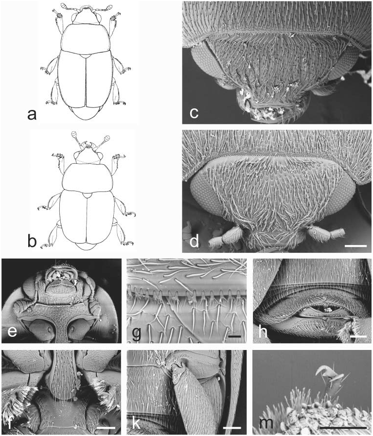
Fig. 17. Aristogethes Audisio & Cline, gen. nov.: a – A. translatus (Grouvelle, 1913); b – A. pecten (Audisio, Kirk-Spriggs & Kirejtshuk, 1998); c, e–k – A. pubescens (Reitter, 1872); d, m – A. marshalli (Grouvelle, 1914). a, b – male habitus (a – length 2.6 mm; b – length 2.4 mm); c, d – dorsal view of head; e – ventral view of head and anterior portion of prosternum; f – prosternal process and mesoventrite; g – anterior portion of scutellum and microsetae on middle of posterior margin of pronotum; h – exposed portion of last visible abdominal ventrite; k – caudal marginal line of metacoxal cavity; m – last tarsomere of middle leg. Scale bars: Figs. d, f, h, k, m = 100 μm; Fig. g = 20 μm.
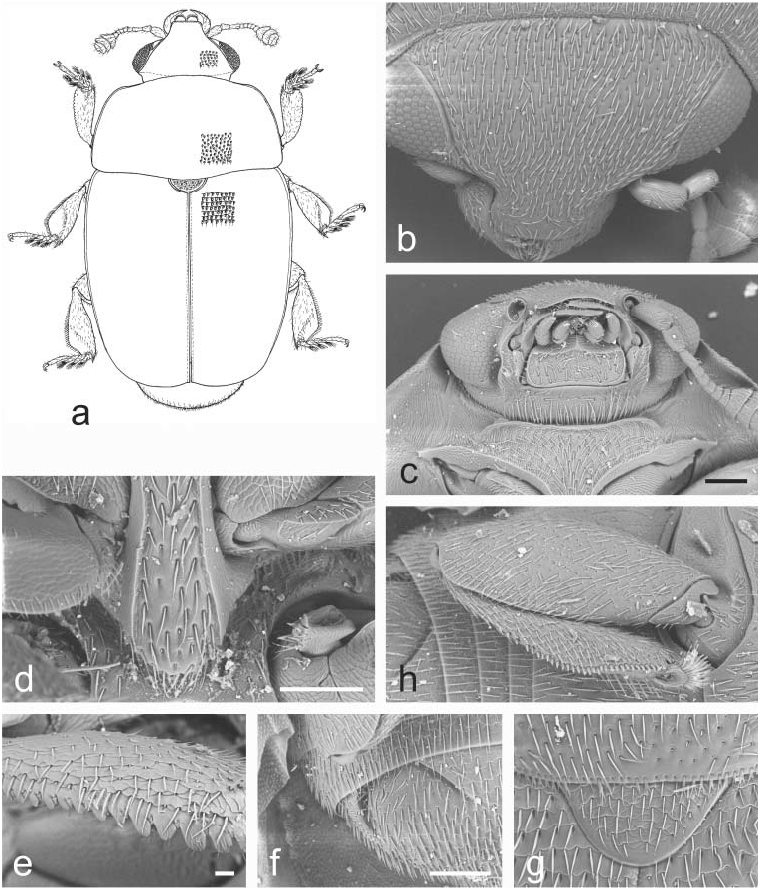
Fig. 19. Astylogethes Kirejtshuk, 1992: a–h – A. subrugosus (Gyllenhal, 1808). a – male habitus (length 2.4 mm); b – dorsal view of head; c – ventral view of head and anterior portion of prosternum; d – prosternal process and mesoventrite; e – outer margin of protibia; f – exposed portion of last visible abdominal ventrite; g – scutellum and microsetae on middle of posterior margin of pronotum; h – caudal marginal line of metacoxal cavity. Scale bars: Figs. c, d, f = 100 μm; Fig. e = 20 μm.

Fig. 21. Paleogethes Audisio & Cline, gen. nov.: a–k – P. wollastoni (Easton, 1950). a – male habitus (length 1.9 mm); b – dorsal view of head; c – protibia; d – ventral view of head and anterior portion of prosternum; e – middle leg with outer margin of mesotibia; f – prosternal process and mesoventrite; g – anterior portion of scutellum and microsetae on middle of posterior margin of pronotum; h – exposed portion of last visible abdominal ventrite; k – ventral view of body. Scale bars: Figs. b, d, e, f, h = 100 μm; Fig. c = 20 μm; Fig. g = 10 μm; Fig. k = 200 μm.
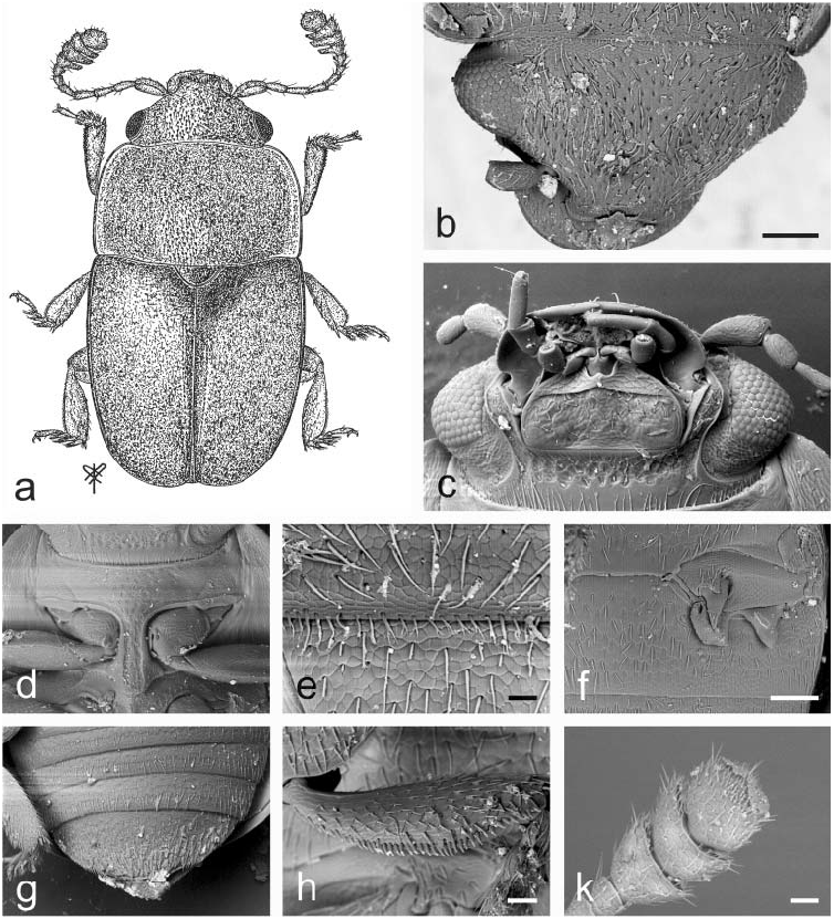
Fig. 37. Pria Stephens, 1830: a–k – P. dulcamarae (Scopoli, 1763).a – male habitus (length 2.0 mm); b – dorsal view of head; c – ventral view of head; d – ventral view of prosternum; e – anterior portion of scutellum and microsetae on middle posterior margin of pronotum; f – caudal marginal line of metacoxal cavity; g – exposed portion of last ventral visible abdominal ventrite; h – middle tibia; k – female antennal club. Scale bars: Figs. b, f = 100 μm; Fig. e = 20 μm; Figs. h, k = 30 μm.
No known copyright restrictions apply. See Agosti, D., Egloff, W., 2009. Taxonomic information exchange and copyright: the Plazi approach. BMC Research Notes 2009, 2:53 for further explanation.
|
Kingdom |
|
|
Phylum |
|
|
Class |
|
|
Order |
|
|
Family |
Cyclogethes Kirejtshuk, 1979
| Audisio, Paolo, Cline, Andrew Richard, Biase, Alessio De, Antonini, Gloria, Mancini, Emiliano, Trizzino, Marco, Costantini, Lorenzo, Strika, Sirio, Lamanna, Francesco & Cerretti, Pierfilippo 2009 |
Cyclogethes
| Kirejtshuk 1979: 359 |