
Crurifarcimen vagans, Enghoff, Henrik, 2011
|
publication ID |
https://doi.org/ 10.5281/zenodo.276689 |
|
DOI |
https://doi.org/10.5281/zenodo.5612652 |
|
persistent identifier |
https://treatment.plazi.org/id/03BC87EA-FFAA-316F-FF22-F922031BFCAE |
|
treatment provided by |
Plazi |
|
scientific name |
Crurifarcimen vagans |
| status |
sp. nov. |
Crurifarcimen vagans View in CoL n. sp.
Figs 1–5, 8 View FIGURE 1 View FIGURES 2 – 3 View FIGURES 4 – 8 , 11 View FIGURES 11 – 12 , 13–27 View FIGURES 13 – 16 View FIGURES 17 – 18 View FIGURE 19 View FIGURES 20 – 23 View FIGURES 24 – 25 View FIGURES 26 – 27 , 84 View FIGURE 84
Pachybolus n. sp. II. Enghoff & Enghoff 1976
HOLOTYPE: 3 TANZANIA, Tanga Region, E. Usambara Mts., Amani 5°06’S 38°38’E, forest clearing at Kwankoro road, ca. 1000 m, on the ground, 1.viii.1974, I. B. Enghoff & H. Enghoff leg. ( ZMUC 00100972).
PARATYPES from TANZANIA, Tanga Region, East Usambara Mts., Amani 5°06’S 38°38’E: 1 Ƥ, 2 juveniles, data as holotype ( ZMUC 00100973- 100974). – 3 3, 1 juvenile data as holotype, but 2. – 7.viii.1974 ( ZMUC 0 0 100982, 0 0 100983, 00100975). – 1 Ƥ, forest house, secondary rainforest, ca. 1000 m, in rotten wood, 29.vii.1974, I. B. Enghoff & H. Enghoff leg. ( ZMUC 00100978). – 1 3, 1 Ƥ, Bomole, ca. 1000 m, 5.viii.1974, I. B. Enghoff & H. Enghoff leg. ( ZMUC 00100979-00100980). – 1 3 TANZANIA, Monga, ca. 1000 m, 6.ii.1977, H. Enghoff, O. Lomholdt, O. Martin leg. ( ZMUC 00100984). – 1 3, 2 ƤƤ, ca. 1000 m, 26.i. –1.ii.1977, H. Enghoff, O. Lomholdt, O. Martin leg. ( ZMUC 0 0 100985, 00100987). – 1 3 1 Ƥ, Kwamkoro, 25.i.1977, H. Enghoff, O. Lomholdt, O. Martin leg. ( ZMUC 00100992). - 2 ƤƤ, 3 juveniles, ca. 1000 m. 1–5.viii.1979, M. Stoltze leg. ( ZMUC 0 0 100988, 00100990). – 1 3 ca. 1000 m, 24.vi.1970, R. Jacobsen leg. ( ZMUC 00100991). – 7 3, 1 Ƥ, 1 subad. 3 TANZANIA, ca. 1000 m, 27.x. – 9.xi. 1995, C. Griswold, N. Scharff & D. Ubick leg. ( CAS).
PARATYPES from TANZANIA, Tanga Region, West Usambara Mts., Mazumbai, 4°49’S 38°30’E: 1 3, 3 juv. 1600 m, in trunk, 1.viii.1980, M. Stoltze & N. Scharff leg. (ZMUC 00100995). – 1 3, 1550 m, 15.xi. 1974, I. Jakobsen leg. (ZMUC 00100996). – 2 ƤƤ, 1600 m, 7.xii.1978, J.B. Rasmussen leg. (ZMUC 00100997). – 1 3, 2 ƤƤ, montane forest, 1600– 1800 m, 10. – 20.xi.1995, C. Griswold, N. Scharff & D. Ubick leg. (CAS).
ADDITIONAL MATERIAL from TANZANIA, Tanga Region, East Usambara Mts., Amani 5°06’S 38°38’E: 3 juv. Monga, ca. 1000 m, on the ground, 5.viii.1974, I. B. Enghoff & H. Enghoff leg. (ZMUC 00100976). – 1 subadult Ƥ, ca. 1000m, arable land, 6.viii.1974, I. B. Enghoff & H. Enghoff leg. (ZMUC 00100977). – 1 Ƥ, 9 juveniles, cultivation in rain forest, ca. 1000 m, on the ground, 26.vii.1974, I. B. Enghoff & H. Enghoff leg. (ZMUC 00100981). – 2 eggs, 1 small juv., lab-reared from parents from Amani (ZMUC 00100993). – 3 juveniles ca. 1000 m, 24.i.1977, in rotten wood, H. Enghoff, O. Lomholdt, O. Martin leg. (ZMUC 00100986). – 1 juvenile, ca. 1000 m. 14.vii.1979, M. Stoltze leg. (ZMUC 00100989).
ADDITIONAL MATERIAL from TANZANIA, Tanga Region, East Usambara Mts: 5 3, 1 Ƥ, Muheza + Korogwe District, East Usambara Mts. 4°56’32”S 38°39’38.5’’E, 940 masl, trapsite 8 plot 59A, 31.i.2001, Frontier Tanzania leg. (ZMUC 00101001). – 1 Ƥ, 1 juvenile Nilo Forest Reserve 4°54’16’’S, 38°39’45’’E, date?, Frontier Tanzania leg. (ZMUC 00101000). – 1 3, 1 Ƥ, 1 subad. Ƥ Muheza District, Magrotto Estate, 5°07 S, 38°45’ E, 2000–3000 ft asl, iii.1981, S.N.Stuart leg. (VMNH.)
ADDITIONAL MATERIAL from TANZANIA, Tanga Region, Lushoto District, West Usambara Mts., Mazumbai, 4°49’S 38°30’E: 16 juveniles 1600 m, in trunk, 1.viii.1980, M. Stoltze & N. Scharff leg. (ZMUC 00100998). – 3 juveniles TANZANIA, 19. – 29.ix.1992, M. Andersen leg. (ZMUC 00100999). – 1 3, 17.vi.1980, S. Mandia leg. (VMNH). – 22 3, 10 ƤƤ, 2 juveniles, S. Mandia leg. (VMNH). – 3 3, 1 Ψ, i.1981, K. M. Howell leg. (VMNH).
ADDITIONAL MATERIAL from TANZANIA, Tanga Region, West Usambara Mts: 1 3, 1 subadult 3, Lushoto District, Mahezangulu Forest Reserve 4° 57' S, 38° 31' E, rain forest, 3000–3500 ft asl, 14–16.iii.1982, S.N. Stuart leg (VMNH). – 5 3 Korogwe District, Ambangulu Estate Rain Forest, 5° 4'51’’ S, 38° 25'54’’ E, 2– 11.iv.1980, S.N. Stuart & J. Simkin leg. (VMNH). –– 3 3 Lushoto District, Balangai Forest Reserve, 4° 55'55’’ S, 38° 37'07’’ E, 11– 13.iii.1981, S.N. Stuart leg. (VMNH).
Etymology. Latin vagans (wandering, itinerant). The full species name thus means “the itinerant sausage with feet”, and this species is indeed the most sausagelike millipede I have seen.
Diagnosis. See that of genus.
Description. Adults with 56 podous rings (exceptionally 54, 55 or 57), no apodous rings. Length 14–16 cm. Width of specimens from East Usambara Mts.: 14.2–16.2 mm (3) / 14.4–16.1 mm (ƤƤ). Width of specimens from West Usambara Mts.: 13.5–14.2 mm (3) / 14.9–15.4 (ƤƤ).
Colour: overall impression light brown, occasionally darker brown; head, collum, antennae, legs and telson brownish yellow; legs occasionally dark caramel; prozona of body rings yellow, mesozona greyish brown, metazona reddish brown ( Fig. 1 View FIGURE 1 ).
Head capsule ( Fig. 2 View FIGURES 2 – 3 ) punctate above eye level, otherwise smooth; occipital furrow extending down between but not beyond eyes; clypeal furrow reaching level of antennal sockets. Area below antennal socket and eyes impressed, forming part of antennal furrow. 2+2 or 3+3 low, broad labral teeth. A row of labral setae present or missing (abraded?). Supralabral setae absent in adults (abraded?), one subadult Ƥ with 1+1 tiny setae. Eyes small, largest diameter 43–47% of interocular space in frontal view; ocelli usually arranged in neat rows, adults with 9–10 RO (see chapter on postembryonic development), 5 horizontal series, 30–39 ocelli per eye. Antennae short, not reaching past collum when stretched back, accommodated in shallow furrow composed of a horizontal segment in the head capsule and a vertical segment in the mandibular cardo and stipes, lateral margins of head capsule also shallowly concave adjacent to mandibular segment of antennal furrow. Antennomeres 1, 3 and 4 as long as broad, 2 longer, 5 and 6 broader, 7 very short, almost contained within subspherical antennomere 6; antennomeres 1–2 glabrous, 3–4 with a few ventral setae, 5–6 densely setose and each with a dorsodistal field of small sensilla; four apical sensilla partly separated by outgrowths from the wall of antennomere 7. Mandibles in part visible in frontal view; cardo and stipes excavated for accomodation of antenna. Cardo with vertical ridge ca. 1/3 from anterior margin. Stipes ventrally excavated in mesal half for accommodation of gnathochilarium. Gnathal lobe (terminology of Enghoff 1979): external tooth with single, subrectangular lobe; internal tooth with four shallow cusps plus an isolated, triangular cusp dorsally; Four pectinate lamellae, plus a short fifth row and a few isolated teeth basal to fifth row; a narrow strip ("fringe") of small spines ventral to pectinate lamellae; a soft, smooth-edged ridge between pectinate lamellae and mola, ridge dorsally with a row of small spines; molar plate with six transverse ridges decreasing in size towards base. Gnathochilarium similar to those of Pelmatojulus tigrinus Hoffman & Mahsberg, 1996 , and Madabolus maximus Wesener et al., 2008 . Mentum transversely wrinkled. Stipites wrinkled parallel to mento-stipital suture, each with 3–5 distal setae. Lamellae linguales each with 3–6 setae arranged in a transverse, distal row or group.
Collum ( Fig. 2 View FIGURES 2 – 3 ) entirely smooth, or with faint short striae along lowermost part of anterior margin, somtimes a weak marginal furrow along lateral parts of anterior margin; lateral lobes rounded/triangular, not extending as far ventrad as body ring 2.
Body rings two to five/six ventrally concave, hence with distinct ventrolateral "corners". Body rings very smooth, dorsal profile linear. "Tergo-pleural" suture visible on pro- and mesozona. Prozona with very fine chagrination. Mesozona ventrally with fine oblique striae, gradually changing into fine punctation laterally and dorsally. Metazona ventrally with fine longitudinal striae, otherwise smooth in 3, finely punctate to coriaceous in ƤƤ. "Pleural" parts of rings with fine oblique striae. Sterna transversely striate. Ozopores from ring 6, situated in mesozona, ca. 1½ pore diameter in front of metazona. Sutures between pro-, meso- and metazona fine but distinct; horizontal suture sometimes distinct on pro- and mesozona, less so on metazona. Posterior margin of body rings with numerous very short longitudinal impressions, giving a crenulate impression; margin itself straight, however; no limbus.
Telson very shallowly punctate, appearing smooth. Preanal ring with slightly concave dorsal profile. Anal valves slightly impressed submarginally; margins hence slightly protruding, liplike. Subanal scale broadly triangular.
Legs: Length of midbody legs 62% of body diameter in 3, 51% of body width in Ƥ. Coxa dorsally angled; prefemur basally constricted; tarsus longer than other podomeres. First and second legs with numerous (35–40) ventral setae, third and following pairs with much fewer setae, numbers of setae reaching constancy from pair 5 or 6, each leg with 1 coxal, 1–2 prefemoral, 1–2 femoral, 2 postfemoral, and 2 tibial setae. Tarsi in 3 with 2 ventral apical and one dorsal apical seta ( Figs 4–5 View FIGURES 4 – 8 ), ƤƤ with 2+2 ventral and one dorsal apical seta, the basal ventral pair smaller than the others.
Male sexual characters. Body more shining than in females, due to absence of punctation on metazona. Tarsus of all legs from third to last pair with large ventral soft pad occupying entire ventral surface ( Figs 4–5 View FIGURES 4 – 8 ). Coxae of fifth and sixth pair of legs with anterior, rounded lobes, slightly larger on fifth ( Fig. 3 View FIGURES 2 – 3 ). Body ring seven ( Fig. 3 View FIGURES 2 – 3 ) entirely fused ventrally, no trace of a suture. Anterior part ventrally clearly delimited as smooth "shelf", contrasting with sculptured areas posteriorly and laterally. Gonopods ( Figs 13–23 View FIGURES 13 – 16 View FIGURES 17 – 18 View FIGURE 19 View FIGURES 20 – 23 ) entirely concealed within body ring seven at rest.
Anterior gonopod sternum concave on oral face, with high, broad, bell-shaped mesal process reaching ca. 3/4 the height of the coxae; right and left halves of process meeting under blunt angle in midline, sometimes forming a midline ridge; tip of process more or less pointed; sternum laterally continuing as narrow band around side of gonopod, reaching well along base of coxa on aboral side, articulating with a narrow sclerite at base of telopodite, the "bride trachéenne” of Demange (1967) ( Fig. 19 View FIGURE 19 ). Coxae in oral view with apical margins oblique, set off from parallel lateral margins by blunt angle somewhat apical to mid-height of coxa; opposing coxae meeting in midline distal to sternum; here with small tooth (cxt on Figs 17, 18 View FIGURES 17 – 18 ) projecting basad-mesad over tip of sternum, sometimes with an impression delimited by a small vertical ridge just distal of cxt; oral surface of coxa slightly concave, i.e., due to rounded lateral ridge occupying ca. half of coxal width; aboral surface of coxa (hidden from view in undissected specimens) broadly swollen mesally, swelling apically connecting with basad-mesad tooth on oral surface. Coxae extending to aboral side of gonopod where they appear as narrow lateral bands. Telopodites ( Figs 14–19 View FIGURES 13 – 16 View FIGURES 17 – 18 View FIGURE 19 ) extending well beyond coxal tips, overall shape ovoid; in situ meeting in midline; posterior face of telopodite with rectangular elevated area, the basal-mesal corner of which meets that of opposite telopodite; opposite corners usually slightly asymmetrical (one telopodite forming short process fitting into concavity on the other); lateral margin of telopodite with sub-semicircular incision, delimiting a subapical lateral rounded-triangular projection.
Posterior gonopods ( Figs 13–14 View FIGURES 13 – 16 , 20–23 View FIGURES 20 – 23 ) in situ completely hidden within anterior ones, connected by median sternal remnant consisting of mesal membranous region flanked by a pair of rounded-triangular sclerites ( Fig. 14 View FIGURES 13 – 16 ). Gonopods oriented with larger surfaces facing aboral-lateral and oral-mesal, respectively, for simplicity these faces are termed aboral and oral below. Each posterior gonopod clearly divisible into four parts:
1. a long, slender, almost straight tracheal apodeme ( Figs 15–16 View FIGURES 13 – 16 , 20 View FIGURES 20 – 23 ), flattened towards its end,
2. a basal, aboral sclerite ( Fig. 21 View FIGURES 20 – 23 ) which articulates with the tracheal apodeme, is roughly pentagonal in outline and has a distinct notch on its distal margin, next to articulation with median sternal sclerite,
3. a smooth, stout, sausagelike intermediate segment delimited against terminal segment by very distinct articulation across lateral margin ( Fig. 20 View FIGURES 20 – 23 , art), by membranes on oral and aboral faces, membrane most extensive on latter face,
4. a complicated terminal segment with smoothly convex lateral margin, this margin with longitudinal concavity next to basal articulation against intermediate segment. Basal part of terminal segment roughly quadrate in outline, extensively covered by membrane on lateral face. Distal part of terminal segment abruptly set off by rectangular notch. Aboral face of terminal segment with longitudinal groove parallel to lateral margin; distalmost part of groove covered by large irregular, ±triangular process; this process forming part of slightly concave apical-mesal margin of gonopod, corners of concavity blunt, slightly protruding. Apical-mesal margin overreached by conspicuous solenomerite emerging from oral face of gonopod, lamellate, longitudinally striate, sinuous, attenuated, ending as curved tip pointing in a mesal-basal direction. Efferent groove only visible on terminal segment of gonopod, running along mesal edge of broad basal part, moving to lateral surface and there running subparallel to distal margin of basal part, further along mesal edge of distal part (where accompanied by narrow, transparent, striate membrane) and onto solenomerite where it ends as an open furrow near the tip.
Female sexual characters. Distal margin of lateral coxosternal extensions projecting as right-angled lobe
( Fig. 11 View FIGURES 11 – 12 ). Vulva ( Figs 11 View FIGURES 11 – 12 , 24–25 View FIGURES 24 – 25 ) in posterior view sausage-shaped. Valves very elongated, slightly curved (their bases
concave), quite asymmetrical and variable in details, margins lobed except distally, lobes interlocked, basally in a
coarse zig-zag pattern. No apical appendix. Crest not protruding.
Postembryonic development. Eggs and first stadium juveniles were obtained in culture (offspring of a female
collected in Amani, E Usambaras, in 1974). Stadia II-X were collected in the field (cumulative material from E and
W Usambaras, 1970–1995). The stadium numbers can be read by counting rows of ocelli (RO). Stadium I has one
row, II has two etc. (cf. Enghoff et al. 1993).
The eggs are placed individually in spherical earthen capsules manufactured by the female ( Fig. 26 View FIGURES 26 – 27 ), as in Epi-
bolus pulchripes ( Dhaenens & VandenSpiegel 2006). The stadium I juveniles ( Fig. 27 View FIGURES 26 – 27 ) remain inside the capsules.
The body ring formulae of the studied specimens are shown in Table 2 View TABLE 2 . Out of 91 specimens, 82 follow the princi-
pal pathway. Whereas not observed formulae can be inferred with some confidence from the preceding stadium
(e.g., 54+3+T in stadium VIII can be inferred from 49+5+T in stadium VII), it is more difficult to infer a not
observed formula preceding an observed one, e.g., 49+5+T in stadium VII could have resulted from 43+6+T or
42+7+T in stadium VI. The course of anamorphosis in C. vagans is almost identical to that observed in two species
of the West African pachybolinine genus Pelmatojulus ( Enghoff et al. 1993) , whereas in Epibolus pulchripes , studied by Dhaenens & VandenSpiegel (2006), the numbers of podous rings in each stadium are somewhat smaller, corresponding with the generally lower number of podous rings (50–53) in adults.
Sexes are separable from stadium IV inclusive. The observed adults belong to stadia IX and X. Immature specimens were observed belonging to both stadium IX and X. However, several stadium X specimens had the tenth row of ocelli represented only by a single ocellus, sometimes even only on one side of the head. Thus, some specimens assigned to stadium IX may in fact belong to stadium X. Likewise, the hypothetical stadium XI may be "cryptically" represented in the sample. Immatures were consistenly slenderer than adults in the same stadium, see Table 3 View TABLE 3 .
Habitat. Montane forest, most often found in decaying wood.
Distribution. E and W Usambara Mts., from 940 to 1600– 1800 m altitude ( Fig. 84 View FIGURE 84 ).
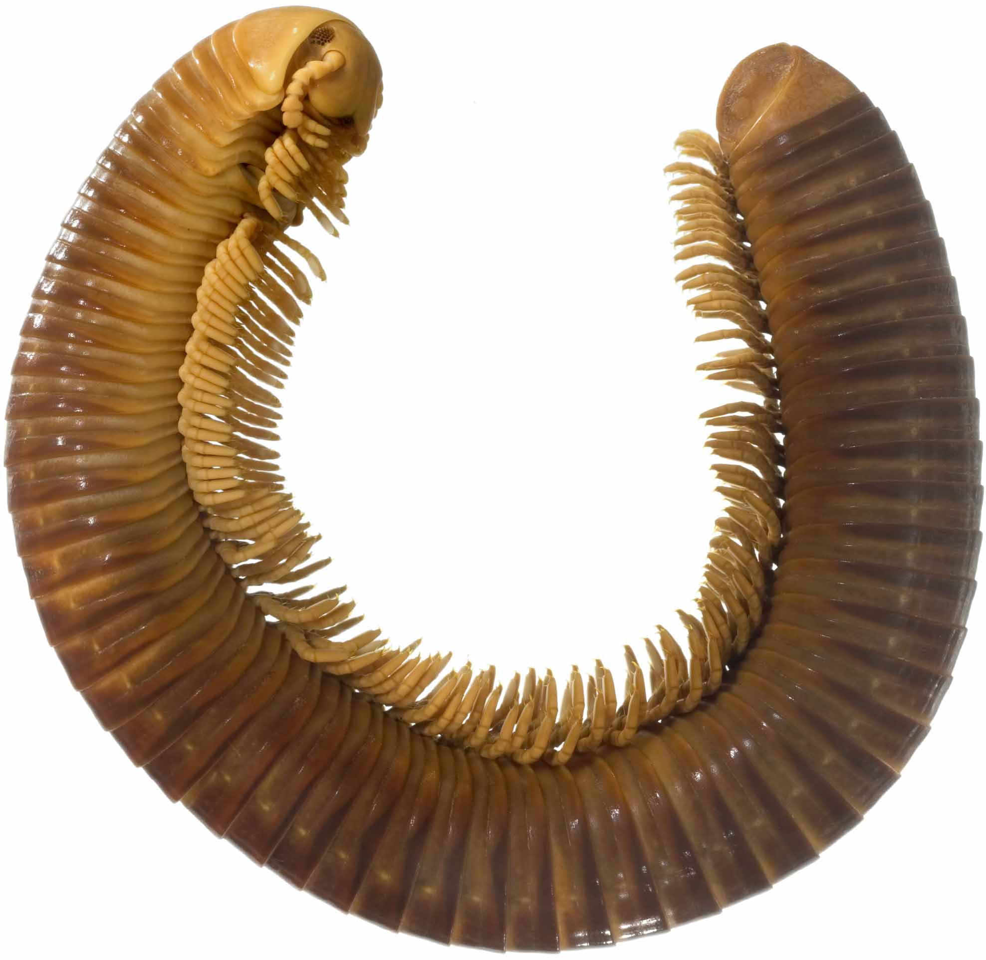
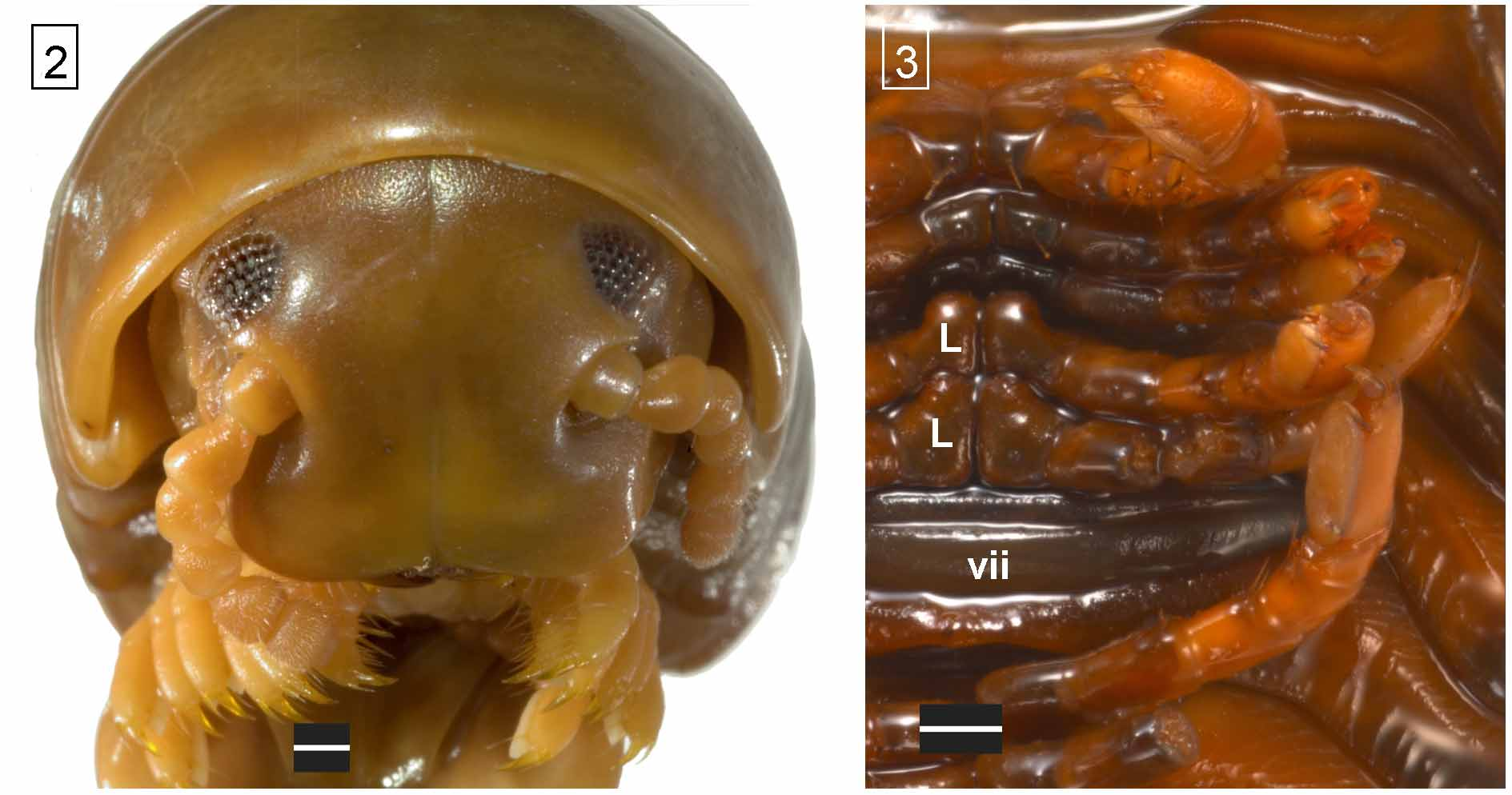
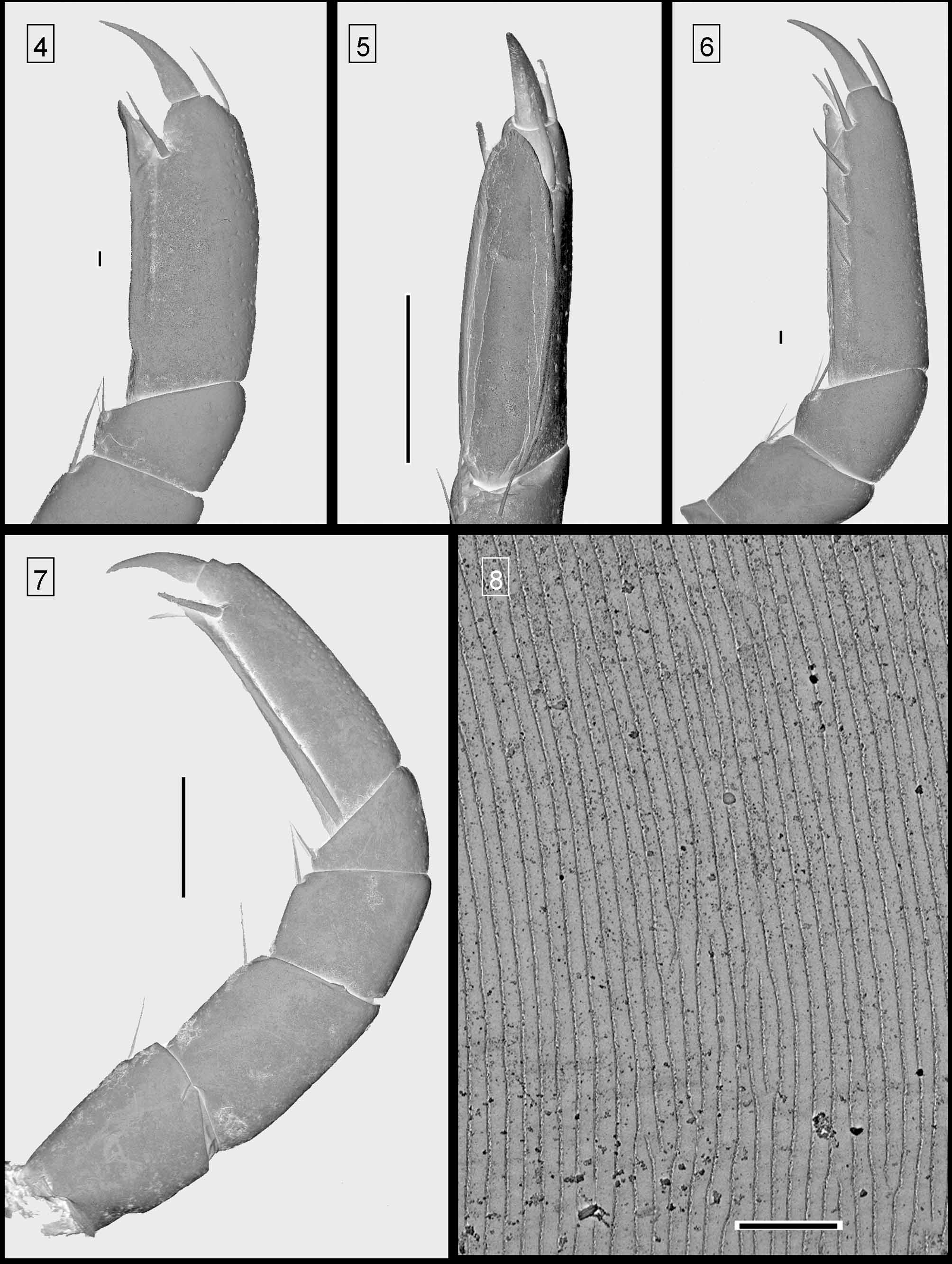
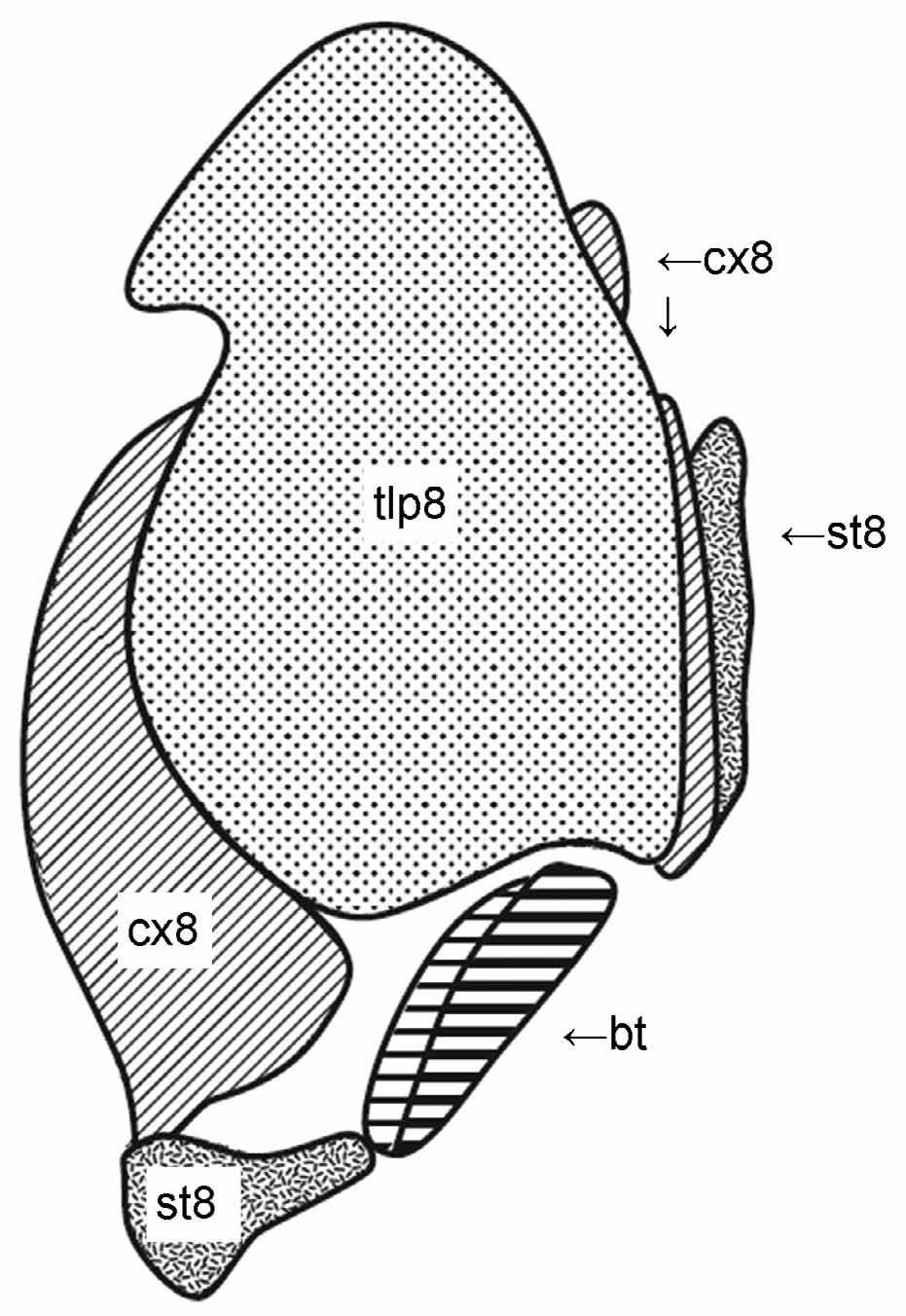
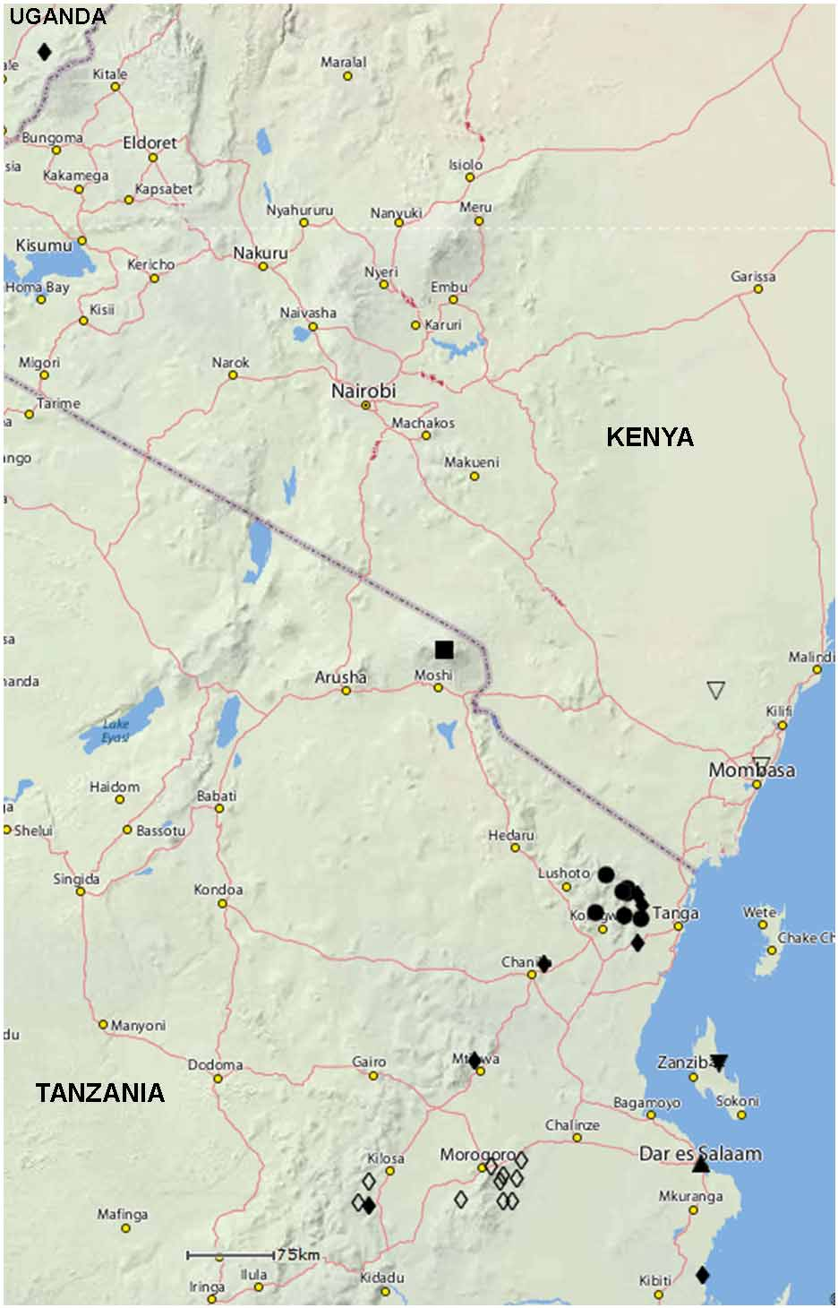

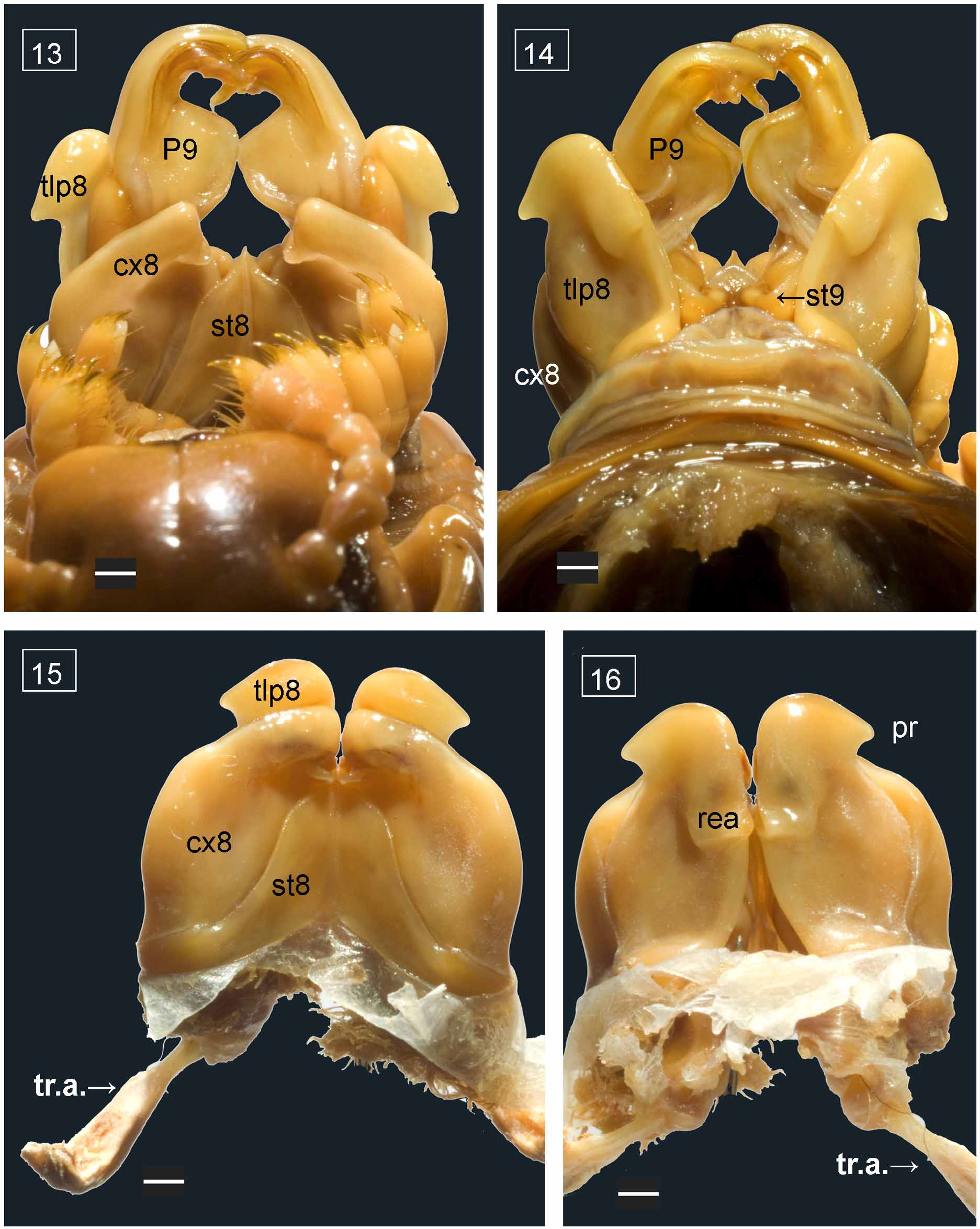
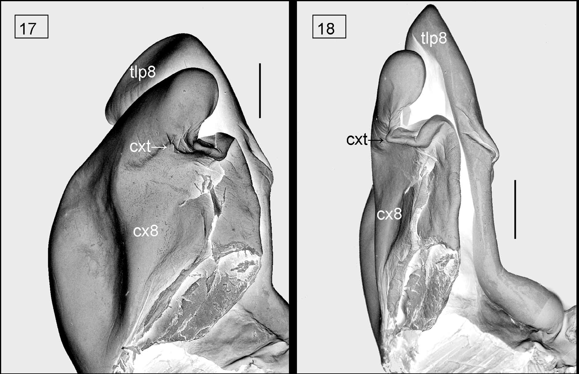
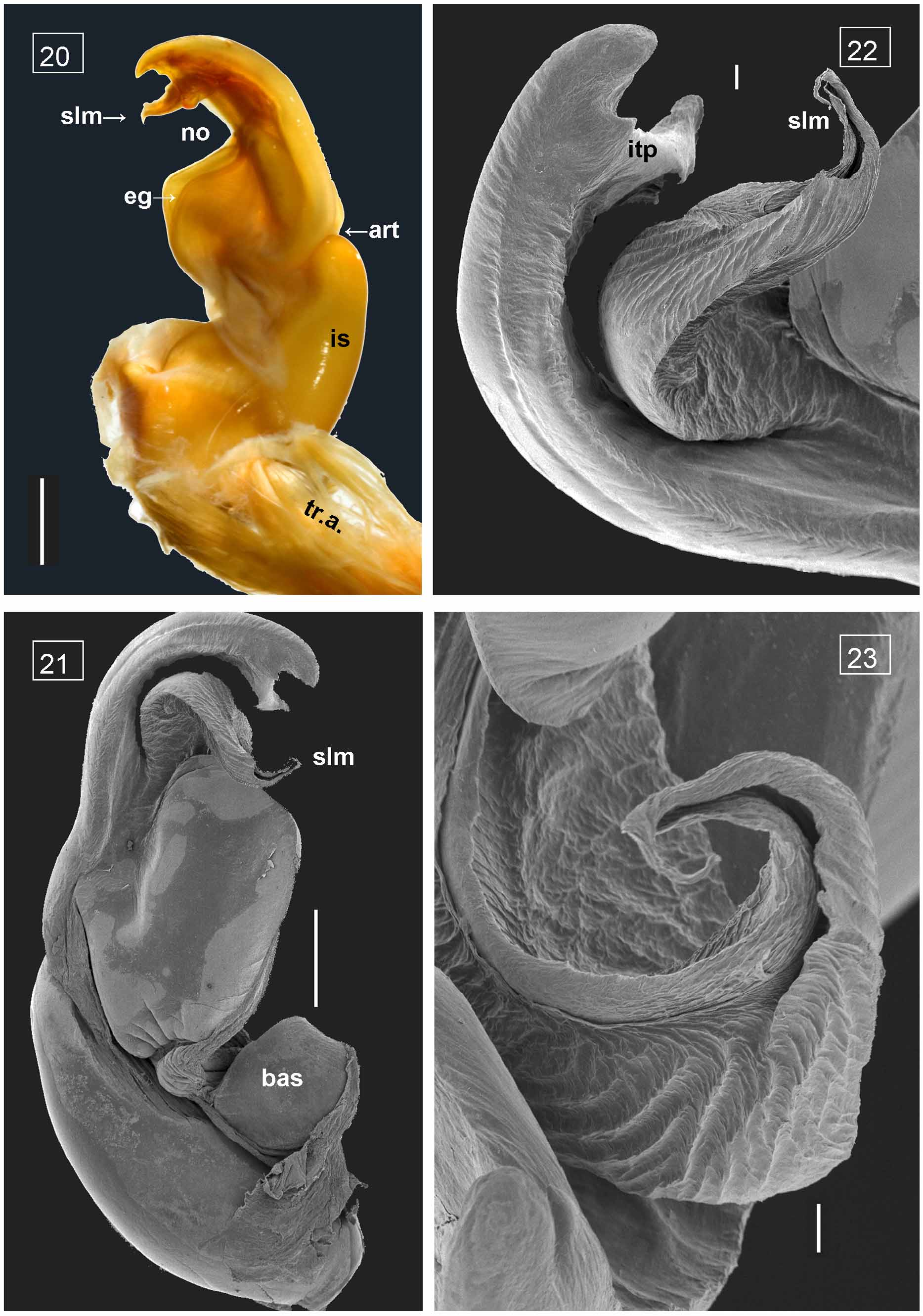
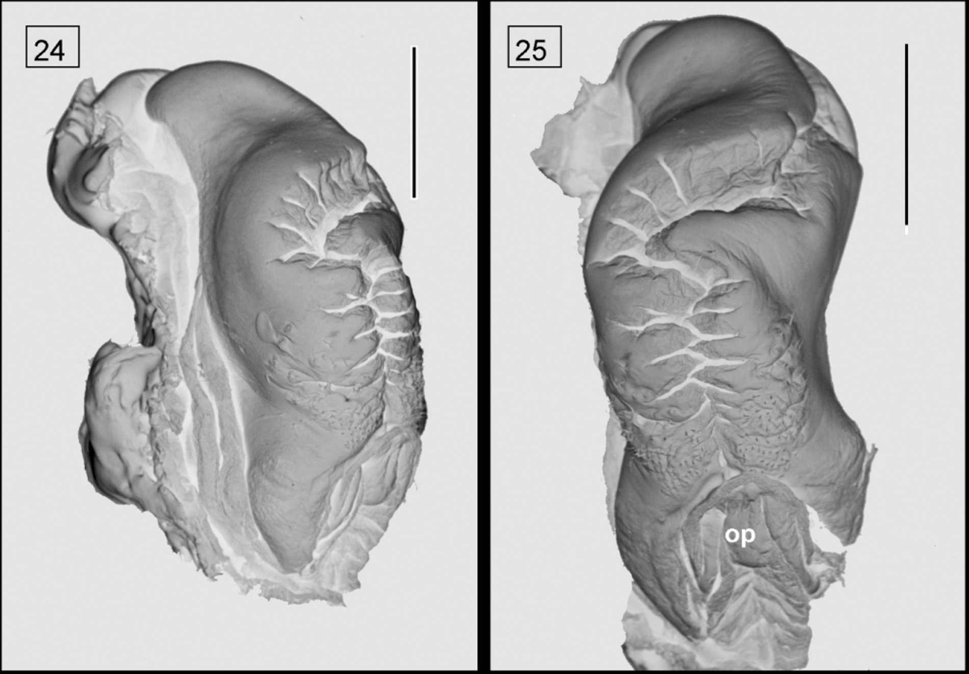
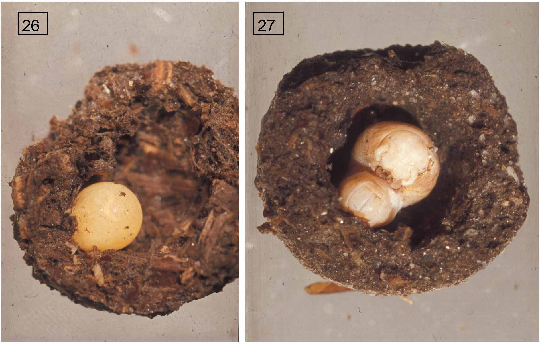
TABLE 2. Postembryonic development of Crurifarcimen vagans based on all examined entire specimens. Entries are body ring formulae sensu Enghoff et al. (1993), i. e., (number of podous rings) + (number of apodous rings) + T [= telson]. Stadium numbers are inferred from counts of rows of ocelli, see further in text. Numbers of specimens in each stadium and with each body ring formula are given in (parentheses) after the formula. Inferred formulae which were not observed, are given in [square brackets]. The single stadium I juvenile was not dissected; it has 6 body rings, a long zone of not fully formed segments, and a telson; the formula 4 + 2 + T is given in analogy with what is known for other pachybolids (Pelmatojulus spp., Enghoff et al. 1993). The principal pathway of postembryonic development is indicated with bold types.
| Stadium | I | 4+2+T (1) |
|---|---|---|
| Stadium | II | 6+18+T (1) |
| Stadium | III | 24+6+T (2) |
| Stadium | IV | 30+6+T (8) |
| Stadium | V | 36+6+T (2) |
| Stadium | VI | 42+6+T (4) |
TABLE 3. Crurifarcimen vagans, comparison of adults and immatures. Entries are horizontal diameters in mm, followed by no. of observed specimens (in parentheses). In each cell, the upper line refers to specimens from the E Usambaras, the lower line to the slightly smaller specimens from the W Usambaras.
| Stadium | Diameter of ad. males | Diameter of juv. males | Diameter of ad. females Diameter of juv. females |
|---|---|---|---|
| IX | 14.3–16.2 (14) 13.5–14.2 (7) | 12.2–13.9 (3) 11.8–11.9 (2) | 14.7–16.0 (5) 12.8 (1) 14.9–15.4 (3) 12.2 (1) |
| X | 14.2–15.2 (6) - | - - | 14.4–16.1 (7) 12.2 (1) 14.9–15.3 (2) - |
No known copyright restrictions apply. See Agosti, D., Egloff, W., 2009. Taxonomic information exchange and copyright: the Plazi approach. BMC Research Notes 2009, 2:53 for further explanation.
|
Kingdom |
|
|
Phylum |
|
|
Class |
|
|
Order |
|
|
Family |
|
|
Tribe |
Pachybolini |
|
Genus |
Crurifarcimen vagans
| Enghoff, Henrik 2011 |
pulchripes
| Dhaenens & VandenSpiegel 2006 |