
Foa brachygramma (Jenkins, 1903)
|
publication ID |
https://doi.org/ 10.5281/zenodo.278368 |
|
DOI |
https://doi.org/10.5281/zenodo.5678710 |
|
persistent identifier |
https://treatment.plazi.org/id/03BC8799-FF9F-FFCE-FF49-041CFC28F865 |
|
treatment provided by |
Plazi |
|
scientific name |
Foa brachygramma (Jenkins, 1903) |
| status |
|
Foa brachygramma (Jenkins, 1903) View in CoL
Figures 1–5 View FIGURE 1 View FIGURE 2 View FIGURE 3 View FIGURE 4 View FIGURE 5 , 13–15 View FIGURE 13 View FIGURE 14 View FIGURE 15 , Tables 1–4 View TABLE 1
Material examined. Holotype Fowleria brachygrammus , USNM 50699 35.1 mm SL, Hawaiian Is., Honolulu, O. P. Jenkins, 1889, coral rocks, x-ray.
Other material. Hawaiian Islands: AMNH 17680 (3, 31-44), 1925. BPBM 6360, (20, 33-64), Oahu, NE side, Kaneohe Bay, Coconut Island swimming pool, 1-30 Apr 1968. BPBM 10955 (1, 29), Oahu, NE side, Kaneohe Bay, reef, 1–3 m, 14 July 1971. BPBM 19661 (3, 36-42), Oahu, SW side, Campbell Industrial Park, tidepool, 0–1 m, 14 Sep 1975. BPBM 15128 (229, 15-64), Oahu, Kaneohe Bay, about 1/ 4 mile N of Coconut Island dock, hole in coral reef, 11 November 1948. BPBM 22651 (9, 27-44), Oahu, Kaneohe Bay, SE of buoy #24, 27 Sep 1968. BPBM 22689 (48, 34-50), Oahu, NE side, Kaneohe Bay, Coconut Island, W side, pond, 13 June 1978. BPBM 34620, (1, 32), Hawaii, E tip, Kapoho, anchialine pool, 0–1.5 m, 21 May 1991. BPBM 34763 (1, 27), Midway Atoll, Sand Island, SE side, off middle of runway, inside reef, 2 m, 10 Sep 1989. BPBM 34785 (1, 20.6), Midway Atoll, Sand Island, SE side, off NOPF, outside reef, coral ledge, 25 m, 13 Sep 1989. BPBM 34794 (1, 40.5), Midway Atoll, lagoon, under cargo pier, 3–5 m, 12 Sep 1991. BPBM 34870 (3, 26-31), Midway Atoll, Sand Island lagoon, wreckage at shore between cargo pier and launching ramp, 3–5m, 18 Sep 1991. BPBM 34919 (1, 21), Maui, Maalaea Bay, silty sand, 11.5 m, 7 Apr 1991. BPBM 37227 (1, 40), Oahu, Kaneohe Bay, 22 Jan 1991, color photo. BPBM 37322 (3, 28-36), Oahu, Pearl Harbor, 18 Sep 1996. CAS (SU) 7633 (26-35), USFC 1901B, Hawaii, Hilo. U.S. Fish Commission 1901. CAS 55143 (1, 44), Kehi weather station, 22 Apr 1971. CAS 80820 (1, 49) G937 Oahu, patch reef north of Sampan Channel opposite red marker #8, 2– 3 m, 20 May1993. CAS 81066 (1, 26) G9322 Oahu, E of marker #3 at Crash Boat Channel, 2 m, 11 Oct 1993. CAS 83092 (2, 39-39), Oahu, shore reefs at Kahala, GVF 0 0 54, 15 Sep 1951. CAS 83107 (1, 22), Oahu, shore reefs at Kahala. W. Gosline, V. Brock, E. Herald GVF 0 0 54, 15 Sep 1951. USMN 6907 (3, 19-34), Oahu. USNM 55200 (1, 30), same data as holotype, in poor condition. USNM 112285 (1, 45), Pearl Harbor, Apr 1950. USNM 38288 (1, 48), Honolulu. USNM 175459 (3, 31-54), Kaneohoe Bay, Oahu, 11 Nov 1948. USNM 206228 (15, 14-51), Moana Reef, Oahu, 0.6 m, 8 Jun 1968. USNM 175462 (19, 13-54), West Loch, Pearl Harbor, 8 Dec 1949 x-ray.
Diagnosis. Pored lateral-line scales usually 9–10; gill rakers and rudiments on first gill arch usually 14–16; body scales with narrow dark margins; body uniform to mottled brownish without darker bars on sides (except at night); second dorsal fin, anal fin and caudal fin pale without darkish banding; no whitish spot at axil of pectoral fin; no whitish spots at base of caudal peduncle or behind posterior base of second dorsal fin; two darkish small marks near base of caudal peduncle.
Description. Range of proportions (as percentage of standard length) in Table 1 View TABLE 1 with holotype first and other material in parentheses.
Dorsal fin IX spines as VII(I)-I,9, third spine longest and strongest, sixth radial free in advance of hidden nubbin representing eighth spine; anal fin II,8; pectoral-fin-rays 12 rarely 11; pelvic fin I,5; principal caudal rays 1- 8+7-1, upper and lower rays unbranched, soft procurrent rays; lateral-line scales 21–22 as 9–10 pored scales and11–12 pitted scales ( Table 4); transverse scale rows above lateral line 1; transverse scale rows below lateral line 6; median predorsal scales 3 or 4; circumpeduncular scale rows 12 as 5+2+5; first gill arch with 14–16 rudiments and rakers, well developed rakers 8–9 ( Table 3), upper arch 2-3+1, lower arch 7-8+3-5, second arch with rudiments on hypobranchial and ceratobranchial with one raker at angle.
Villiform teeth in band on premaxilla and on dentary; 2–3 rows on vomer; 2–3 rows on the palatine; none on ectopterygoid, endopterygoid or basihyal.
Vertebrae 10+14; five free hypurals; one pair of short, slender uroneurals; three epurals, the first two expanded; a free parhypural; three supraneurals, no procumbent spines (spurs); two supernumerary spines on first dorsal pterygiophore, no procumbent spines (spurs); basisphenoid reduced to upper portion (meningost); supramaxilla thin and reduced in length; posttemporal smooth on posterior margin; preopercle smooth on vertical and horizontal edges, ridge smooth; infraorbitals smooth; infraorbital shelf present on third bone; interhaemal gap 2+4 (Table 2).
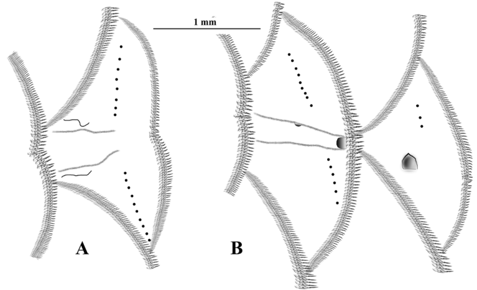
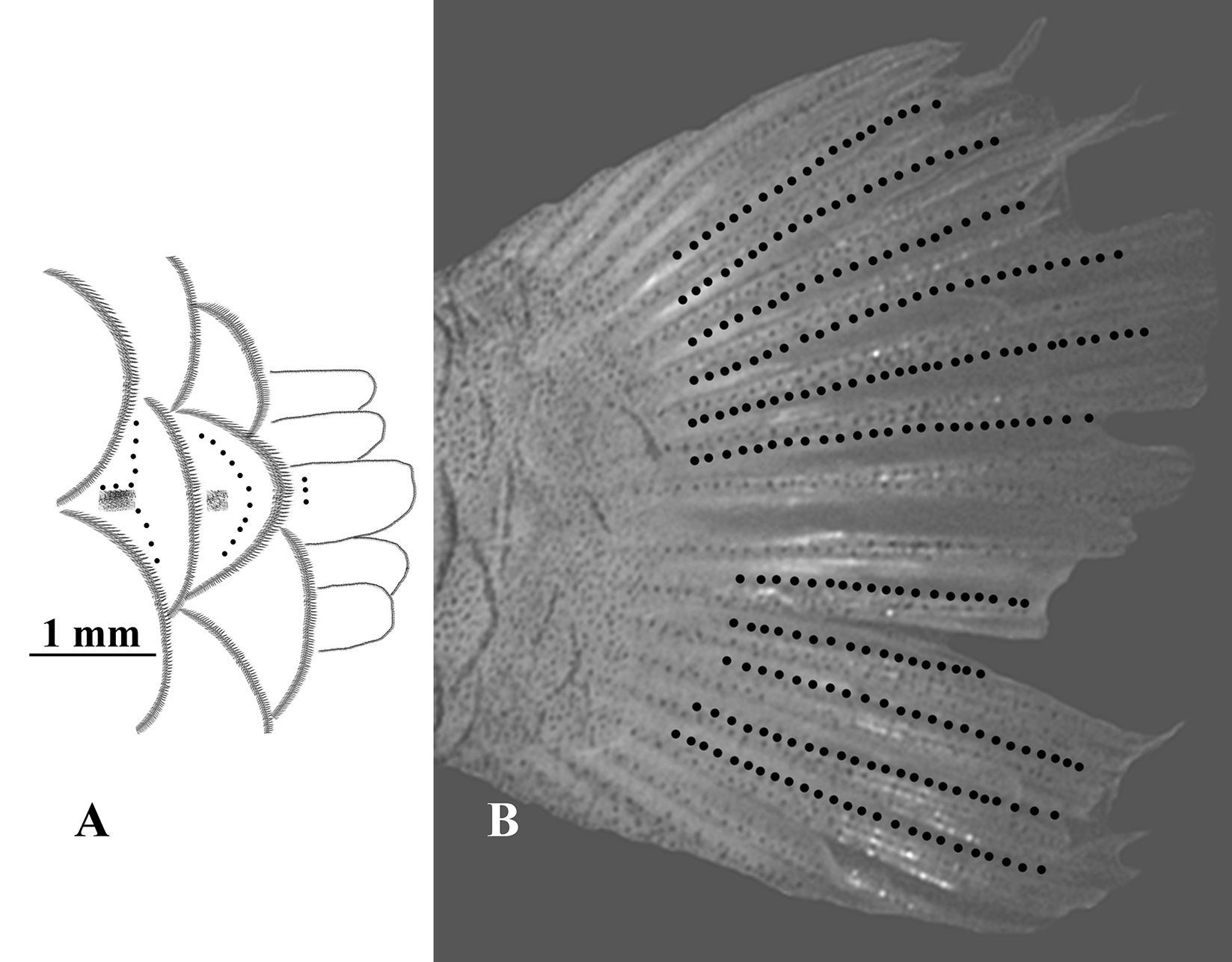
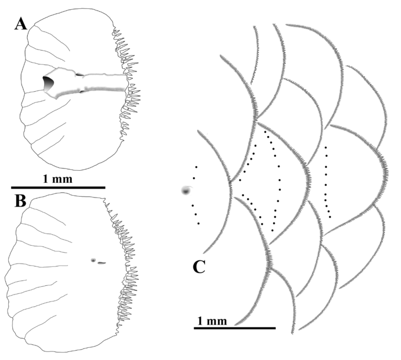
Peripheral ctenoid scales (see Roberts, 1993) on cheek, subopercle, opercle, isthmus, nape, predorsal, base of pectoral fin, behind pectoral fin and on body; base of pelvic fin with two large scales, both weakly ctenoid; no rudimentary or developed axillary scale; fourth pored lateral-line scale with one pore above and one below main canal; scales on base of caudal fin cycloid ( Fig 5 View FIGURE 5 A).
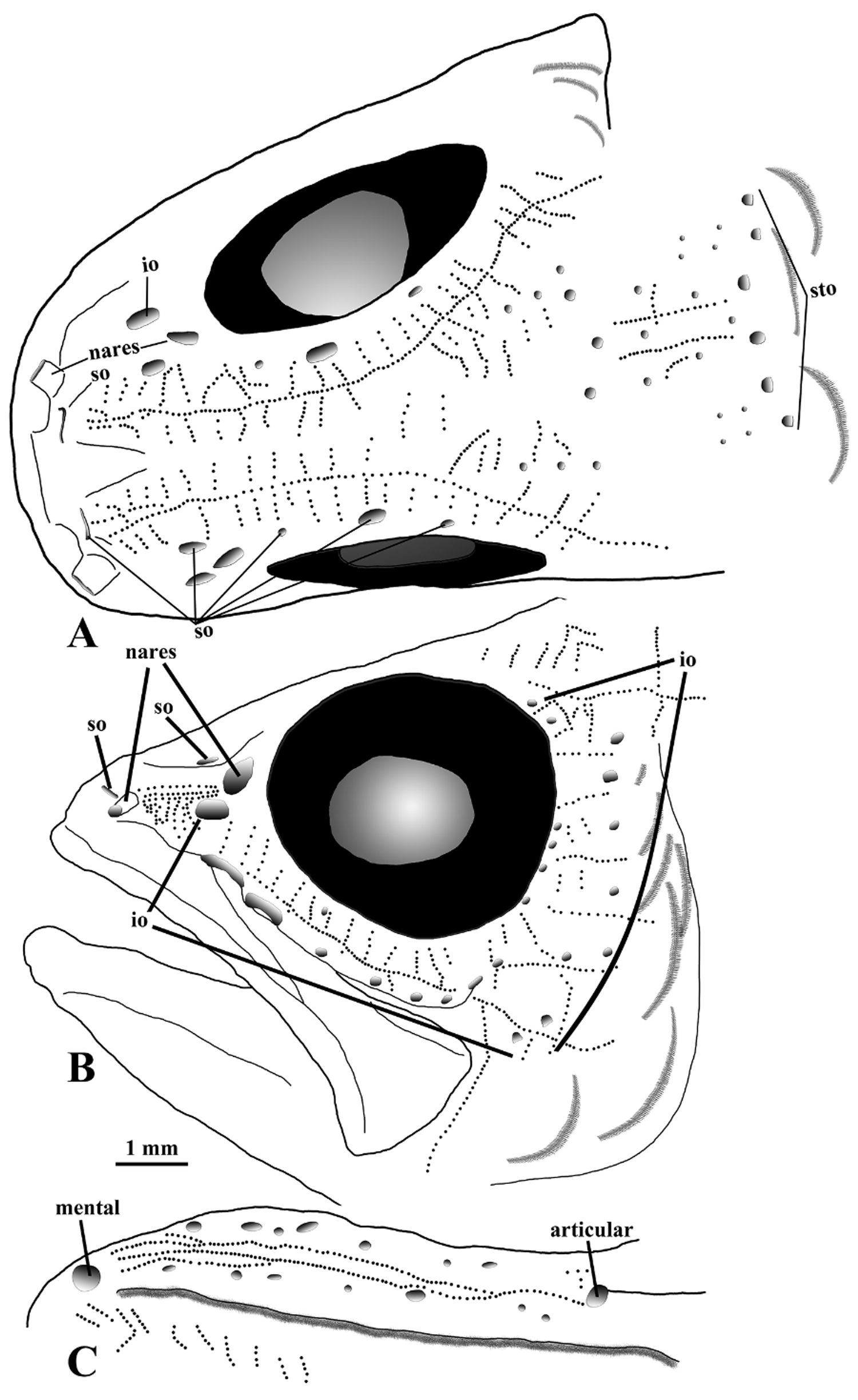
Head with many small pores ( Fig. 2 View FIGURE 2 ); anterior end of supra-orbital canal as a broad slit pore back from edge of upper lip with a cleft-like distal edge open, four other supra-orbital pores, one near posterior nare and three over eye, small pores on head from simple projection off supra-orbital canal and central commissure; lachrymal with large anterior pore near flat posterior nare opening, two large ventral slit pores along edge of lachrymal and other small pores near lower edge of infra-orbitals below eye, small infra-orbital pores along posterior edge of eye and onto cheek; preopercle with two large pores on ventral edge, smaller pores off edges and a series of tiny pores vertically along preopercular ridge ( Fig. 3 View FIGURE 3 ); anterior portion of dentary with dentary (anterior, not shown) and mental (ventral) pores, posterior with a large articular pore, small pores near margins; supratemporal canal with short posterior canals onto nape ending in slightly larger pores than present anteriorly.
Free neuromasts on snout, interorbit and temporal areas in ( Fig. 2 View FIGURE 2 A), a split line of neuromasts becoming one line from snout over eye continuous to below posttemporal, short lines generally perpendicular to long line, short medial lines associated with commissure region, two medial longitudinal neuromast lines on supratemporal; several rows of neuromasts between nares and anterior infraorbital oriented horizontally and vertically, a long linear line of neuromasts from near first infraorbital (lachrymal) to near posterior corner of maxilla dividing on to lower cheek horizontally as well as downward, short lines of neuromasts radiate from ventral and posterior edge of eye ( Fig. 2 View FIGURE 2 B); three or four rows of neuromasts on anterior third of lower jaw grading posteriorly to two rows, then one row to edge of articular pore ( Fig. 2 View FIGURE 2 C), gular free neuromasts in chevron rows except for anterior two rows; preopercle with single long vertical line of neuromasts on upper arm from posterior part of lower arm, two linear rows on lower arm, lower row near anterior end of preopercle over first pore bending up to mid-portion of upper row, short semi-vertical rows on lower arm ( Fig. 3 View FIGURE 3 A); opercle with short vertical row of neuromasts on upper anterior scale ( Fig. 3 View FIGURE 3 B); pored lateral-line scales ( Fig. 4 View FIGURE 4 A) with vertical rows of neuromasts above and below canal pore;, first scale with pit ( Fig. 4 View FIGURE 4 B) with a reduced row above pit, no free neuromasts on other pit scales until at base of caudal fin on last two pit scales and small caudal scale, no free neuromasts observed on other basicaudal scales ( Fig. 5 View FIGURE 5 A); and, free neuromasts associated with 11 principal caudal fin-rays in single linear lines on ventral edges of upper branched rays (3–8) and on dorsal edges of lower branched rays (11–15), middle two caudal rays (9–10), upper unbranched and branched caudal rays (1–2), and lower unbranched and branched caudal rays (16–17) lack free neuromasts (fig. 5B).
Anterior nare tubular, posterior nare flat.
Caudal fin truncate or slightly rounded; second dorsal and anal fin with rounded distal edges.
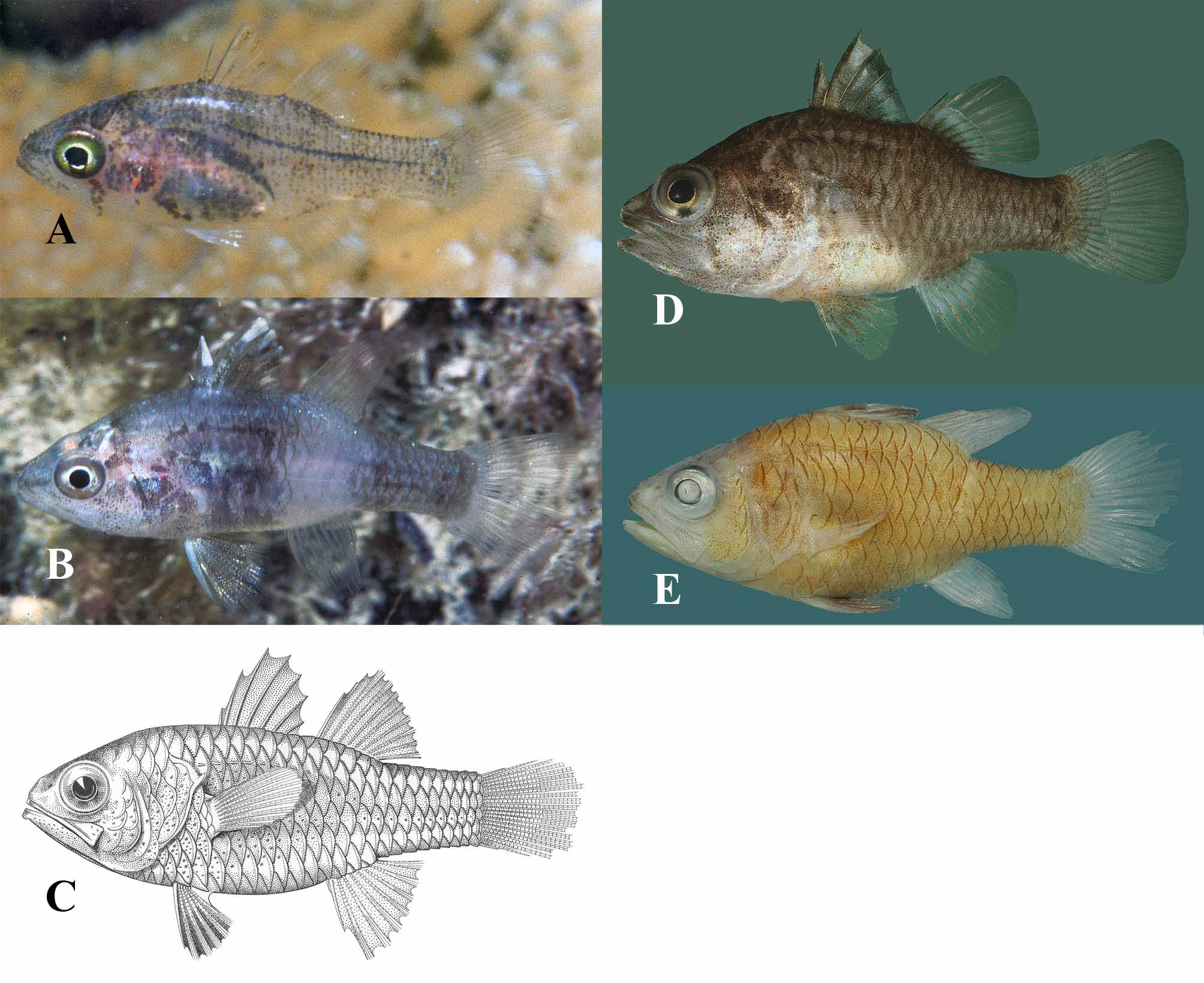
Life colors. Day, Figure 1 View FIGURE 1 A: Head with faint, narrow brown mark on snout, broad, brown interorbit marks, narrow, brown cheek mark, broad, brown opercle mark; body tranlucent with brown markings along side of abdomen, small, brownsh, linear-like spots throughout rest of body; first dorsal fin with a few spots in membranes, but mostly pale, second dorsal fin, caudal fin, anal fin and pelvic fin pale. Night, Figure 1 View FIGURE 1 B: Head with faint mark on snout, a high post-ocular mark, cheek with small spots; body steel gray with broad pale band from second dorsal fin to anal fin, body scales sharply edged dark margins, narrow dark line along first eight lateral-line scales, darkish areas on side of abdomen up to level of pectoral fin, darkish lines on caudal peduncle, two small peduncular spots; tips of second to fourth dorsal spines whitish with small melanophore spots below; second dorsal fin, caudal fin and anal fin pale; pelvic fin with whitish spine, small melanophores throughout soft rays.
Preserved color pattern. Post-mortem ( Figure 1 View FIGURE 1 D) color brown on head and body with whitish areas on side of abdomen, lower portions of opercle and cheek; lips brown without alternating whitish bands, snout brown, faint post-ocular marks down to tip of maxilla, broad mid-line on opercle and above level of pupil; narrow dark body scale margins, pored lateral-line scales with lighter areas, two small peduncular spots; membrane behind second and third dorsal spines light tannish to whitish, membrane behind fourth and fifth spines darkish distally with whitish area below extending to seventh spine, many small darkish spots proximal to whitish band; second dorsal fin, caudal fin and anal fin pale; pelvic fin with small melanophores on fin-rays. Alcohol preserved ( Figure 1 View FIGURE 1 E) head and body brown to light tan; markings on head faint or absent; head and body with narrow dark scale margins; first dorsal fin and pelvic fin with dark areas, pectoral fin, second dorsal fin, caudal fin and anal fin pale, without any marks. Juveniles (14 mm) and some adults (up to about 40 mm) exhibit the faint bar under the second dorsal fin and the bar on the anterior part of the caudal peduncle.
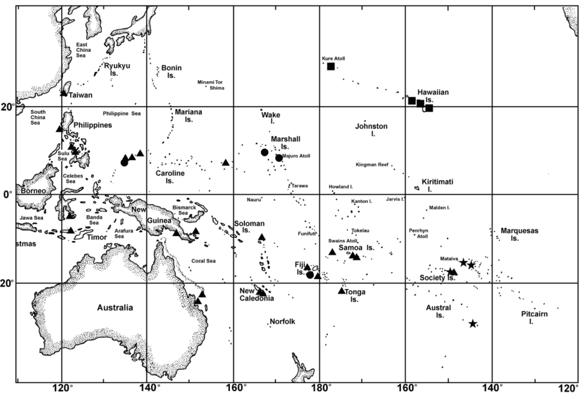
Distribution. Foa brachygramma is treated as endemic to the Hawaiian Islands ( Fig 14 View FIGURE 14 ). No collections of any species of Foa were found in museum collections (or listed in online catalogs) at Johnston Atoll, Kingman Reef, Palmyra Atoll, Teraina (Washington) Island or Tabuaeran (Fanning) Island.
Remarks. Color pattern, gill arch, number of mandibular pores, mandibular free neuromast and pored lateralline scales comparisons should be used for positive identification of this species. There is a relationship between the size of the species and the number of pored lateral line scales ( Fig 15 View FIGURE 15 ). This relationship may be helpful for the identification of the species of Foa on the Pacific Plate. Material reported in Fraser (1972) represents specimens of Foa fo and F. madagascariensis not F. brachygramma .
The Hawaiian Islands have only 10 species of cardinalfishes ( Randall, 1998), four of which are endemic (compared to 25% endemism for the total shore fish fauna of the islands). The Line Islands, all low atolls or low islands, have a total of 18 apogonids (one identified only to family, and two only to genus), none in Foa , and none endemic ( Mundy et al., 2010) with their caveat about collecting cryptic species. Therefore, the precursor to Foa brachygramma most likely came from the western Pacific via a gyre from the Kuroshio Current than from the south.
The presence of eleven rows of free neuromasts on the caudal fin of Foa brachygramma exceeds the eight rows previously reported for a gobioid fish ( Ahnelt and Göschl, 2004).


No known copyright restrictions apply. See Agosti, D., Egloff, W., 2009. Taxonomic information exchange and copyright: the Plazi approach. BMC Research Notes 2009, 2:53 for further explanation.