
Foa nivosa, Fraser, Thomas H. & Randall, John E., 2011
|
publication ID |
https://doi.org/ 10.5281/zenodo.278368 |
|
DOI |
https://doi.org/10.5281/zenodo.5678716 |
|
persistent identifier |
https://treatment.plazi.org/id/03BC8799-FF88-FFDF-FF49-0635FE65F853 |
|
treatment provided by |
Plazi |
|
scientific name |
Foa nivosa |
| status |
sp. nov. |
Foa nivosa View in CoL new species
Figures 9 View FIGURE 9 , 11–15 View FIGURE 11 View FIGURE 12 View FIGURE 13 View FIGURE 14 View FIGURE 15 , tables 1–4
Type material. Holotype BPBM 39658, 26 mm SL, Majuro Atoll, lagoon off Uliga, 7°06'27.4" N 171°22'07"E, 24 m, 9 Jan 2005, underwater color photograph.
Paratypes: Marshall Islands: BPBM 39703 (1, 26.8), Kwajalein Atoll, Bigej-Meck reef, 25 m, 29 Dec 2004, color photo. BPBM 28761 (2, 28–29), Kwajalein Atoll, Bigej-Meck reef, 8°58'N 167°44'E, 17–27 m, 19 Sep 1982, color photo. BPBM 40434 (1, 19), Majuro Atoll, lagoon side, 15 m, 3 Jan 2006. BPBM 41055, (4, 17–23), same data as holotype, cleared and counter stained (1, 21). Palau: CAS 83093 (1, 25), G/V Gaines, 5657 #302, 45– 48 m, 1956. ROM 77135 (5, 17–27), Ngeruketabel I., RW 04-13, 20 Jul 2005. Fiji: USNM 245649 (2, 16–20), Viti Levu, Naqara (Nangara) Passage, 18°11'15"S 178°17'E, VGS 82- 41, 40 m, 3 Jun 1982.
Diagnosis. Pored lateral-line scales usually 3–7; gill rakers and rudiments usually 12–14; body scale margins uniform; body pale yellowish brown, suffused with red especially on head and ventrally on body, with numerous white spots outlined in red, no head, body or peduncular bars; no spot on axil of pectoral fin; three or more whitish basicaudal spots; second dorsal, anal and caudal fins pale reddish with small white spots, the membranes translucent.
Description. Range of proportions (as percentage of standard length) in Table 1 View TABLE 1 .The holotype is described, except as noted in parentheses, and variation in counts for paratypes is given in Tables 2–4. Internal characteristics are described from a clear and counter-stained paratype. One paratype was used to describe the free neuromasts on the head.
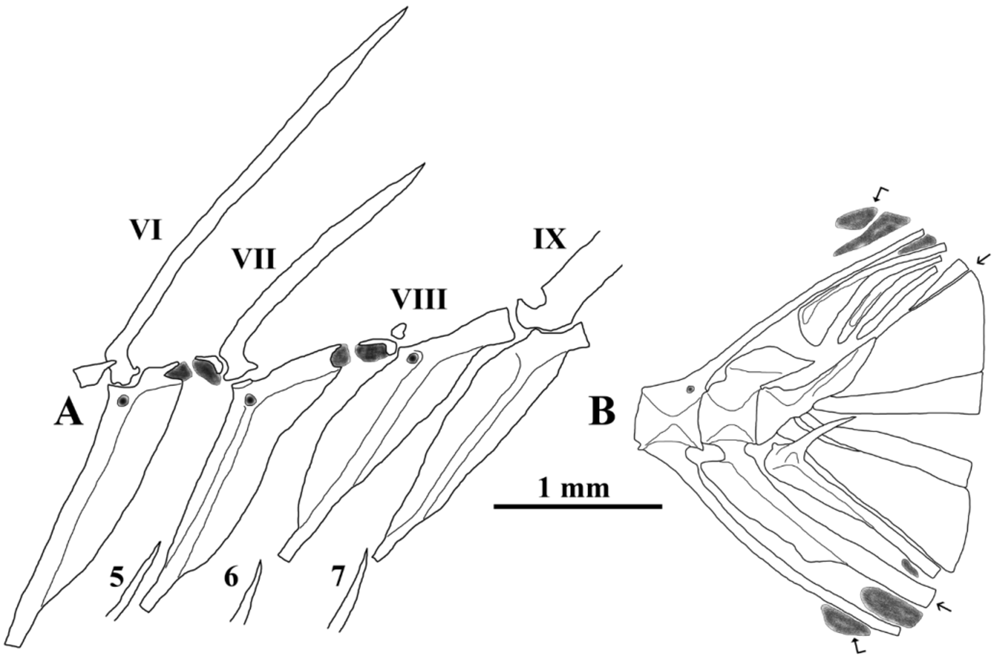
Dorsal fin IX spines as VII(I)-I,9 (rarely VII-I,8), third spine longest and strongest, eighth spine as a hidden nubbin; sixth radial free from pterygiophore under closely associated eighth spine ( Fig 12 View FIGURE 12 A); anal fin II,8; pectoralfin-rays 12; pelvic fin I,5; longest procurrent caudal rays segmented, principal caudal rays 1+8+7+1; some pitted lateral-line scales missing, 6 scales pored, 21–22 total as 3–7 pored scales and 14–18 pitted scales, ( Table 4); one large scale rows above lateral line with interdigitated small scale abutting base of first dorsal fin; transverse scale rows below lateral line 6; median predorsal scales 3 (4); circumpeduncular scale rows 12 as 5+2+5; total gill rakers and rudiments 13 (12–14, Table 3), well developed 7 (6–8), upper arch 2+1, lower arch 6+4 (1-2+1, 5-7+3-5); second arch with tooth patches and rudiments; interarcual cartilage extending from uncinate process of first epibranchial to lateral tip of second pharyngobranchial; first epibrachial with a thin cartilage ending with ossified first pharyngobranchial..
Villiform teeth in band on premaxilla and on dentary; 1–3 rows on vomer; 1–3 rows on the palatine; none on ectopterygoid, endopterygoid or basihyal.
Vertebrae 10+14; eight epineurals on first eight vertebrae; eight ribs on third to tenth vertebrae, last rib expanded; five free hypurals; no uroneurals; three epurals, the first expanded; a free parhypural ( Fig. 12 View FIGURE 12 B); three thin supraneurals, first one curved, second and third straight, no procumbent spines (spurs); two supernumerary spines on first dorsal pterygiophore, no procumbent spines (spurs); interhaemal gap 1+5 (Table 2); basisphenoid absent; very thin supramaxilla (not visible except by clearing and staining); neurocranium similar to Foa fo ( Fraser, 1972, plate 28); posttemporal smooth on posterior margin; preopercle smooth on both edges, two to three tiny serrae at angle, ridge smooth; infraorbitals smooth; a shelf present on third infraorbital; a ring of scleral cartilage, no ossification present; seven branchiostegals; ceratohyal notched, suture smooth with epihyal; urohyal without anterior process;
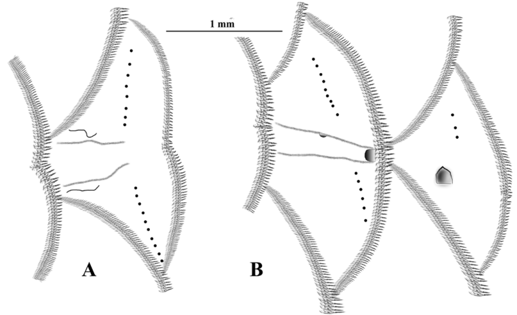
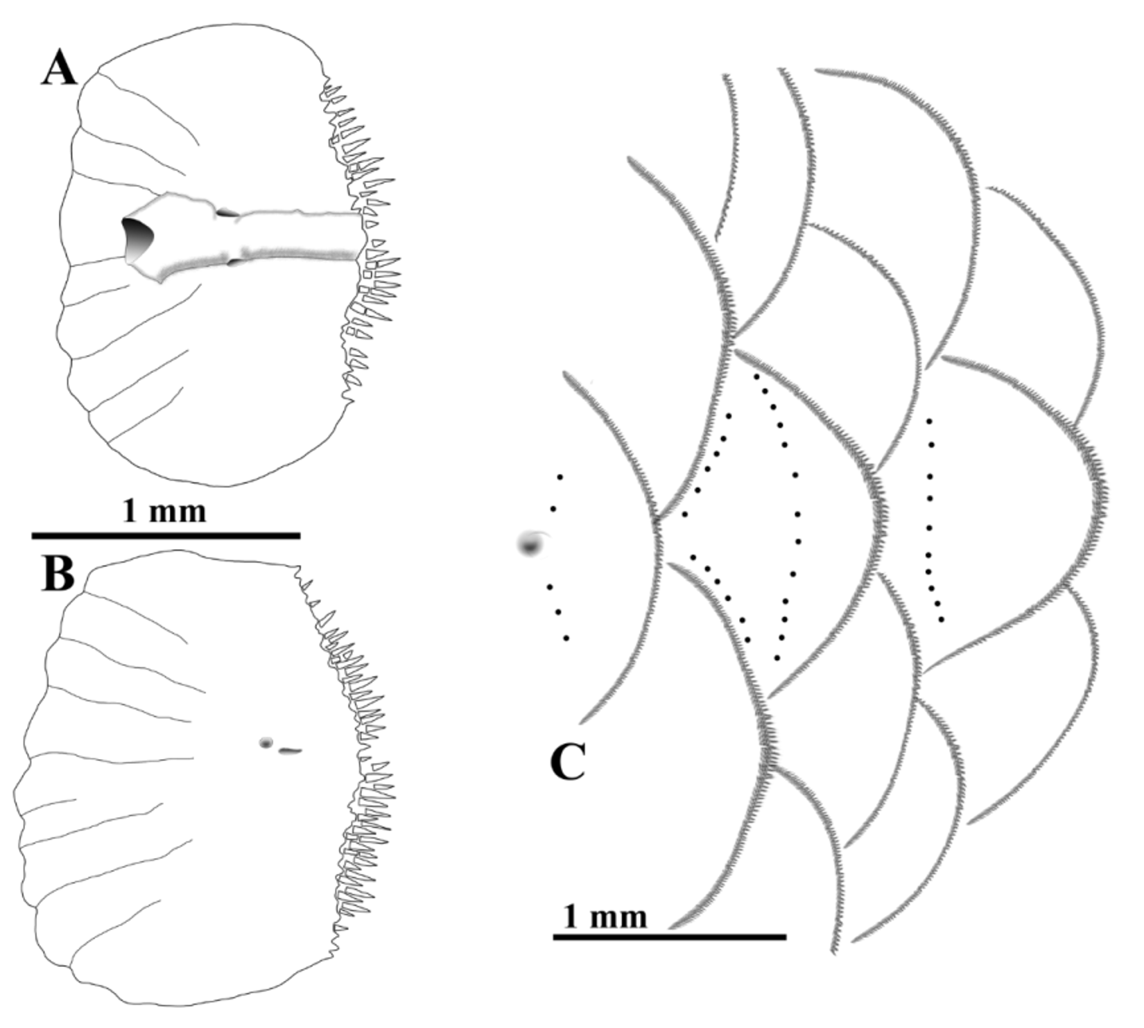
Peripheral ctenoid scales (see Roberts, 1993) on cheek, subopercle, opercle, isthmus, nape, predorsal, base of pectoral fin, behind pectoral fin and on body; two large scales on base of pelvic fin, cycloid (smaller fish) or becoming ctenoid (larger fish), posterior scale elongate; no axillary scale; pored lateral-line scales short, fourth scale with one pore above and one below main canal ( Fig 13 View FIGURE 13 A), remaining lateral-line scales pitted to base of caudal fin ( Fig 13 View FIGURE 13 B); last few scale rows on caudal fin ctenoid ( Fig. 13 View FIGURE 13 C); a single row of detached cteni with a few truncated cteni.
Head with many small pores; anterior end of supraorbital canal as a broad slit pore back from edge of snout, one large pore over eye, lachrymal with large anterior pore near flat posterior nare opening, two large ventral slit pores along edge of lachrymal; anterior portion of dentary with anterior and mental pores, posterior with articular pore; supratemporal canal with four pores across head, posterior canals over scales with pores not far onto nape or predorsal scales; lateral line scales with a single row of ctenii ( Fig. 13 View FIGURE 13 ), pored scale with small foramina for nerve endings leading to single rows of free neuromasts ( Fig. 13 View FIGURE 13 A), pitted scales without such foramina ( Fig. 13 View FIGURE 13 B), last pitted scale and last two scales with free neuromasts ( Fig. 13 View FIGURE 13 C).
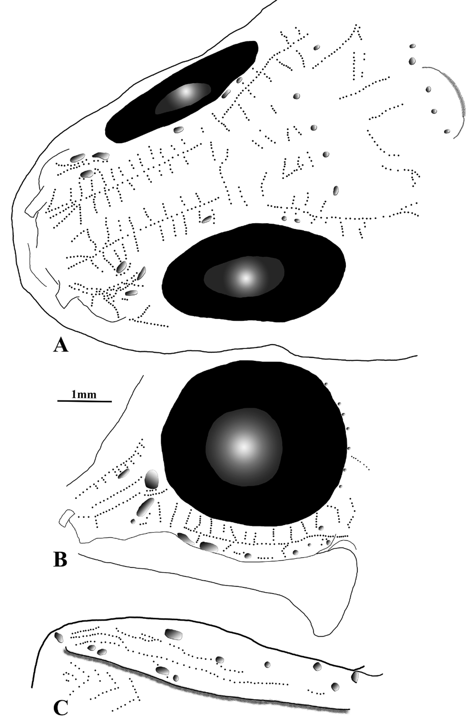
Free neuromasts on snout, interorbit and temporal areas in ( Fig. 11 View FIGURE 11 A) a split line becoming one line of neuromasts from snout interrupted over eye, offset, not overlapping, continuing onto side of head below posttemporal, short lines generally perpendicular to long lines, short medial lines associated with commissure region, two supratemporal rows of neuromasts; several rows of neuromasts between nares and anterior infraorbital oriented horizontally and vertically; a long linear line of neuromasts from first infraorbital (lachrymal) to corner of maxilla, short lines of neuromasts radiate from ventral and posterior edge of eye ( Fig. 11 View FIGURE 11 B); three rows of neuromasts on anterior third of dentary grading posteriorly to one row near edge of articular pore ( Fig. 11 View FIGURE 11 C), gular free neuromasts as chevrons; free neuromasts on pored lateral line scales in single lines similar to Fig. 4 View FIGURE 4 , pit scales without free neuromasts, last two scales with free neuromasts ( Fig. 13 View FIGURE 13 C); caudal fins of all specimens incomplete for presence of free neuromasts, two paratypes with 5 and 6 lines on lower rays, upper rays with at least 5 lines; incomplete information available for other free neuromast patterns.
Anterior nare tubular, posterior nare flat.
Caudal fin slightly rounded; second dorsal and anal fin with rounded distal edges.
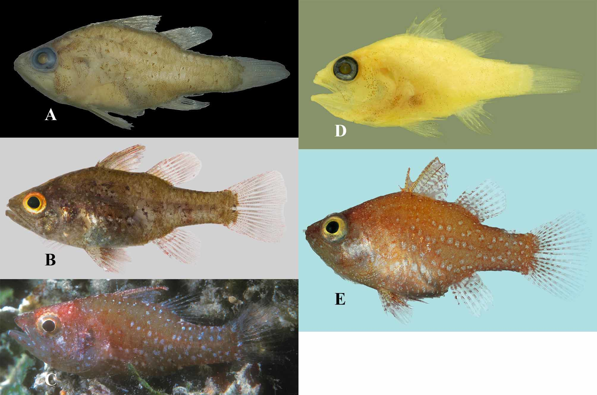
Life colors. Figure 9 View FIGURE 9 C, holotype: Head and body reddish to brownish red with whitish spots of variable cluster sizes and distribution; head with small whitish spots on snout, interorbit, larger spots on nape; alternating whitish and reddish markings on outer rim of eye, iris yellowish brown; pupil edge slightly angled anteriorly; cheek with coalescing whitish spots forming a mark extending downward halfway toward preopercle, some small melanophores near vertical and horizontal edges of preopercle; lower jaw with three narrow whitish marks alternating with broad reddish marks; upper jaw reddish with two narrow whitish marks in line with two posterior whitish lower jaw marks; opercle with tiny whitish spots above angle of preopercle, larger whitish spots on opercle near angle of preopercle; whitish spots on branchiostegal membranes; body with small whitish spots outlining edges of predorsal and nape scales becoming larger, single spots on scales below first dorsal fin; pored lateral-line scales with one or two small whitish spots; body just behind opercle, below lateral line and above pectoral fin mostly reddish with two faint clusters of whitish spots; base of pectoral fin with whitish cluster at upper edge, a smaller cluster midlower fin base; side of body from behind pectoral fin to mid-dorsal fin base with large clusters of whitish spots on to base of caudal peduncle; small whitish spots on reddish to translucent background on first dorsal fin; second dorsal fin with reddish spine interrupted with several narrow whitish marks, rest of fin with small whitish spots, base of last fin ray with large whitish spot; caudal fin translucent except at or near fin base with dorsad whitish spot, mid-line spot and ventrad spot, several smaller whitish clusters and single spots near base of fin; anal fin not clearly shown but large whitish spot at base of last fin ray; pelvic fin not clearly shown, but with some alternating whitish markings.
Preserved color pattern. Figure 9 View FIGURE 9 D post mortem and 9E preserved: Holotype head and body with uniform yellowish tan ground color, scattered brown melanophores on head, body and pelvic fins; snout with few melanophores, premaxilla edged with melanophores; cluster of melanophores on cheek bounded by preopercle ridge and posterior infraorbitals; scattered melanophores on branchiostegal membranes; few melanophores on opercle; posttemporal area and upper body with tiny melanophores; cluster of melanophores below pectoral fin on side of abdomen; rest of side and caudal peduncle with scattered tiny melanophores; dorsal fins, caudal and anal fins uniformly pale, pelvic fins with few linear melanophores on fin rays. Paratypes: Similar to holotype.
Etymology. The Latin adjective nivosus meaning snowy, here referring to numerous whitish spots on the body as nivosa .
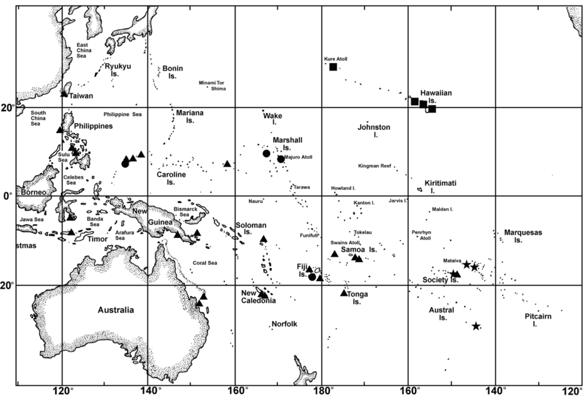
Distribution. Known from Palau, the Marshall Islands and Fiji. We expect this species to be found elsewhere in the Caroline Islands and from Indonesia, New Guinea and other islands to Fiji ( Fig 14 View FIGURE 14 ).
Remarks. All of the specimens from the Marshall Islands were collected in Halimeda beds in 15–27 meters. One collection included the presence of the algal genera Padina , Caulerpa , and Dictyota . In Palau, the new species can be distinguished from Foa hyalina by whitish spots on a reddish body versus 4–5 irregular reddish bars on a reddish body and conspicuous lack of marks on the head versus cheek, snout and post-ocular markings. Deeper sea grass meadows, Halimeda beds and other areas with abundant macroalgae have only recently been subjected to more rigorous fish sampling. Such field work may lead to more record of Foa throughout the Indo-Pacific and possibly other undescribed species.

No known copyright restrictions apply. See Agosti, D., Egloff, W., 2009. Taxonomic information exchange and copyright: the Plazi approach. BMC Research Notes 2009, 2:53 for further explanation.