
Potamotrygon limai, sp.
|
publication ID |
https://doi.org/ 10.11646/zootaxa.3765.3.2 |
|
publication LSID |
lsid:zoobank.org:pub:62224480-B209-4D05-90F4-0739FBC44F3B |
|
DOI |
https://doi.org/10.5281/zenodo.5624284 |
|
persistent identifier |
https://treatment.plazi.org/id/03BA8C6C-FFA9-6205-48D2-7C60FE5AAA97 |
|
treatment provided by |
Plazi |
|
scientific name |
Potamotrygon limai, sp. |
| status |
sp. |
Potamotrygon limai, sp. View in CoL nov.
( Figures 1−17 View FIGURE 1 View FIGURE 2 View FIGURE 3 View FIGURE 4 View FIGURE 5 View FIGURE 6 View FIGURE 7 View FIGURE 8 View FIGURE 9 View FIGURE 10 View FIGURE 11 View FIGURE 12 View FIGURE 13 View FIGURE 14 View FIGURE 15 View FIGURE 16 View FIGURE 17 ; Tables 1−2 View TABLE 1 View TABLE 2 )
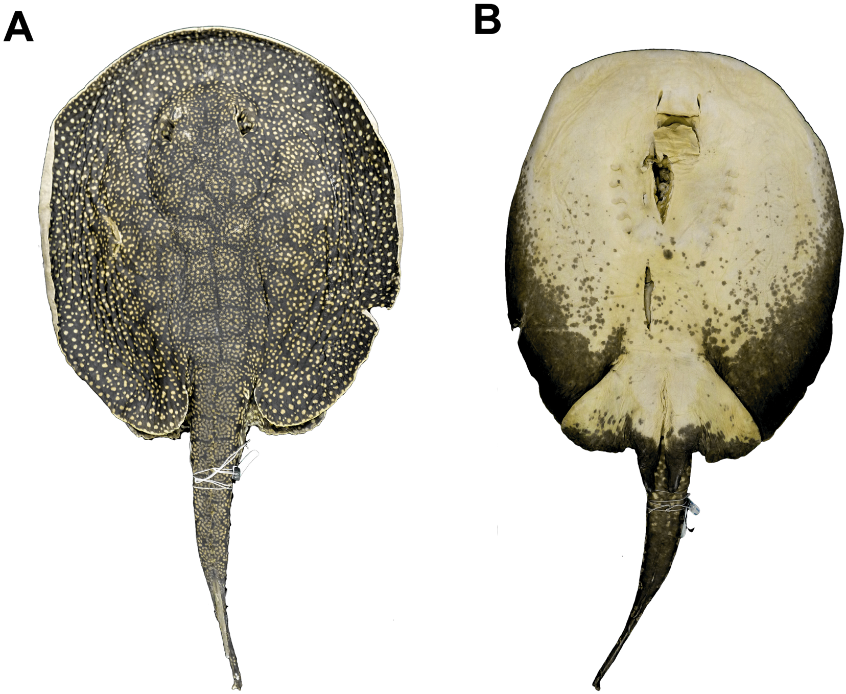
Holotype. MZUSP 103031 (adult male, 498 mm DW), Madeira River basin, Jamari River, Represa Samuel, municipal district of Itapoã do Oeste, upper Amazon Basin, state of Rondônia, Brazil, 9º4’33.96” S, 63º18’17.64” W, May 2006, coll. F. P. L. Marques, N. M. Luchetti, V. M. Bueno and T. Loboda ( Figure 1 View FIGURE 1 ).
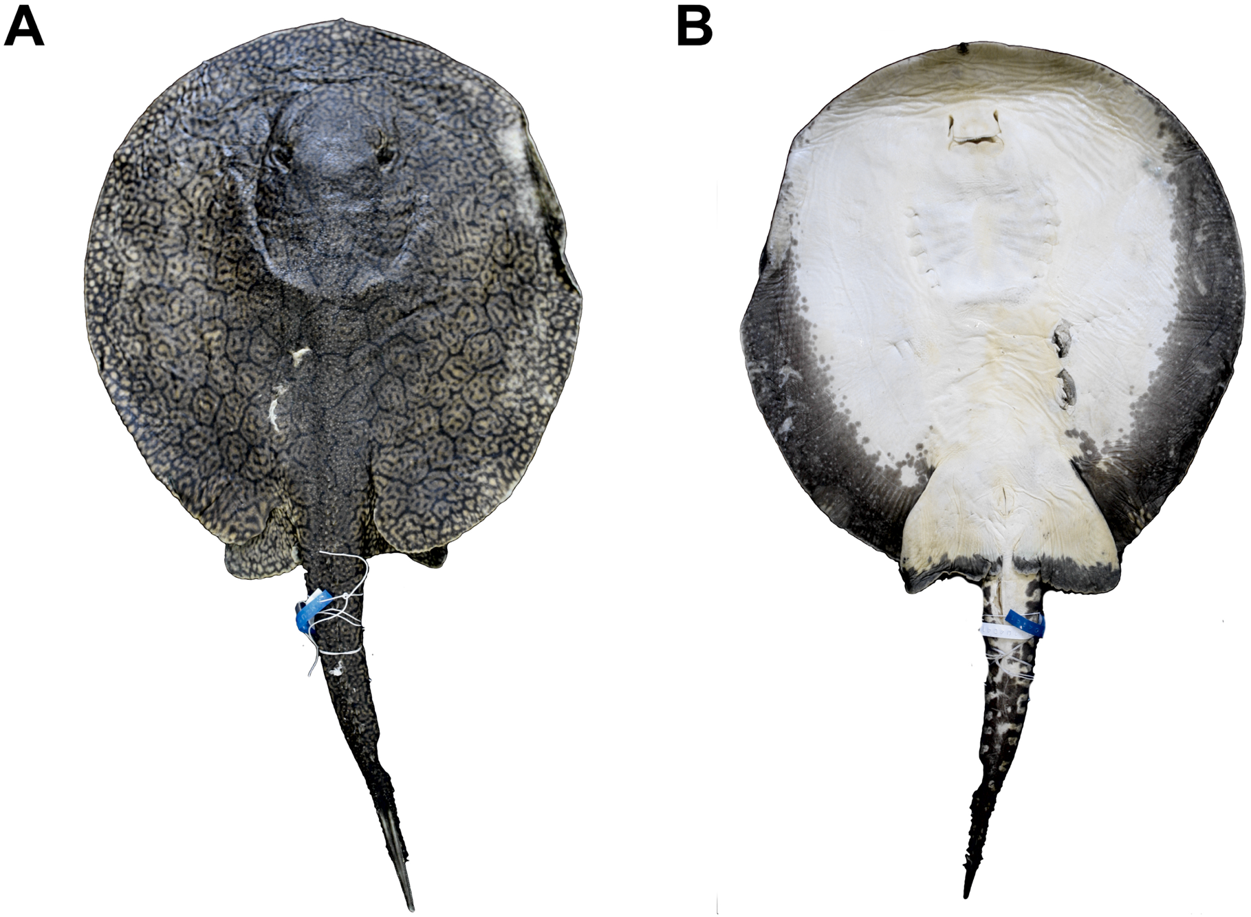
Paratypes. MZUSP 104039 (adult male, 349 mm DW), Madeira River basin, Jamari River, Represa Samuel, municipal district of Itapoã do Oeste, upper Amazon Basin, state of Rondônia, Brazil, 9º4’33.96” S, 63º18’17.64” W, May 2006, coll. F. P. L. Marques, N. M. Luchetti, V. M. Bueno and T. Loboda ( Figure 2 View FIGURE 2 ); MZUSP 104040 (preadult male, 356 mm DW), same data as MZUSP 104039; MZUSP 104041 (?preadult female, 356 mm DW), same data as MZUSP 104039 ( Figure 3 View FIGURE 3 ).
Non type-material. MZUSP 104032 (adult female, 447 mm DW), Madeira River basin, Jamari River, Represa Samuel, municipal district of Itapoã do Oeste, state of Rondônia, Brazil, 9º4’33.96” S, 63º18’17.64” W, May 2006, coll. F. P. L. Marques, N. M. Luchetti, V. M. Bueno and T. Loboda; MZUSP 104046 (juvenile male, 290 mm DW), same data as MZUSP 104032; MZUSP 104060 (juvenile male, 316 mm DW), same data as MZUSP 104032; MZUSP 104036 (adult female, 478 mm DW), same data as MZUSP 104032; MZUSP 104058 (adult female, 429 mm DW), same data as MZUSP 104032; MZUSP 104062 (juvenile male, 306 mm DW), same data as MZUSP 104032; MZUSP 104067 (juvenile male, 276 mm DW), same data as MZUSP 104032; MZUSP 104066 (not measured), same data as MZUSP 104032; MZUSP 104076 (adult female, 380 mm DW), same data as MZUSP 104032; MZUSP 104069 (juvenile male, 277 mm DW), same data as MZUSP 104032; MZUSP 104072 (adult female, 414 mm DW), same data as MZUSP 104032; MZUSP 104059 (preadult male, 365 mm DW), same data as MZUSP 104032; MZUSP 104065 (juvenile male, 300 mm DW), same data as MZUSP 104032; MZUSP 104034 (adult female, 541 mm DW), same data as MZUSP 104032; MZUSP 104073 (adult female, 500 mm DW), same data as MZUSP 104032; MZUSP 104035 (adult female, 500 mm DW), same as MZUSP 104032; MZUSP 104074 (adult female, 648 mm DW), same data as MZUSP 104032; MZUSP 104042 (juvenile female, 295 mm DW), same data as MZUSP 104032; MZUSP 104044 (juvenile male, 298 mm DW), same data as MZUSP 104032; MZUSP 104068 (juvenile male, 219 mm DW), same data as MZUSP 104032; MZUSP 104033 (adult male, 359 mm DW), same data as MZUSP 104032; MZUSP 104071 (not measured), same data as MZUSP 104032.
Diagnosis. Potamotrygon limai, sp. nov., is distinguished from other Potamotrygon species by the following combination of characters: dorsal disc with a dark brownish background, covered with beige to whitish, closely packed small spots roughly arranged in small concentric patterns, these wider toward disc margins, without ocelli; whitish spots may be closely set forming vermicular patterns; lower back portion of disc with a characteristic roughly polygonal pattern, interspersed with small light spots; rostral dermal denticles fairly simple, composed of a single central crown and star-shaped base, with central and posterior disc denticles presenting star-shaped crown ridges, mostly with anterior and lateral dichotomies; two to three irregular rows of hook-shaped spines on dorsal tail midline. Compared to other amazonian Potamotrygon species, Potamotrygon limai, sp. nov., is distinguished from P. motoro (Müller & Henle, 1841) , P. boesemani Rosa, Carvalho & Wanderley, 2008 , P. signata Garman, 1913 , P. leopoldi Castex & Castello, 1970 , P. henlei (Castelnau, 1855) , and the doubtful Potamotrygon ocellata (Engelhardt, 1912) by lacking any form of discrete dorsal ocelli or circular or irregular spots larger than eyediameter. From P. schroederi Fernandez-Yépez, 1958 and P. tigrina Carvalho, Sabaj Pérez & Lovejoy, 2011 , P. limai , sp. nov., is distinguished by presenting small dorsal beige to white spots arranged in concentric patterns, slightly greater than eye-diameter, and without banded pattern of alternating yellow and dark brown bands on tail (vs. much larger spots in vivid yellow with black pigmentation pattern and evidently banded tail). From P. orbignyi (Castelnau, 1855) and P. h um ero s a Garman, 1913, P. limai , sp. nov., is distinguished by its characteristic dorsal color pattern composed of small spots forming whitish polygonal patterns, especially on lower back region (vs. a broad, dark reticulate pattern spread over most of disc, usually without accessory spots). A relatively smaller head region, longer tail, and also by the lack of disc tubercles (sometimes present in latter species). From P. falkneri Castex & Maciel, 1963 and P. tatianae Silva & Carvalho, 2011 , P. limai , sp. nov., is differentiated by a darker brown dorsal backgroung color, its characteristic polygonal vermicular pattern on posterior disc, and shorter tail (means of 86.3% DW, against 93.5% DW for P. falkneri and 109% DW for P. tatianae ). Potamotrygon limai, sp.
nov., is further distinguished from P. s c o b i n a Garman, 1913 by the lack of dorsal ocelli, by a much shorter tail (means of 86.3% DW vs. 121.5% DW), by a broader tail base (means of 14.8% DW vs. 13.4% DW), and by presenting 2−3 rows of midtail spines (against 1−2 rows in P. s c o b i n a). Furthermore, apart from P. scobina , P. limai sp. nov., is the only species of Potamotrygon with three angular cartilages (all other species have one or two).
Description. (Morphometric and meristic data presented in Tables 1−2 View TABLE 1 View TABLE 2 ).
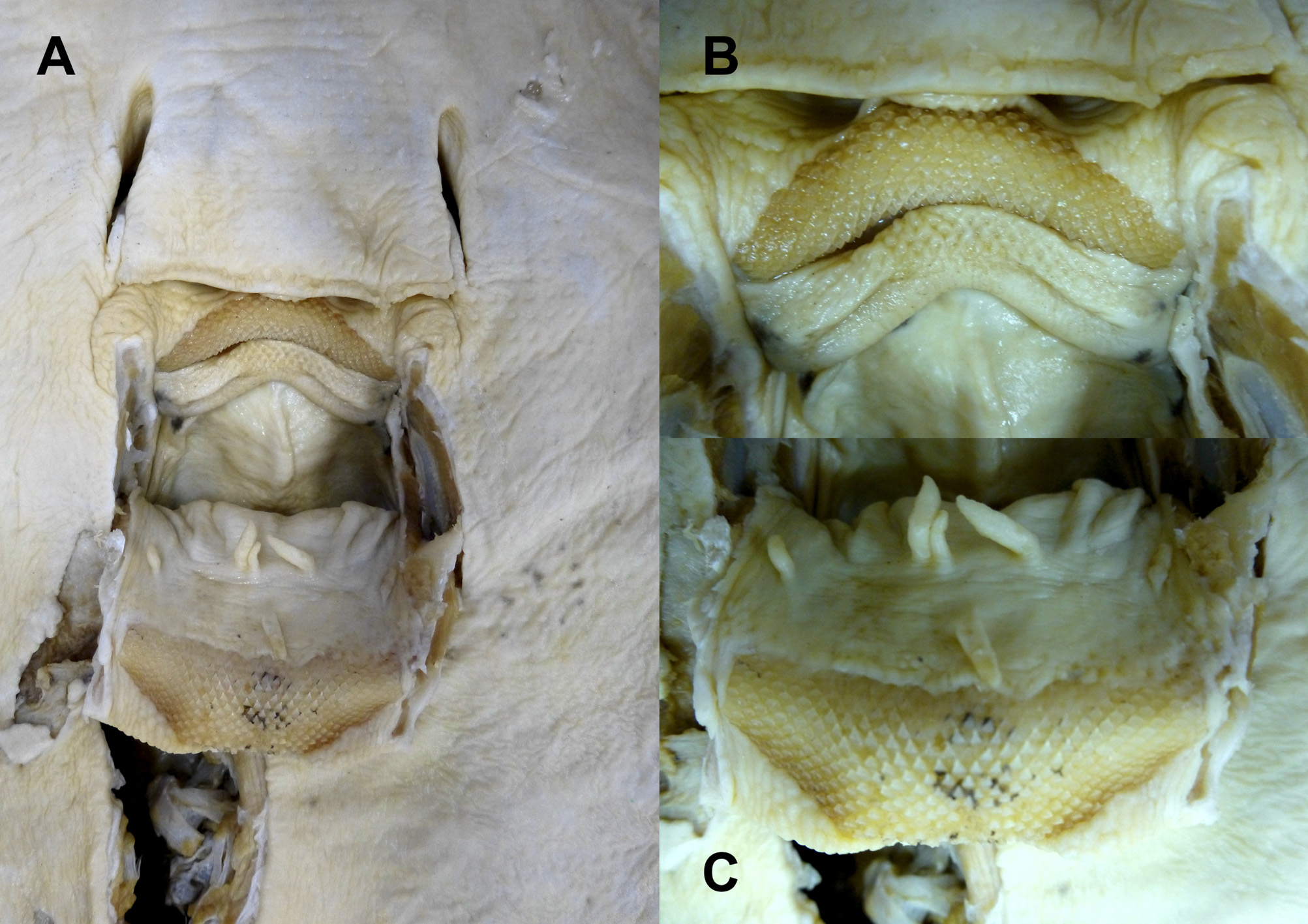
External morphology. Disc oval, slightly longer than wide (DL varying from 98 to 107.8% DW) ( Figures 1−3 View FIGURE 1 View FIGURE 2 View FIGURE 3 ). Rostral portion of disc convex, presenting a small round protuberance on snout. Disc dorsoventrally compressed, with margins much more slender than disc center. Eyes small and oval, around 2 to 2.5 times smaller than spiracles; spiracles oblique and markedly oval or trapezoidal ( Figure 4 View FIGURE 4 a). Head region approximately one-third of disc length, with interorbital distance ranging from 14.3 to 17.4% DW, and interspiracular distance ranging from 15.0 to 19.2% DW ( Figure 4 View FIGURE 4 a). Nasal curtain partially covering mouth and posteriorly fringed ( Figure 5 View FIGURE 5 a). Mouth small and lightly undulated (mouth width ranging from 7.0 to 10.8% DW), the same width as the internasal space ( Figure 5 View FIGURE 5 a). No labial folds or ridges present. Five buccal papillae present, two posterior and three anterior ( Figure 6 View FIGURE 6 a). Branchial basket wider than long, with space between first branchial slits 22.8 to 29.2% DW, and distance between fifth branchial slits 16.6 to 21.7% DW. Teeth small and numerous in each jaw ( Figures 6 View FIGURE 6 b, 6c), wider than long and set in quincunx in a narrow, arched upper tooth plate and a wide and trapezoidal lower tooth plate. Tooth rows varying from 36 to 48 in both arches, with usually the same number of rows for both jaws in each specimen examined. Adult males presenting a single central pointed cusp on central teeth of both jaws, but more predominant on lower jaw. Laterally, juvenile male and female teeth simple, presenting a single rounded cusp.
Pelvic fins wide (ranging from 47.8 to 62.6% DW), subtriangular, with a rounded and undulated posterior margin ( Figure 5 View FIGURE 5 b). Pelvic fins usually exposed in dorsal view from under the disc. Length of anterior margins of pelvic fins ranging from 22.9 to 29.2% DW. Claspers dorsoventrally depressed, robust, presenting a rounded tip ( Figure 5 View FIGURE 5 c). Dorsal pseudosiphon narrow and set dorsolaterally on clasper. Ventral pseudosiphon in terminal position. Clasper groove long, running obliquely to pelvic fins, and curving medially posterior to dorsal pseudosiphon.
Tail width ranging from 12.0 to 19.1% DW ( Figures 4 View FIGURE 4 b, 4c). Tail long, narrowing considerably after origin of caudal sting, but not abruptly. Internal cartilaginous rod elongate, originating at level of caudal sting origin. Tail presenting abundant but somewhat slender dorsal enlarged spines, especially on anterior portion (see below); lateral caudal spine rows starting from level of caudal sting insertion ( Figure 4 View FIGURE 4 d). Distal tail with dorsal and ventral membranous caudal folds. Caudal stings varying in length from 17.4 to 29.2% DW.
Coloration. Dorsal disc background generally dark brownish, with different patterns of spots and dorsal markings, but always presenting the same pattern over lower back, anterior dorsal and lateral portions of tail. Dorsal pattern consisting of concentric or polygonal arrangements formed by very small whitish spots, and with a small central unpigmented region. These sets represent small reticular or convoluted patterns, roughly resembling honeycombs, organized in pairs, side by side; paired arrangements markedly organized and symmetrical at caudalbase region. Some specimens, however, may present a variant pattern with paired arrangements separated longitudinally by a single hexagonal pattern. Color pattern of rest of disc variable, with light irregular colored spots, radially set, without a darker halo, larger than eyes in diameter. Innermost portion of disc presenting irregular light colored spots, smaller than marginal disc spots, forming closely packed rosettes. Disc sometimes completely covered by characteristic whitish pattern over caudal-base region with radially set spots visibly larger and presenting more vibrant tones on central disc region. Marginal portion of disc with small irregular spots forming vermicular patterns, with small region without unpigmented area at center.
Ventral disc with lateroposterior margins covered by dark blotches, with some adults presenting light colored spots over dark blotches. Dark color pattern does not reach anterior margin of disc, extending to level of first pair of branchial slits. Older (larger) specimens more intensely pigmented on ventral disc.
Tail presents a dark dorsal background, equivalent to disc. Anteriormost tail presents characteristic polygonal reticular pattern of light colored markings. Posteriorly on tail, small groupings composed of small light colored spots present, without any apparent concise organization. On lateral tail, small loose groupings formed by very small light colored irregular spots present over dark background. Ventrally, tail with a light background, covered by dark blotches, almost completely covering ventral surface. Dark blotches become larger from lateral tail to tail center. Older (larger) specimens more pigmented.
Pelvic fins present on dorsal surface same pattern seen on dorsal margins of disc, except for a small light colored posterior margin. On ventral side, pelvic fins light colored with posterior dark margin. Claspers also light colored but with dark blotches posteriorly.
Dermal denticles. Disc covered with differently shaped dermal denticles in three distinct regions ( Figure 7 View FIGURE 7 ). Rostral region presenting smaller and simpler denticles, with a unique pointed crown. Basal plate star-shaped, composed of five basal ridges, converging at central crown but not symmetrically ( Figures 7 View FIGURE 7 a, 7b). Cranial and middisc region with star-shaped denticles, composed of anteroposteriorly arranged crown plates with a small pointed cusp on posterior extremity. Two posterior coronal ridges present, sometimes with a coronal dichotomy. Anterior to crown plate, two coronal ridges laterally placed and opposite to each other ( Figure 7 View FIGURE 7 c). Each of these ridges may present a tiny terminal coronal dichotomy ( Figure 7 View FIGURE 7 d). Denticles on this region numerous and variously sized and oriented. Caudal region with denticles presenting anteroposteriorly arranged and prominent crown plates ( Figure 7 View FIGURE 7 e), with cusps opposing two converging posterior coronal ridges; very small terminal dichotomies sometimes present. Two simple and opposite coronal ridges facing cusps posteriorly. Coronal ridges always significantly smaller than central coronal plate ( Figure 7 View FIGURE 7 f).
Ventral lateral line canals. Hyomandibular canal originating anteriorly to nasal curtain posterior margin, extending almost linearly to anterior margin of disc ( Figures 8 View FIGURE 8 a, 8b). Hyomandibular canal curves laterally, running parallel to anterior margin of disc where many short and straight anterior subpleural tubules project from hyomandibular canal. Hyomandibular canal then extends posteriorly, in an almost 90 ° angle, as the subpleural component of hyomandibular canal, visibly straight with few undulations, extending to level of last branchial slit. Subpleural loop, the posteriormost part of hyomandibular canal, occurs at posterior disc. Posterior subpleural tubules apparently absent. Jugular component projects anteriorly towards branchial basket, and deflects at an approximately 60 ° angle externally, just posterior to level of fifth branchial slit. The angular component of hyomandibular canal external to branchial slits, with a light undulation at level of first branchial slit. Jugular canal extends medially, in a 75 ° angle, presenting intense undulations and reaching the posterior jugular loop. From this loop, a short and straight nasal canal projects medially. Orbitonasal component of supraorbital canal highly undulated, projecting medially, with accentuated prenasal loop at its extremity. Infraorbital canal parallel to jugular canal, projecting linearly anteriorly and curving medially, parallel to hyomandibular canal. Suborbital component of infraorbital canal elongate and slightly curved towards middle of disc, projecting from suborbital loop in an angle over 120 °. Suborbital component then deflects in a 175 ° angle to extend to anterior disc margin, undulated. Next to it, subrostral component of supraorbital canal present as, a straight segment extending towards mouth and terminating as the nasoinferior loop. Prenasal component parallel to subrostral component.
Neurocranium. Neurocranium slightly elongated, with elliptical and ventrolaterally expanded nasal capsules (nc) ( Figures 9–11 View FIGURE 9 View FIGURE 10 View FIGURE 11 ). Anterior margins of nasal capsules oval and convex, presenting a medial notch. An internal, ventromedial septum divides nasal capsules. Preorbital processes (prp) well developed, nearly triangular and curved posteriorly, situated just behind condyle for antorbital cartilage. Postorbital processes (pop) shelf-like, anterolaterally expanded and located on dorsolateral corners of otic region. Supraorbital processes (sp) small and triangular, situated slightly anterior to postorbital processes and laterally projected. Supraorbital crests (soc) originating posteriorly to preorbital processes and extending posteriorly to level of postorbital processes. Antorbital cartilage condyles (anc) on posterolateral extremities of nasal capsules. Nearly circular precerebral fontanelle (pcf) and cone-like frontoparietal fontanelle (fpf) partially separated by a moderately developed transverse bridge, the epiphysial bar. Together, fontanellae keyhole-shaped, wide and rounded anteriorly and narrowing posteriorly. Hyomandibular facet (hmdf) located on ventrolateral corner of otic region, corresponding to an oval, horizontally positioned depression. Dorsally, neurocranium presents anterior foramen for preorbital canal (afpc) inside an anteriorly oriented fossa at base of preorbital process; several diminute foramina for superficial ophthalmic nerve (sup) pierce supraorbital crest. Paired internal carotid artery foramina (icaf) ventrally situated near lateral edges of neurocranium, just under level of supraorbital processes. Laterally, neurocranium with nearly rounded eye-stalk (es) extending from lateral orbital wall, just posterior to wide and oval optic nerve foramen (II). Oculomotor nerve foramen (III) anterodorsal to the eye-stalk. Trochlear nerve foramen (IV) just above optic nerve foramen. A bridgelike lateral commissure (lc) present external to circular aperture for hyomandibular branch of facial nerve foramen (VII). Posterior foramen for preorbital canal (pfpc) also located dorsally, at junction of nasal capsule and orbit. Anterior cerebral vein foramen (acvf) located anterior to optic nerve foramen. The efferent spiracular artery foramen (esaf) anteroventral to eye-stalk. Relatively wide and circular orbital fissure (obf) located above and slightly anterior to hyomandibular branch of facial nerve foramen (VII). Foramen magnum (fm) ovoid. Deep articular surface (as) and narrow and horizontally arranged occipital condyles (oc), for articulation with synarcual, ventral to foramen magnum. Vagus nerve foramen (X) lateral to foramen magnum, close to its anterior margin; glossopharyngeal nerve foramen (IX) dorsolateral in relation to occipital condyles. Two pairs of lymphatic foramina present dorsally in otic region, the anterior and wider endolymphatic foramen (elf) and posterior perilymphatic foramen (plf).
Jaws and hyomandibular arch. Hyomandibular cartilages (hyo) elongated, anteroposteriorly compressed, articulating with hyomandibular facet on otic region of neurocranium, from where they project anterolaterally ( Figures 12 View FIGURE 12 , 17 View FIGURE 17 ). Hyomandibula articulates with Meckel’s cartilage (Mc) by means of three angular cartilages embedded within robust hyomandibular-Meckelian ligament. Anterior angular cartilage (aac) wider and robust compared to other two angular cartilages. Posterior angular cartilage (pac) visibly slender compared to anterior angular cartilage (about one-fourth of its width). Lateral angular cartilage (lac) small (about one-third of pac), nearly square and associated with outer edge of posterior angular cartilage. Meckel’s cartilage and palatoquadrate (pq) dorsoventrally flattened; antimeres of both arches not fused symphysially, separated by short space containing strong horizontal ligaments. Laterally, between palatoquadrates and Meckel’s cartilages, occurs a circular aperture formed by inferior surface of palatoquadrate and superior portion of Meckel’s cartilage. Palatoquadrate slightly arched, relatively straight on dorsal border and with pronounced convex curvature on ventral margin. Posterior triangular projections (ptp) present close to lateral edges of palatoquadrates. Palatoquadrates become progressively wider in direction of midline. Meckel’s cartilages more arched than palatoquadrates, anteroposteriorly wide, with marked convex posterior margins, and with a pronounced concavity on inner corners that articulate with palatoquadrates. Another marked convex curvature present on external aspect of Meckel’s cartilage for attachment of robust hyomandibular ligament. Ventrolateral processes (vlp) nearly rectangular and projecting posteroventrally from both extremities of Meckel’s cartilages.
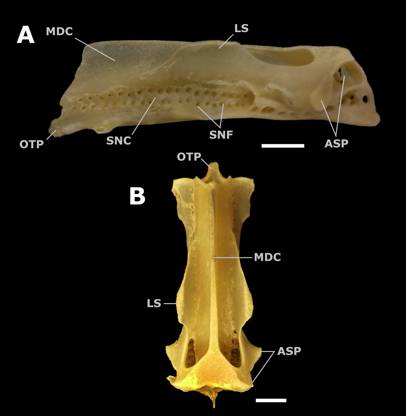
Synarcual cartilage. The cervicothoracic or anterior synarcual articulates with neurocranium between occipital condyles by a median, anterior protuberance at anteroventral extremity of synarcual, the odontoid process (otp) ( Figure 13 View FIGURE 13 ). Synarcual higher anteriorly and decreasing in height towards posterior region. Medial crest (mdc) corresponds to an anteroposterior longitudinal dorsal crest present on synarcual. Spinal nerve foramina (snf) present along lower portion of synarcual, adjacent to lateral bulge of spinal nerve canal (snc). A nearly trapezoidal lateral stay (ls) projects dorsally from lateral wall of synarcual, ceasing at the height of medial crest. Suprascapulae firmly fused posterolaterally on synarcual through a circular socket and oval condyle (asp) for articulation with distal portion of scapular processes.
Pectoral girdle. Coracoid bar dorsoventrally flattened, anterodorsally straight and laterally expanded, located ventral to synarcual ( Figures 14 View FIGURE 14 , 17 View FIGURE 17 ). Four fenestrae for muscles, nerves and condyles for the pectoral pterygia located on lateral surface of pectoral girdle. Pectoral condyles situated along horizontal axis of scapular cartilage. Procondyle (pc) widest pectoral condyle, slender and elliptical, vertically disposed at anterior lateral face of scapula. Mesocondyle (msc) smallest pectoral condyle, oval and horizontally arranged. Metacondyle (mtc) nearly ovoid and situated on lateral aspect of posterior scapula. Anterior fenestrae wider than posterior ones. Nearly triangular anterodorsal fenestrae (adf) dorsal to condyles, and nearly oval and horizontally arranged anteroventral fenestrae (avf) ventral to condyles. Posterodorsal (pdf) and posteroventral (pvf) fenestrae rounded and approximately equal in width, situated dorsal and ventral to posterior margin of mesocondyle, respectively. Scapular processes, comprising dorsal tips of pectoral girdle, firmly articulated with suprascapulae on lateral aspects of synarcual.
Pelvic girdle. Puboischiadic bar (pib) slightly arched, with anterior margin slightly convex and posterior margin markedly concave ( Figures 15 View FIGURE 15 , 17 View FIGURE 17 ). Lateral extremities wide close to four obturator formanina (of). Median prepelvic process (ppp) consists of an anterior medial elongated projection almost reaching the pectoral girdle anteriorly and culminating in a pointed tip. Robust and posterodorsally directed iliac processes (ip) present on posterolateral extremities of puboischiadic bar. These processes pierced by a medial foramen on their proximal portions. Triangular and well developed posteromedially directed isquial processes (isp) located on outer corners of posterior concave margin of pelvic girdle. Triangular, short, robust and anterolaterally directed lateral prepelvic processes (lpp) present on each anterolateral corner of puboischiadic bar. Two condyles on lateral extremity of puboischiadic bar; first condyle wider and oval and second condyle rounded, shorter and just ventral to the first condyle.
Clasper skeleton. First basal segment (b1) wide and nearly square, connecting to basipterygium and second basal segment (b2), and dorsally associated with proximal part of beta cartilage (be). Second basal segment rectangular and linked to proximal part of axial cartilage. Beta cartilage long and narrow, originating at first basal segment and distally articulated with dorsal marginal cartilage. Axial cartilage (ax) somewhat straight, depressed anteriorly and distally cylindrical, tapering toward extremity, and closely associated to ventral margininal and dorsal marginal cartilages. Dorsal marginal cartilage (dm) nearly rectangular, with an oblique anterior margin and a medial notch on its posterior margin. Inner edge of posterior portion of dorsal marginal forming dorsal pseudosiphon externally. Dorsal terminal 2 elongate (dt2), relatively narrow and oval-shaped, with a short ridge present along its proximal portion ( Figures 16 View FIGURE 16 , 17 View FIGURE 17 ). Outer edges of dorsal marginal and dorsal terminal 2 forming clasper groove externally. Accessory terminal (at) elongated and fusiform, underlying dorsal terminal 2, and forming ventral pseudosiphon externally from its outer edge. Ventral marginal cartilage (vm) long and narrow, with prominent anterior tip. Ventral terminal (vt) broad, long and oval, associated dorsally to accessory terminal and posterior portion of ventral marginal.
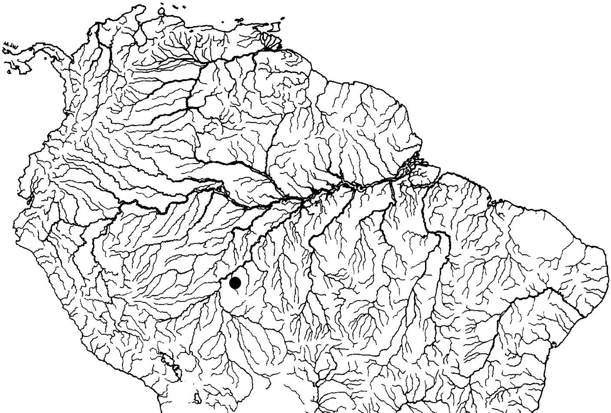
Geographic distribution. Potamotrygon limai is currently known only from the Jamari River, upper Madeira River basin, state of Rondônia, Brazil ( Figure 18 View FIGURE 18 ).
Etymology. This new species is named after Dr. José Lima de Figueiredo, kindly known as Zé Lima, a highly esteemed Brazilian ichthyologist who has contributed immensely to the development of ichthyology in South America. A former researcher and curator of the fish collection of MZUSP, Zé Lima has been a valued mentor and friend of the authors.


| MZUSP |
Museu de Zoologia da Universidade de Sao Paulo |
No known copyright restrictions apply. See Agosti, D., Egloff, W., 2009. Taxonomic information exchange and copyright: the Plazi approach. BMC Research Notes 2009, 2:53 for further explanation.
|
Kingdom |
|
|
Phylum |
|
|
Class |
|
|
Order |
|
|
Family |
|
|
Genus |