
Raveniola micropa ( Ausserer, 1871 )
|
publication ID |
https://doi.org/10.5852/ejt.2018.399 |
|
publication LSID |
lsid:zoobank.org:pub:E836E138-D6E2-4F62-B4B3-CE2E073F2B24 |
|
DOI |
https://doi.org/10.5281/zenodo.5980300 |
|
persistent identifier |
https://treatment.plazi.org/id/03B9B44C-566B-0C39-517E-8068FD354FEE |
|
treatment provided by |
Plazi |
|
scientific name |
Raveniola micropa ( Ausserer, 1871 ) |
| status |
|
Raveniola micropa ( Ausserer, 1871) View in CoL
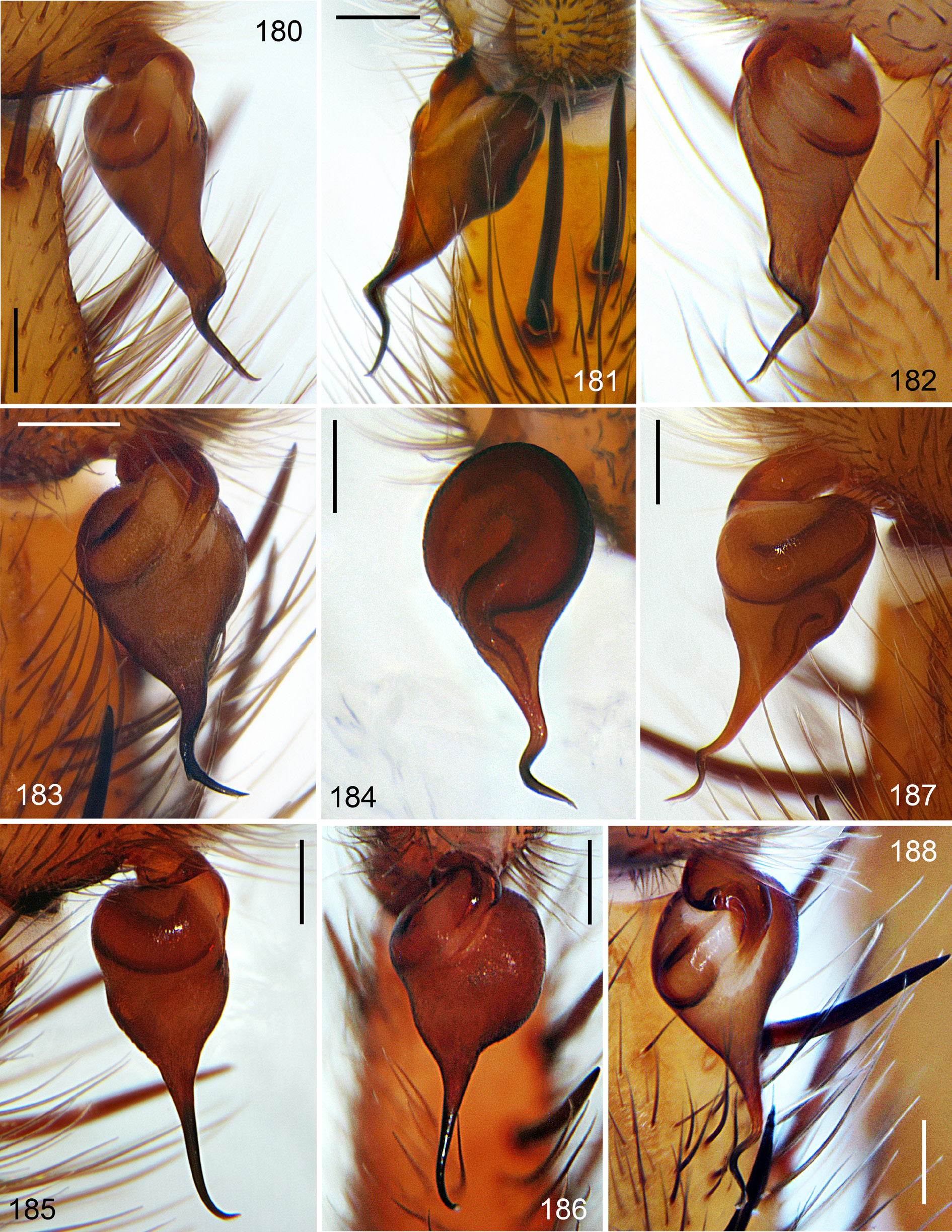
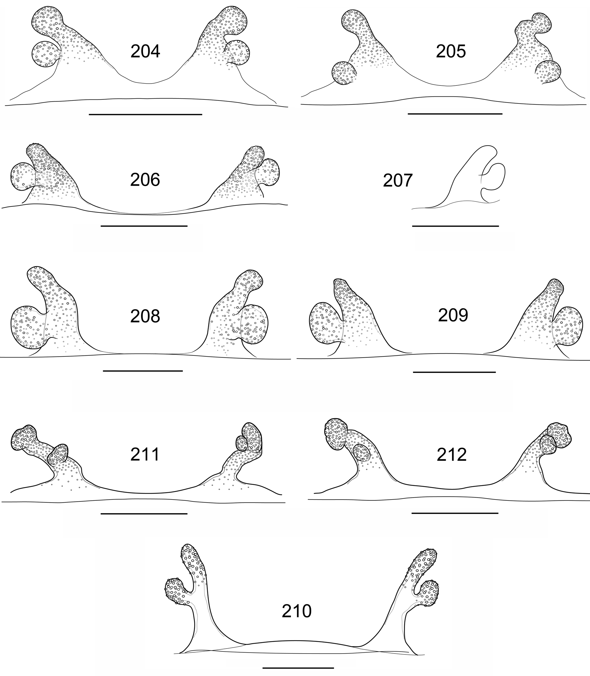
Figs 1, 2 View Figs 1–6 , 11 View Figs 7–12 , 16–17 View Figs 13–18 , 23–24 View Figs 19–24 , 32, 36 View Figs 31–36 , 53–54 View Figs 52–58 , 78 View Figs 74–78 , 91–92 View Figs 90–95 , 111 View Figs 102–116 , 124–125 View Figs 117–128 , 139 View Figs 137–140 , 159 View Figs 157–160 , 187–188 View Figs 180–188 , 206–207 View Figs 204–212 , 221 View Figure221 , 239–246 View Figs 236–241 View Figs 242–247
Brachythele micropa Ausserer, 1871: 177 (♀).
Brachythele micropa – Reimoser 1919: 11. — Roewer 1942: 196. — Bonnet 1955: 912. — Zonstein 1985: 161.
Raveniola microps – Zonstein 1987: 1015 (an unjustified emendation). — Platnick 1989: 90.
Raveniola micropa View in CoL – Le Peru 2011: 86.
Diagnosis
In the structure of its embolus, this species is similar to Raveniola arthuri , but differs from the latter by having a considerably longer and less wide proximal part of the embolus ( Figs 187–188 View Figs 180–188 ; cf. Fig. 184 View Figs 180–188 ). In the shape of their spermathecae, however, females of R. micropa resemble those of R. zaitzevi from another species group (the female of R. arthuri is unknown), differing from them in having noticeably broader spermathecal bases and shorter median and smaller lateral receptacles ( Figs 206–207 View Figs 204–212 ; cf. Figs 208–209 View Figs 204–212 ).
Material examined
Holotype TURKEY: ♀, labelled: “ Brachythele micropa Auss. Brussa A D. 1863 , A.N. I. 16 leg. Mann, 1 ex.” = environs of Bursa ( NMW 58 ).
Additional material (2 ♂♂, 4 ♀♀, 3 juvs)
TURKEY: 1 juv., Bursa Province, Oylat, 39°55′59″ N, 29°35′20″ E, 620 m, 23 Sep. 2010, S.L. Zonstein leg. ( TAU); GoogleMaps 2 ♂♂, 4 ♀♀, 2 juvs, same province, Keles district, Uludağ Mts, vicinity of Baraklı Lake , 39°58′02″ N, 29°15′19″ E, 1050 m, 25 Sep. 2010, K.B. Kunt, Yu.M. Marusik, E.A. Yağmur and S.L. Zonstein leg. ( AUZM, TAU). GoogleMaps
Description
Male (Uludağ Mts near Bursa; here described for the first time)
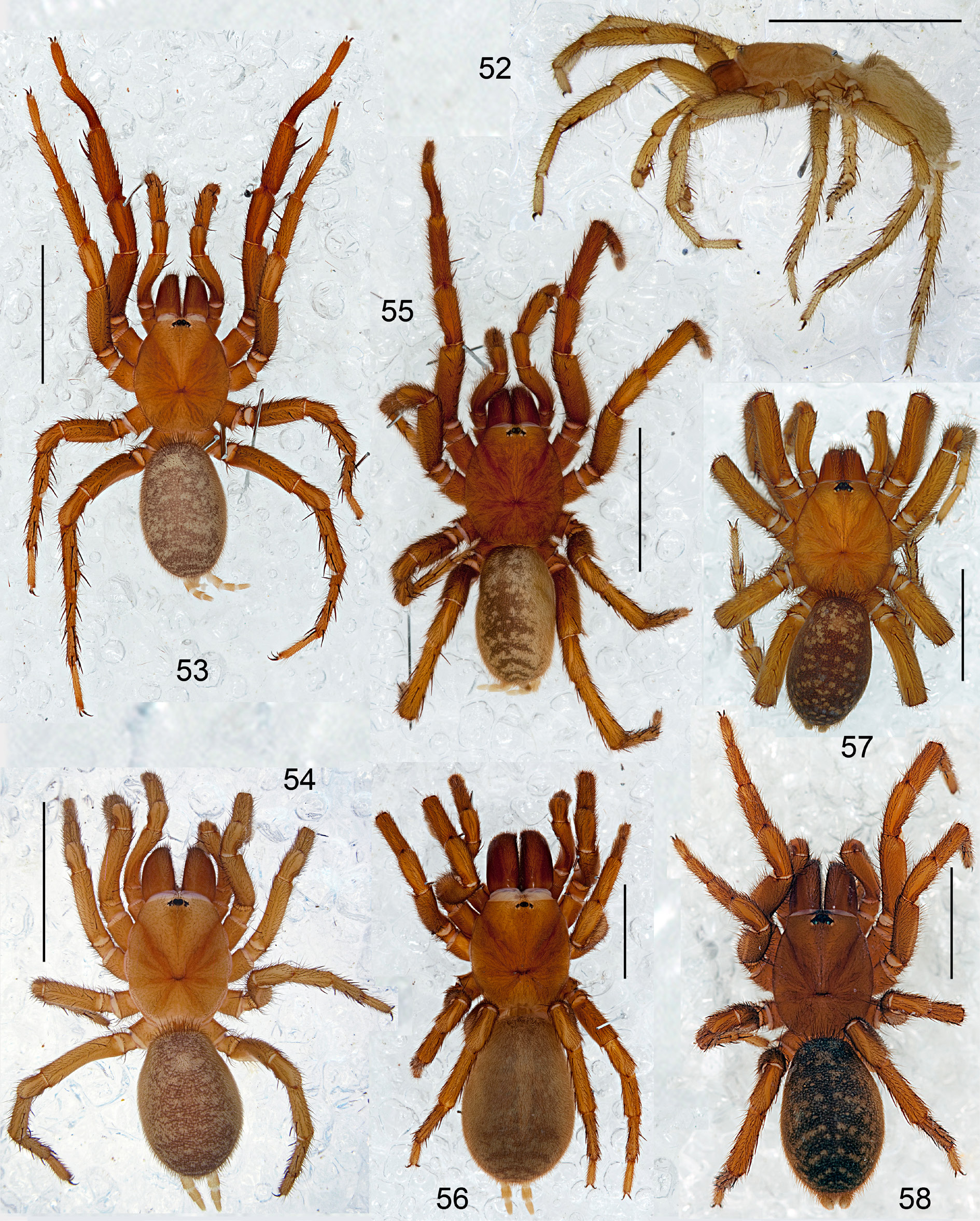
HABITUS. See Fig. 53. View Figs 52–58
MEASUREMENTS. TBL 11.07, CL 4.34, CW 3.60, LL 0.38, LW 0.74, SL 2.17, SW 1.98.
COLOUR. Carapace, chelicerae, palps and first pair of legs dorsally light rufous brown; palp and leg I slightly darker in colour; eye tubercle with dark brown spots surrounding AMEs and lateral eyes; chelicerae light brownish red; sternum, labium, maxillae and legs II–IV light brownish reddish; abdomen and spinnerets pale yellowish brown; diffuse darker dorsal pattern consisting of partially fused transverse brown fasciae mixed with numerous small irregular lighter spots.
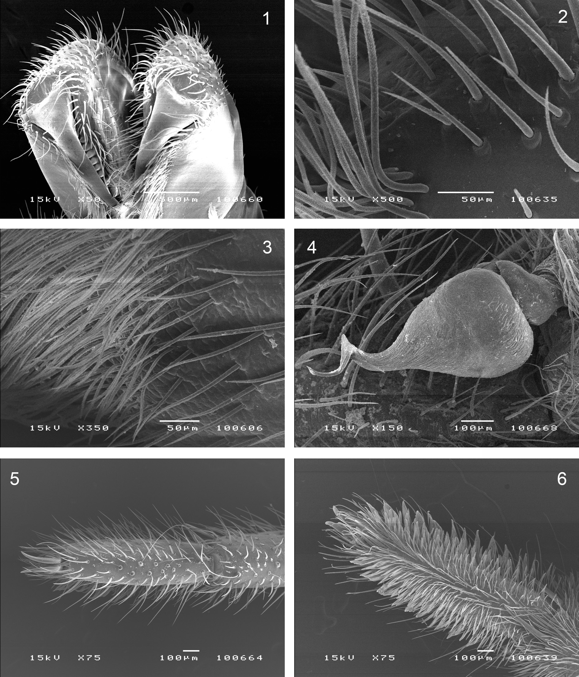
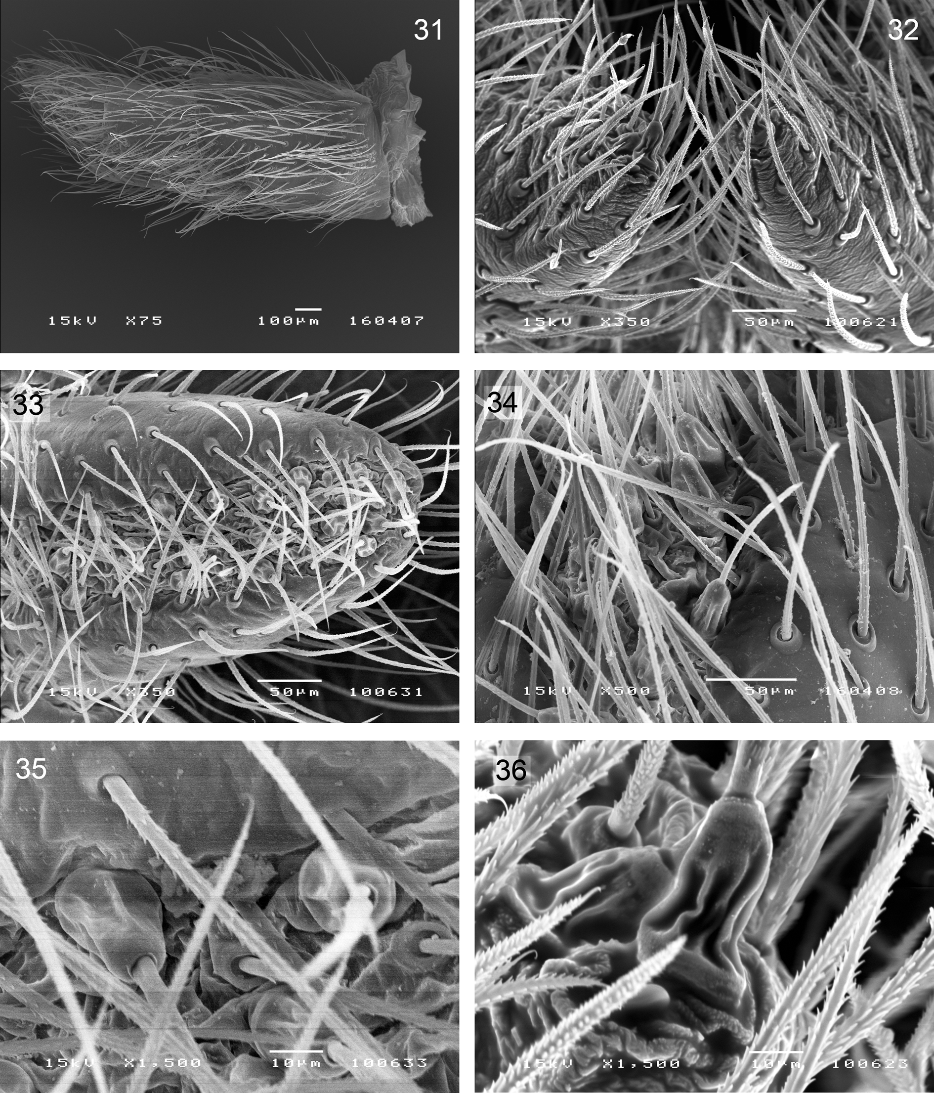
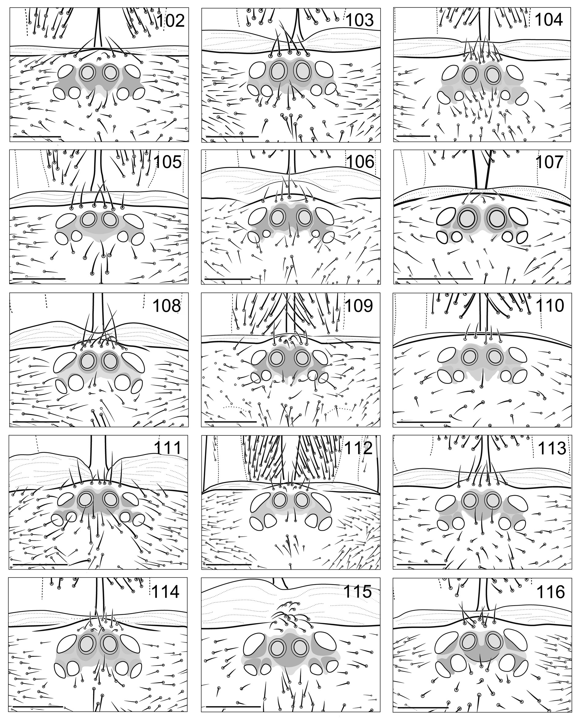
PROSOMA. Carapace and chelicerae as shown in Fig. 78 View Figs 74–78 . Clypeus and eye group as in Fig. 111 View Figs 102–116 . Eye diameters and interdistances: AME 0.12(0.16), ALE 0.17, PLE 0.13, PME 0.09, AME–AME 0.12(0.08), ALE–AME 0.07(0.05), ALE–PLE 0.08, PLE–PME 0.04, PME–PME 0.40. Each cheliceral furrow with 10 promarginal teeth and 5–6 mesobasal denticles. Maxillae with 7–8 cuspules each.
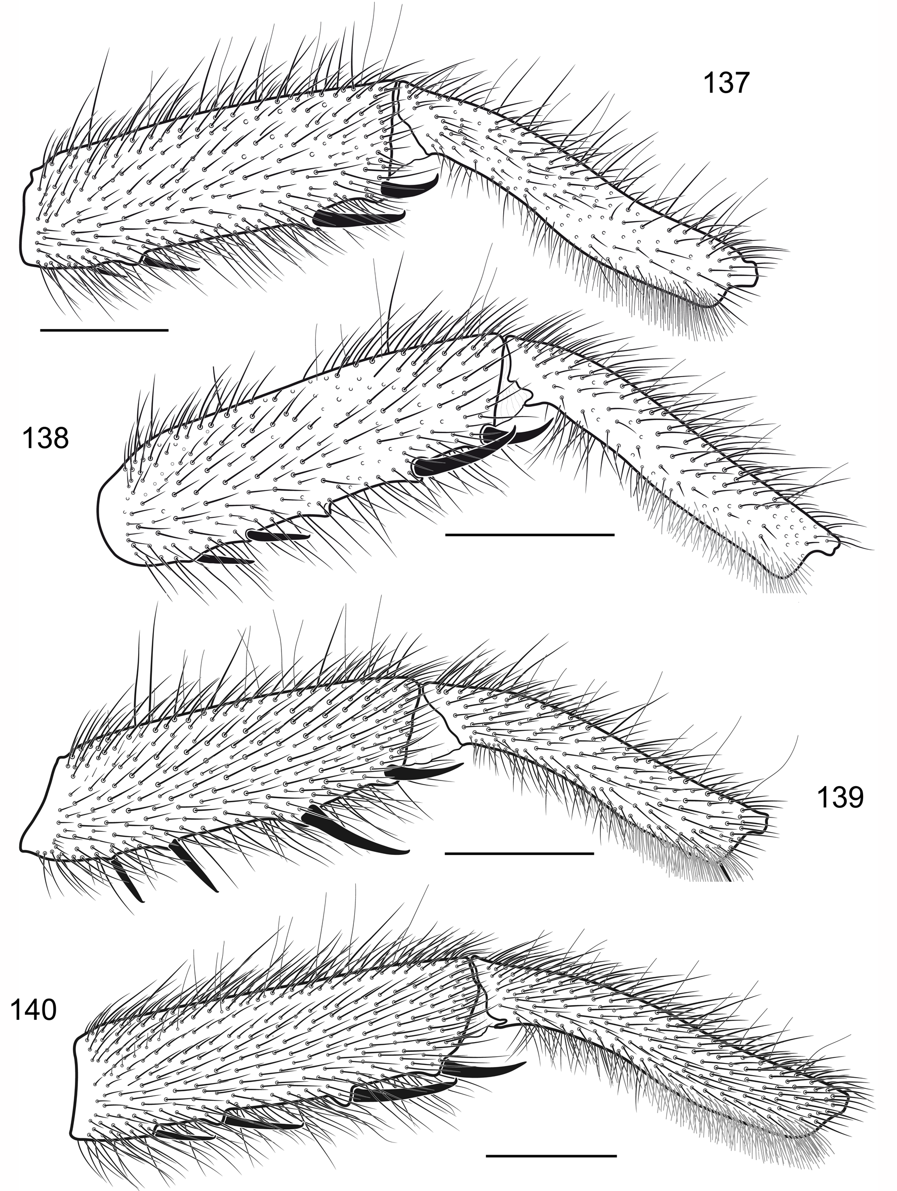
LEGS. Tibia and metatarsus I as shown in Fig. 139 View Figs 137–140 . Scopula: long, very scarce and distal on metatarsus I, long, entire and rather thin on tarsi I and II; elsewhere absent. Trichobothria: 2 rows of 8–9 each on tibiae, 9–10 on metatarsi, 9–10 on tarsi, 7 on cymbium. Paired claws: inner and outer margins each with 6–7 teeth on legs I and II, with 7–8 teeth on legs III and IV.
SPINATION. Palp: femur d1–1–1–1, pd1; patella p1; tibia d1–1, p1–1–1, r0–0–2(1), v3(2)–2–2; cymbium d5. Leg I: femur d1–1–1–1, pd0–0–1; tibia p1–1(0)–0, pv1–1(0), rv0–1–m–m; metatarsus v1(0). Leg II: femur d1–1–1–1, pd1–1–1; tibia p1–1–1, v1(2)–2–3; metatarsus p1, v1–2–2–3. Leg III: femur d1–1–1–1, pd1–1–1, rd1–1–1; patella p1, r1; tibia d1–1–0, p1(0)–1–1, r1–1(0)–1, v2–2–3; metatarsus d0–1–0, p1–1–1–1, r1–1–1, v2–2–3. Leg IV: femur d1–1–1–1–1, pd0–0–1, rd0–0–1; patella r1; tibia d1–1–0, p1–1–1, r1–1–1, v2–2–3; metatarsus pd1–1–0, p1–1–1–1, r1–1–1–1, v2–1(2)–2–3. Patellae I and II aspinose.
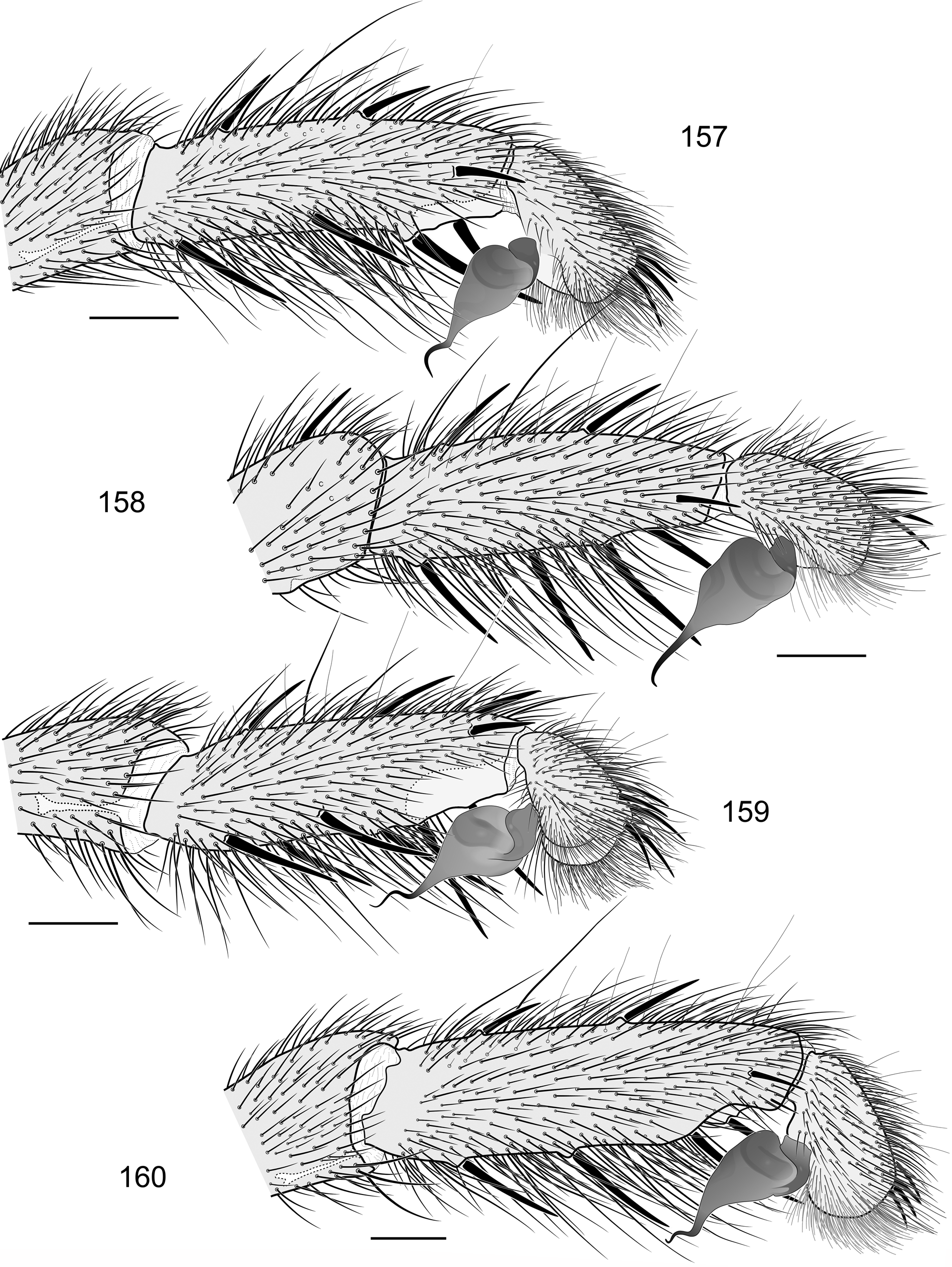
PALP. Tibia, cymbium and palpal organ as shown in Fig. 159 View Figs 157–160 . Embolic keel lost; embolus corkscrewshaped, with bend located subapically ( Figs 187–188 View Figs 180–188 ).
SPINNERETS. PMS: length 0.35; diameter 0.17. PLS: maximal diameter 0.35; length of basal, medial and apical segments 0.58, 0.43, 0.55; total length 1.56; apical segment shortly digitiform.
LEG MEASUREMENTS. ♂(♀).
| Femur | Patella | Tibia | Metatarsus | Tarsus | Total | |
|---|---|---|---|---|---|---|
| Palp | 2.25 (2.20) | 1.33 (1.40) | 1.78 (1.77) | – | 0.79 (1.45) | 6.15 (6.82) |
| Leg I | 3.46 (3.20) | 2.15 (2.10) | 2.66 (2.35) | 2.55 (2.20) | 1.56 (1.28) | 12.38 (11.13) |
| Leg II | 3.17 (2.85) | 1.87 (1.90) | 2.24 (1.95) | 2.08 (2.15) | 1.51 (1.28) | 10.87 (10.13) |
| Leg III | 3.20 (2.20) | 1.60 (1.55) | 1.92 (1.73) | 2.63 (2.67) | 1.59 (1.45) | 10.49 (9.60) |
| Leg IV | 4.35 (3.35) | 1.93 (1.95) | 2.78 (2.67) | 3.74 (3.30) | 1.80 (1.75) | 13.74 (13.02) |
Female (holotype)
HABITUS. Habitus of females from Uludağ Mts as shown in Figs 54 View Figs 52–58 , 244–246. View Figs 242–247
MEASUREMENTS. TBL 10.40, CL 4.75, CW 3.97, LL 0.47, LW 0.95, SL 2.33, SW 2.03.
COLOUR. Carapace, palps and legs light brown, chelicerae light reddish brown, sternum pale yellowish brown; abdomen yellowish grey with brown dorsal pattern consisting of medial lanceolate stripe connected with 3 pairs of inclined and poorly developed lateral chevron-like bands.
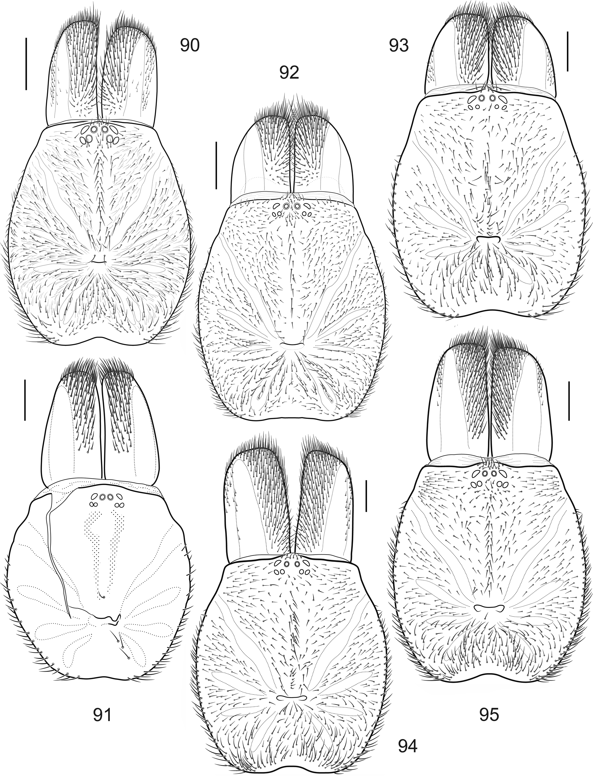
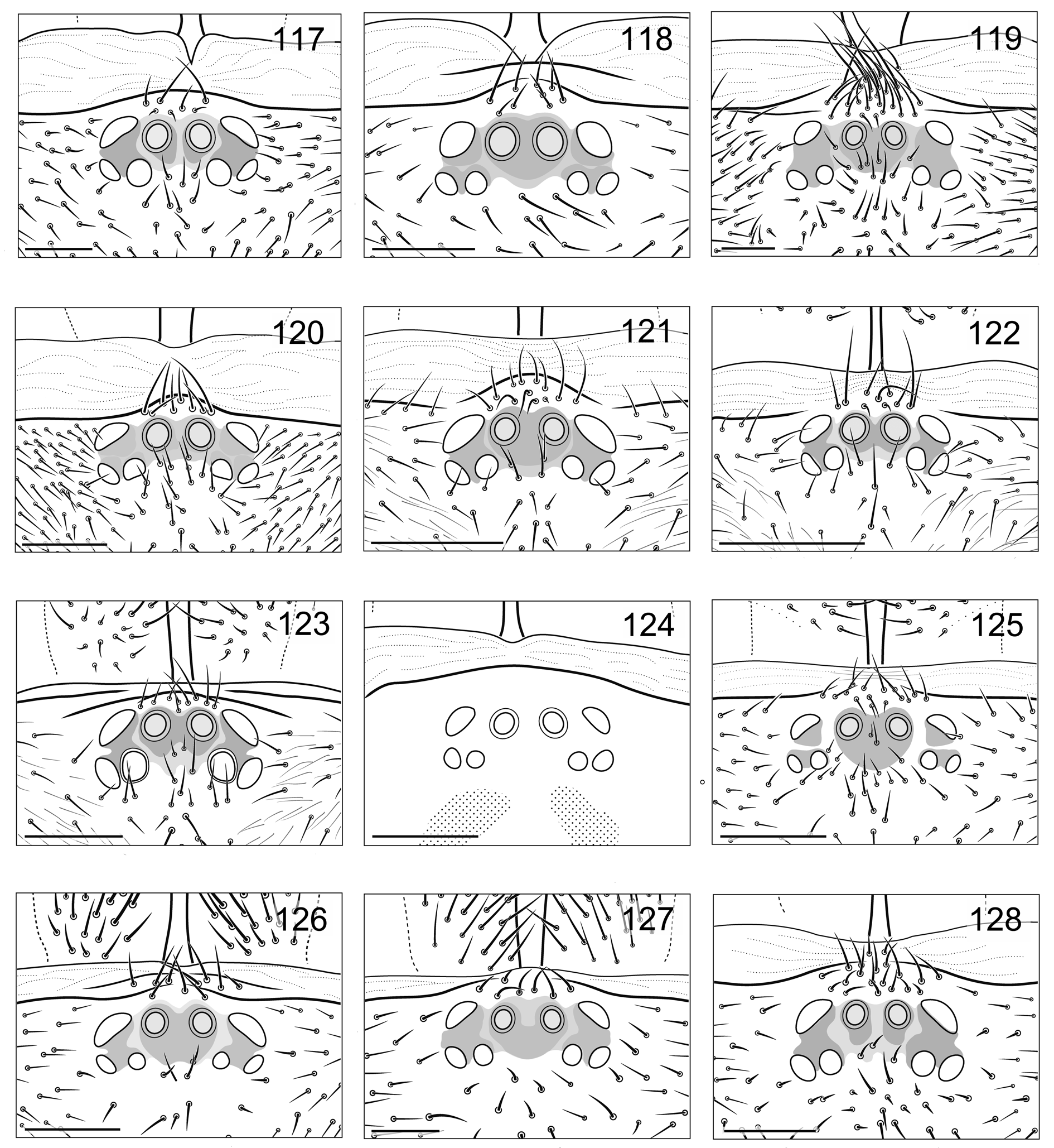
PROSOMA. Carapace and chelicerae as shown in Fig. 91 View Figs 90–95 (conspecific female shown in Fig. 92 View Figs 90–95 ). Clypeus and eye group as in Fig. 124 View Figs 117–128 (conspecific female, Fig. 125 View Figs 117–128 ). Eye diameters and interdistances: AME 0.13(0.17), ALE 0.22, PLE 0.16, PME 0.11, AME–AME 0.16(0.12), ALE–AME 0.16(0.14), ALE– PLE 0.14, PLE–PME 0.02, PME–PME 0.31. Each cheliceral furrow with 9–10 promarginal teeth and 3 mesobasal denticles. Maxillae with 5–6 cuspules each.
LEGS. Scopula: entire on distal metatarsus I and tarsi of palp and leg I, narrowly divided on tarsus II. Trichobothria: 2 rows of 7 each on tibiae, 9–11 on metatarsi, 7–8 on tarsi. Paired tarsal claws with outer and inner teeth rows consisting of 5–6 teeth each. Palpal claw with 5 promarginal teeth.
SPINATION. Femora I–IV dorsally with 3 bristles; patellae I, II, IV and palpal patella aspinose. Palp: femur d1, pd1; tibia v2–2–4; tarsus v3–2. Leg I: femur pd1; tibia v1–1–2; metatarsus v2–2–2. Leg II: femur pd1; tibia v1–1–3; metatarsus v2–2–3. Leg III: femur pd1; patella p1, r1; tibia d1, p1, r1–1, v2–2–3; metatarsus p1–2–2, r1–1–1, v2–2–3. Leg IV: femur pd1; tibia d1, p1–1–1, r1, v2–2–3; metatarsus p1– 1–1–1, r1(0)–1–1, v2–2–3.
SPERMATHECAE. As shown in Fig. 207. View Figs 204–212
SPINNERETS. PMS: length 0.35, diameter 0.15. PLS: maximal diameter 0.40; length of basal, medial and apical segments 0.63, 0.33, 0.37, respectively; total length 1.33; apical segment shortly digitiform.
Variation
Carapace length in males varies from 4.34 to 4.85, in females from 3.97 to 4.69. Spermathecae show a quite insignificant variation (see Figs 206–207 View Figs 204–212 ).
Ecology
The spiders collected in the surroundings of Oylat and Baraklı Lake were found hiding in soil cavities under stones in broad-leaved mountain forest dominated by Quercus sp. ( Figs 239–246 View Figs 236–241 View Figs 242–247 ).
Distribution
Western Turkey (Bursa Province). See Fig. 221 View Figure221 .


| TAU |
Israel, Tel Aviv, Tel Aviv University |
No known copyright restrictions apply. See Agosti, D., Egloff, W., 2009. Taxonomic information exchange and copyright: the Plazi approach. BMC Research Notes 2009, 2:53 for further explanation.
|
Kingdom |
|
|
Phylum |
|
|
Class |
|
|
Order |
|
|
Family |
|
|
Genus |
Raveniola micropa ( Ausserer, 1871 )
| Zonstein, Sergei, Kunt, Kadir B. & Yağmur, Ersen A. 2018 |
Brachythele micropa
| Ausserer 1871: 177 |
Brachythele micropa
| Ausserer 1871 |