
Metamastigophorophyllon lamellohirsutum, Antić, Dragan Ž. & Makarov, Slobodan E., 2016
|
publication ID |
https://doi.org/10.11646/zootaxa.4211.1.1 |
|
publication LSID |
lsid:zoobank.org:pub:6B86C6BA-6AFE-4AAD-870D-04794C138D47 |
|
DOI |
https://doi.org/10.5281/zenodo.6074447 |
|
persistent identifier |
https://treatment.plazi.org/id/03B7878C-FFA8-CDBD-FF4C-ECD9B53AEE08 |
|
treatment provided by |
Plazi |
|
scientific name |
Metamastigophorophyllon lamellohirsutum |
| status |
sp. nov. |
Metamastigophorophyllon lamellohirsutum View in CoL sp. nov.
Figs 105–109 View FIGURE 105 View FIGURE 106 View FIGURE 107 View FIGURE 108 View FIGURE 109
Diagnosis. Differs from the other Metamastigophorophyllon species by the presence of a highly hairy sternal lamella with a raised, medial, bifurcated part and by the presence of strongly developed, wide and lamellar outer branches of the lateral part of the anterior gonopods or by the presence of certain characteristics on male leg-pair 7: a coxal cavity, a prefemoral, posteromesal, acuminate process or a femoral, short, basal process.
Etymology. An adjective; to emphasize the presence of a strongly developed, wide and very hairy anterior sternal lamella of the anterior gonopods.
Material studied (total: 32 males, 18 females). Holotype. GEORGIA: male, Mukhura, ca 15 km E of Tkibuli, 700–800 m asl, Castanea , Fagus , Carpinus , etc. forest, litter, under bark and stones, 7–9 May 1987, S. Golovatch & K. Eskov leg. ( ZMUM ρ3370).
Paratypes (total: 31 males, 18 females). All from GEORGIA, Mukhura, ca 15 km E of Tkibuli: 23 males , 15 females (ZMUM ρ3371), 8 males, 3 females ( IZB), same data as holotype.
Type locality. GEORGIA: Mukhura , ca 15 km E of Tkibuli, 700–800 m asl, Castanea , Fagus , Carpinus , etc. forest, litter, under bark and stones.
Description. Body with 31 segments (including telson) in adults.
MEASUREMENTS. Males 13.5–14.5 mm long, vertical diameter of the largest pleurotergite 1–1.05 mm. Females 14–15.5 mm long, vertical diameter of the largest pleurotergite 1.2–1.25 mm.
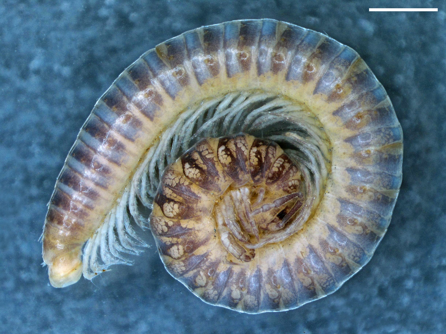
COLORATION ( Fig. 105 View FIGURE 105 ). Dorsal and lateral sides of prozonites greyish, dorsolateral sides with yellowish spots, ventrolateral sides yellowish. Metazonites brownish to greyish, with dorsolateral yellowish spots, ventrolateral sides yellowish. First few body segments darker.
HEAD. Flattened in males. Labrum with three medial teeth and 6+5 labral and 2+2 supralabral setae.
Promentum triangular, without setae. Lingual plates with 8+7 setae, on each plate arranged in two irregular rows. Stipites with ca 25+25 setae. Antennae 1.66 mm long in holotype. Length of antennomeres (in mm): I (0.08), II (0.2), III (0.4), IV (0.24), V (0.4), VI (0.18), VII (0.14) and VIII (0.02). Length/breadth ratios of antennomeres I– VII: I (1), II (1.7), III (4.5), IV (2.4), V (3.1), VI (1.5) and VII (1.5). Antennomeres II, IV, V, VI and VII with one, three, one, four and one sensillum, respectively. Number of ocelli 18–22, arranged in 5 rows in males; 19–23 in 5– 6 rows in females.
COLLUM. Narrower than head, with six macrochaetae. Anterior edge semi-circular, posterior margin gently concave.
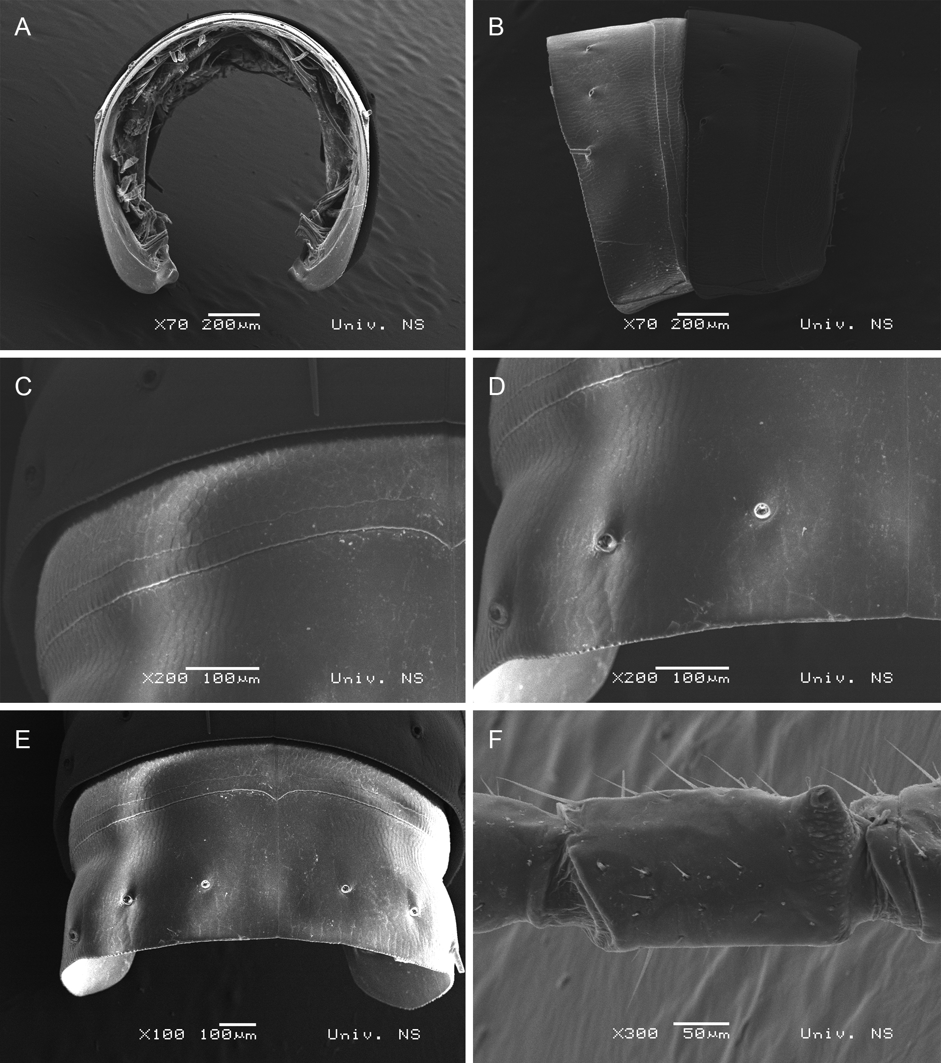
BODY SEGMENTS ( Figs 105 View FIGURE 105 , 106 View FIGURE 106 A–E). Lateral keels not developed; only barely visible lateral swellings present. Macrochaetae short, straight and trichoid, outer somewhat longer than inner and medial. CIX (pleurotergite 15) ~ 0.75; MIX (pleurotergite 15) ~ 0.85; PIX (pleurotergite 15) ~ 0.2; MA (pleurotergite 15) ~ 155˚.
TELSON. Epiproct with a pair of spinnerets and 3+3 setae (1+1 paramedian, 2+2 marginal). Hypoproct with 1+1 apical setae. Paraprocts with 3+3 marginal setae.
WALKING LEGS. In both sexes, leg-pairs 1 and 2 with tarsal combs; prefemora with several long and robust setae; femora and postfemora with a group of several long and robust setae.
MALE SEXUAL CHARACTERS ( Figs 106 View FIGURE 106 F, 107). Leg-pairs 3–7 enlarged. Leg-pairs 3 and 4 each with all podomeres swollen; prefemur with a basal exterior protrusion. Leg-pair 5 with an oral protrusion on prefemur. Legpair 6 without peculiarities. Leg-pair 7 with a coxal cavity; prefemora with a strongly developed, basal, posteromesal, acuminate process, femora with a short basal process. Leg-pair 10 with coxal glands and with coxal horns orientated caudad. Leg-pair 11 with coxal glands, no other peculiarities.
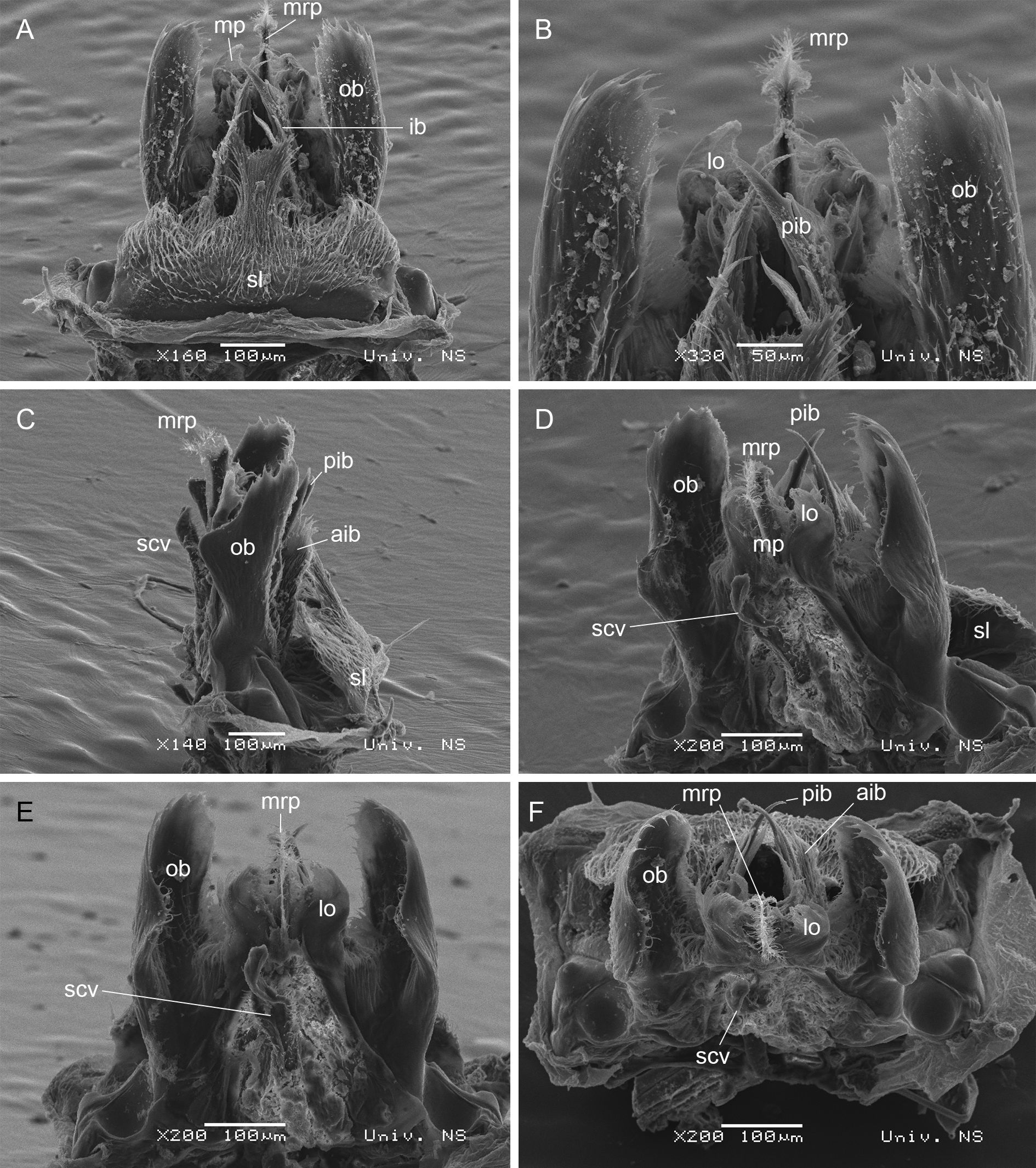
ANTERIOR GONOPODS ( Figs 108 View FIGURE 108 A–C, 109). One of the most striking structures of the anterior gonopods is a strongly developed, very wide and highly hairy anterior sternal lamella ( sl) [= S sensu Mauriès (1982): 387, figs 2 and 3] which represents the widest part of the anterior gonopods. This shield is characterized by one medial, bifurcated and raised part. Outer branches ( ob) [= T sensu Mauriès (1982): 387, figs 1–3] of lateral coxal processes strongly developed, in the form of a shield, lamellar structures, apically with denticles, medial part laterally expanded posteriorly and clothed with setae; setae also present on anterior basal parts of outer branches. Inner branches ( ib) [= T’ sensu Mauriès (1982): 387, figs 1–3] of lateral coxal processes consisting of an anterior part ( aib) in the form of a hairy lamella and a posterior part ( pib) in the form of an acuminate process with anterior denticles and a posterior, small, triangular tooth. Medial part ( mp) [= K sensu Mauriès (1982): 387, figs 1–3] of coxal processes well-developed, characterized by two lateral lobes ( lo) and a medial raised part ( mrp) [= k sensu Mauriès (1982): 387, figs 1 and 3]. Both lateral lobes and raised medial part clothed with setae. The posteriormost part is a medial tongue possibly representing syncoxal vesicle ( scv).
POSTERIOR GONOPODS ( Fig. 108 View FIGURE 108 D). Coxites ( c) divided. Telopodites ( t) present on posterolateral side; clothed with setae. Distal part of coxal processes ( cp) simple, curved mesally and posteriory. Coxal vesicles ( cv) present on anterior side.
Distribution. Georgia (Mukhura, ca 15 km E of Tkibuli) ( Fig. 166 View FIGURE 166 , blue circle).


| ZMUM |
Zoological Museum, University of Amoy |
No known copyright restrictions apply. See Agosti, D., Egloff, W., 2009. Taxonomic information exchange and copyright: the Plazi approach. BMC Research Notes 2009, 2:53 for further explanation.
|
Kingdom |
|
|
Phylum |
|
|
Class |
|
|
Order |
|
|
Family |
|
|
Genus |