
Pseudoflagellophorella mirabilis
|
publication ID |
https://doi.org/ 10.11646/zootaxa.4211.1.1 |
|
publication LSID |
lsid:zoobank.org:pub:6B86C6BA-6AFE-4AAD-870D-04794C138D47 |
|
DOI |
https://doi.org/10.5281/zenodo.6074490 |
|
persistent identifier |
https://treatment.plazi.org/id/03B7878C-FF7B-CD91-FF4C-EA32B51BED8C |
|
treatment provided by |
Plazi |
|
scientific name |
Pseudoflagellophorella mirabilis |
| status |
|
Pseudoflagellophorella mirabilis View in CoL gen. et sp. nov.
Figs 144–146 View FIGURE 144 View FIGURE 145 View FIGURE 146
Diagnosis. The new species differs from the other two species of this genus by the highly unusual anterior gonopods which show fimbriated lateral parts with numerous single, bi- or three-ramous fringes/fingers orientated posteriorly.
Etymology. An adjective; to emphasize the presence of very unusual and magnificent anterior gonopods.
Material studied (total: 1 male, 4 females). Holotype. ABKHAZIA: male, Myussera Nature Reserve , 20–130 m asl, mixed deciduous forest ( Castanea , Alnus , etc.), litter, under bark and stones, 8–10 Apr. 1983, S. Golovatch leg. ( ZMUM ρ3427).
Paratypes (total: 4 females). All from ABKHAZIA: 4 females, same data as holotype ( ZMUM ρ3428).
Type locality. ABKHAZIA: Myussera Nature Reserve.
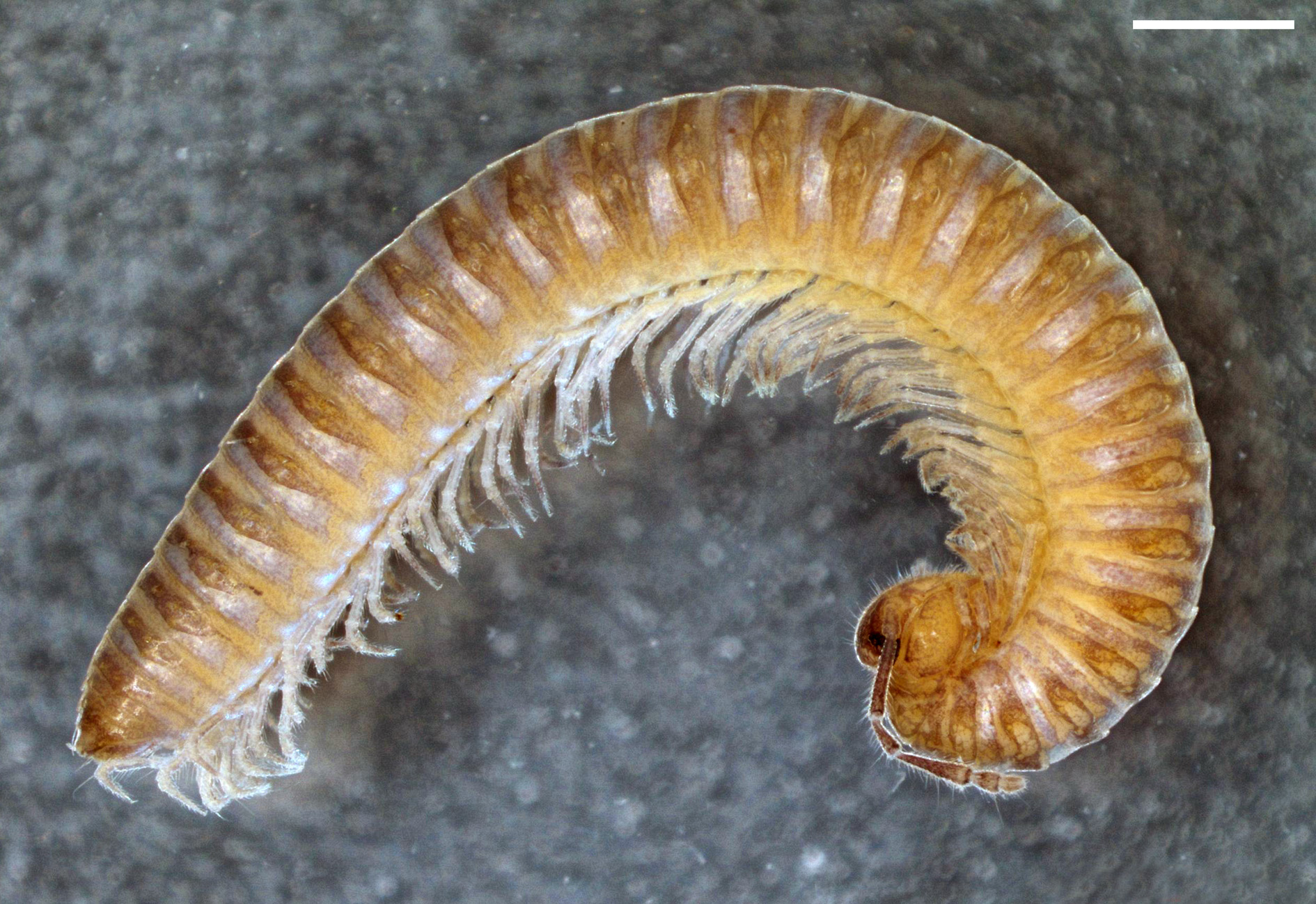
Description. Body with 31 segments (including telson) in adults.
MEASUREMENTS. Holotype male 12.5 mm long, vertical diameter of the largest pleurotergite 1.1 mm. Females 10.5–11.5 mm long, vertical diameter of the largest pleurotergite 1–1.1 mm.
COLORATION ( Fig. 144 View FIGURE 144 ). Prozonites greyish with yellowish ventrolateral spots. Metazonites brownish with yellowish spots on lateral keels.
HEAD. Flattened in male. Labrum with three medial teeth and 4+4 labral and 2+2 supralabral setae. Promentum triangular, without setae. Lingual plates with 6+5 setae, on each plate arranged in 1–2 rows. Stipites with ca 30+30 setae. Antennae 1.9 mm long in holotype. Length of antennomeres (in mm): I (0.08), II (0.12), III (0.51), IV (0.24), V (0.5), VI (0.18), VII (0.16) and VIII (0.02). Length/breadth ratios of antennomeres I–VII: I (0.9), II (1.7), III (5.7), IV (2.2), V (3.8), VI (1.4) and VII (1.6). Antennomeres II, IV, V, VI and VII with one, three, one, four and one sensillum, respectively. Number of ocelli 16–18, arranged in 5 rows in holotype; 14–17 in 5–6 rows in females.
COLLUM. Narrower than head, with six macrochaetae. Anterior edge semi-circular, posterior margin gently concave.
BODY SEGMENTS ( Fig. 144 View FIGURE 144 ). Lateral keels like lateral swellings. Macrochaetae long and trichoid. CIX (pleurotergite 15) ~ 0.8; MIX (pleurotergite 15) ~ 1.3; PIX (pleurotergite 15) ~ 0.7; MA (pleurotergite 15) ~ 85˚.
TELSON. Epiproct with a pair of spinnerets and 3+3 setae (1+1 paramedian, 2+2 marginal). Hypoproct with 1+1 apical setae. Paraprocts with 3+3 marginal setae.
WALKING LEGS. In both sexes, leg-pairs 1 and 2 with tarsal combs; prefemora with several long and robust setae; femora and postfemora with a group of several long and robust setae.
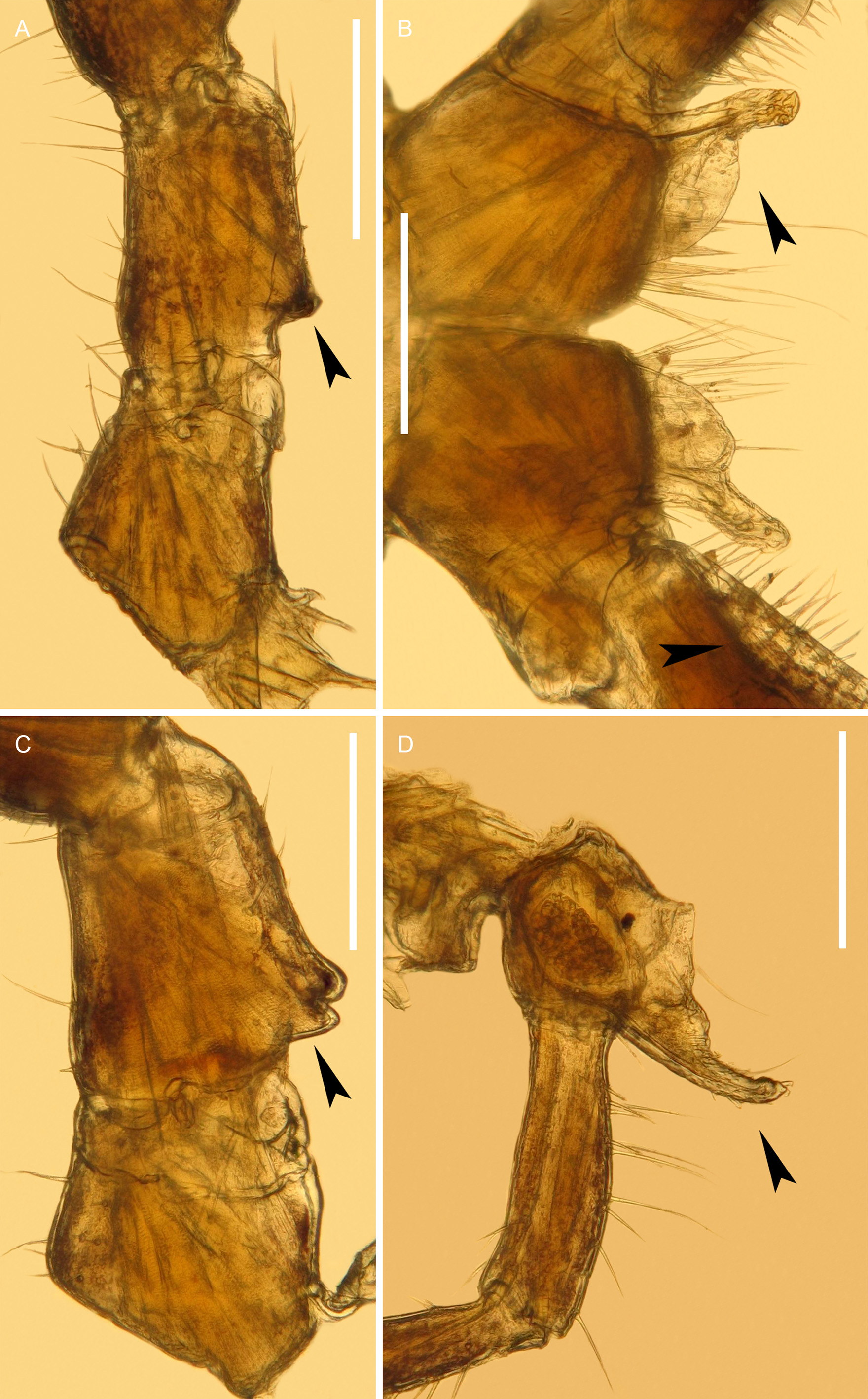
MALE SEXUAL CHARACTERS ( Fig. 145 View FIGURE 145 ). Leg-pairs 3–7 enlarged. Leg-pairs 3 and 4 each with a basal exterior protrusion on prefemur. Leg-pair 5 with a basal oral protrusion on prefemur. Leg-pair 6 with enlarged podomeres, without other peculiarities. Leg-pair 7 with a posterior coxal process, first wide and thin at base, then spatulated, curved orally and outside; prefemur with a longitudinal mesal cavity with numerous setae. Leg-pair 10 with coxal glands and with a well-developed coxal horn, clothed with setulae and with one long seta. Leg-pair 11 with coxal glands, without other peculiarities.
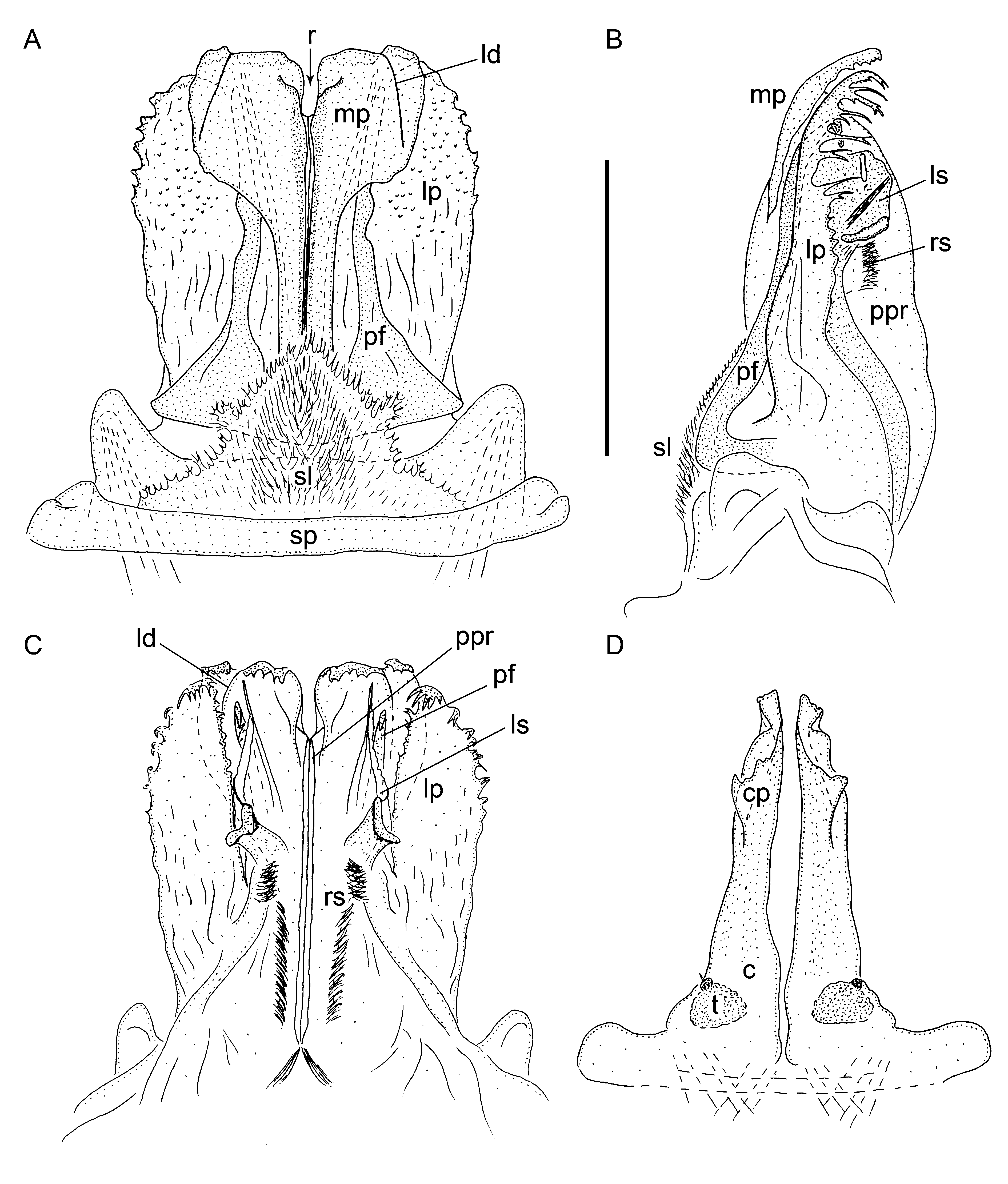
ANTERIOR GONOPODS ( Fig. 146 View FIGURE 146 A–C). Sternal plate (sp) with an anterior, medial, hairy, sternal lamella (sl). Coxal processes with a distal rift (r), connected to each other by longitudinal lamellae, these forming posteriorly a projection (ppr) almost as high as entire anterior gonopods. Gonopods consisting of medial parts (mp) with longitudinal posterior lamellae and of two lateral parts (lp). Medial parts distally with denticles curved posteriorly. Lower half of medial parts narrow, while upper halves much wider and sublaterally with a longitudinal division (ld). Lateral parts lamellar, wide and fimbriated with simple, bi- or three-ramous fringes/fingers curved posteriorly. Both lateral parts anteriorly with a long and relatively slender process (pf) with its apical part sunk below upper halves of medial part. These pairs of processes orientated posteriorly with a downward curved apex. Posteriorly, anterior gonopods with a longitudinal lamellar structure (ls) and rows of setae (rs).
POSTERIOR GONOPODS ( Fig. 146 View FIGURE 146 D). Simple. Coxited (c) divided. Telopodites (t) present on posterior side. Coxal processes (cp) lamellar. Coxal vesicles present anteriorly.
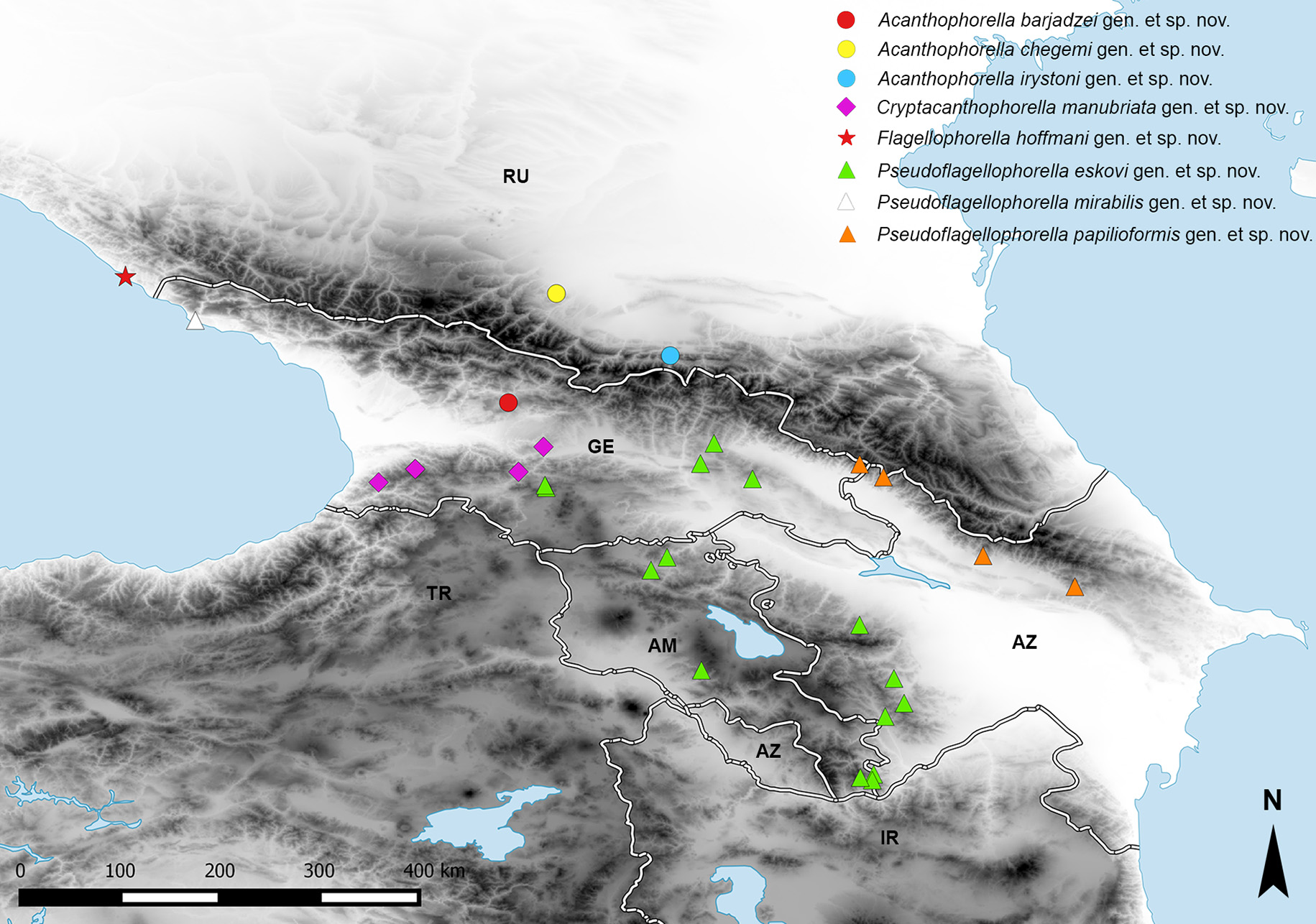
Distribution. Abkhazia (known only from type locality) ( Fig. 169 View FIGURE 169 , white triangle).
| ZMUM |
Zoological Museum, University of Amoy |
No known copyright restrictions apply. See Agosti, D., Egloff, W., 2009. Taxonomic information exchange and copyright: the Plazi approach. BMC Research Notes 2009, 2:53 for further explanation.
|
Kingdom |
|
|
Phylum |
|
|
Class |
|
|
Order |
|
|
Family |
|
|
Genus |