
Flagellophorella hoffmani
|
publication ID |
https://doi.org/10.11646/zootaxa.4211.1.1 |
|
publication LSID |
lsid:zoobank.org:pub:6B86C6BA-6AFE-4AAD-870D-04794C138D47 |
|
DOI |
https://doi.org/10.5281/zenodo.6074482 |
|
persistent identifier |
https://treatment.plazi.org/id/03B7878C-FF77-CD9C-FF4C-ED9BB0E1E899 |
|
treatment provided by |
Plazi |
|
scientific name |
Flagellophorella hoffmani |
| status |
|
Flagellophorella hoffmani View in CoL gen. et sp. nov.
Figs 134–138 View FIGURE 134 View FIGURE 135 View FIGURE 136 View FIGURE 137 View FIGURE 138
Diagnosis. As for the genus.
Etymology. The new species is named after late Dr. Richard Hoffman, one of the most knowledgeable diplopodologists ever.
Material studied (total: 4 males). Holotype. RUSSIA: male Sochi, Khosta, Taxus & Buxus grove, Apr. 2006, Y. Chumachenko leg. ( ZMUM ρ3410).
Paratypes (total: 3 males). All from RUSSIA: 1 male, same data as holotype ( IZB) ; 2 males, same data as holotype, except: Mar. 2006 ( ZMUM ρ3411).
Type locality. RUSSIA: Sochi , Khosta, Buxus & Taxus grove.
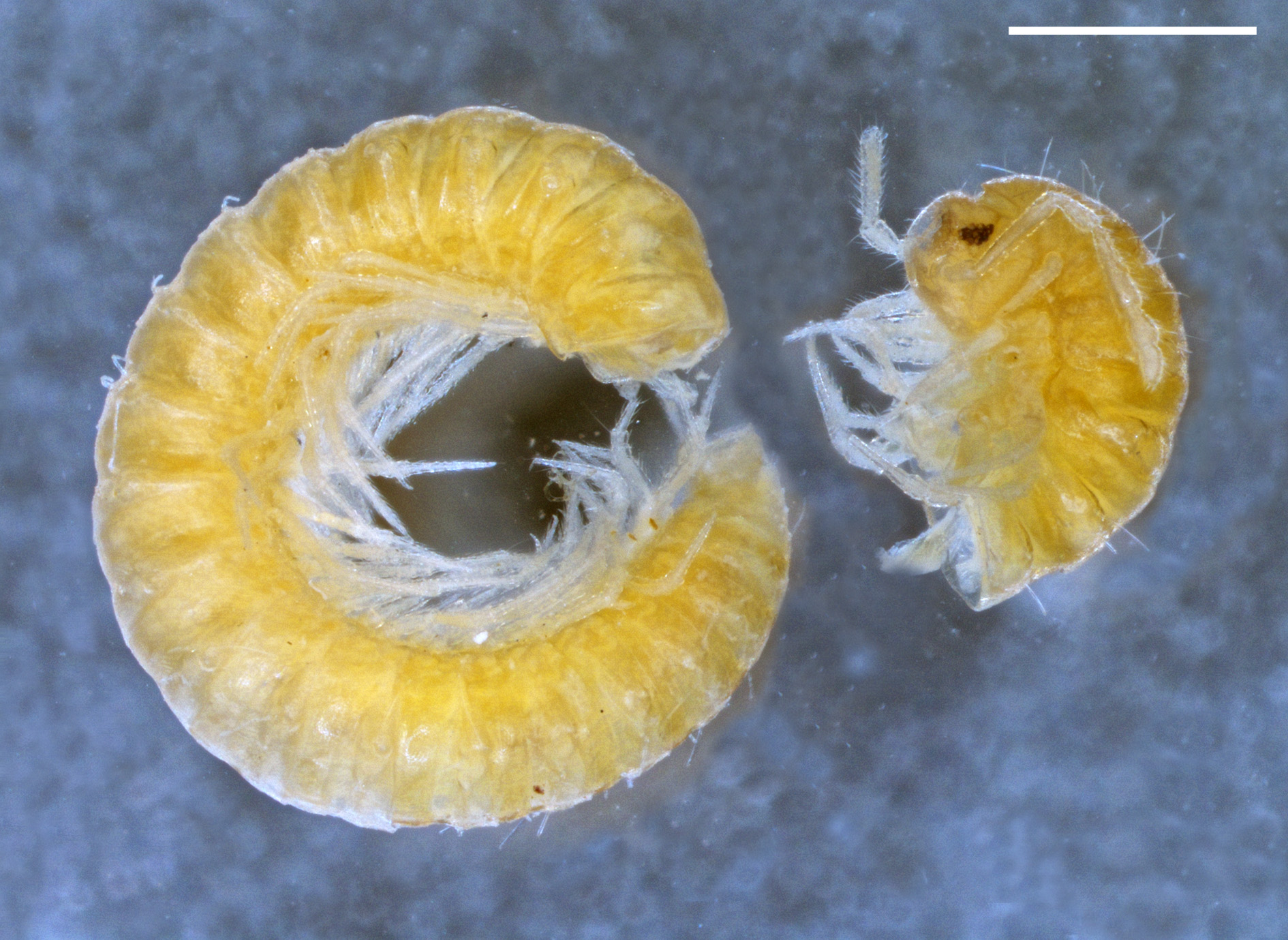
Description. Body with 29 segments (including telson) in adults.
MEASUREMENTS. Holotype male 8.5 mm long, vertical diameter of the largest pleurotergite 0.7 mm.
COLORATION ( Fig. 134 View FIGURE 134 ). Yellowish brown.
HEAD. Flattened in males. Labrum with three medial teeth and 4+4 labral and 2+2 supralabral setae. Promentum triangular, without setae. Lingual plates with 2+2 setae in one row on each plate. Stipites with ca 20+20 setae. Antennae 1.45 mm long in paratype male. Length of antennomeres (in mm): I (0.08), II (0.16), III (0.42), IV (0.16), V (0.35), VI (0.13), VII (0.13) and VIII (0.02). Length/breadth ratios of antennomeres I–VII: I (1), II (1.6), III (5.2), IV (2), V (3.2), VI (1.2) and VII (1.5). Antennomeres II, IV, V, VI and VII with one, three, one, four and one sensillum, respectively. Number of ocelli 13–14, arranged in 4–5 rows in males.
COLLUM. Narrower than head, with six macrochaetae. Anterior edge semi-circular, posterior margin gently concave.
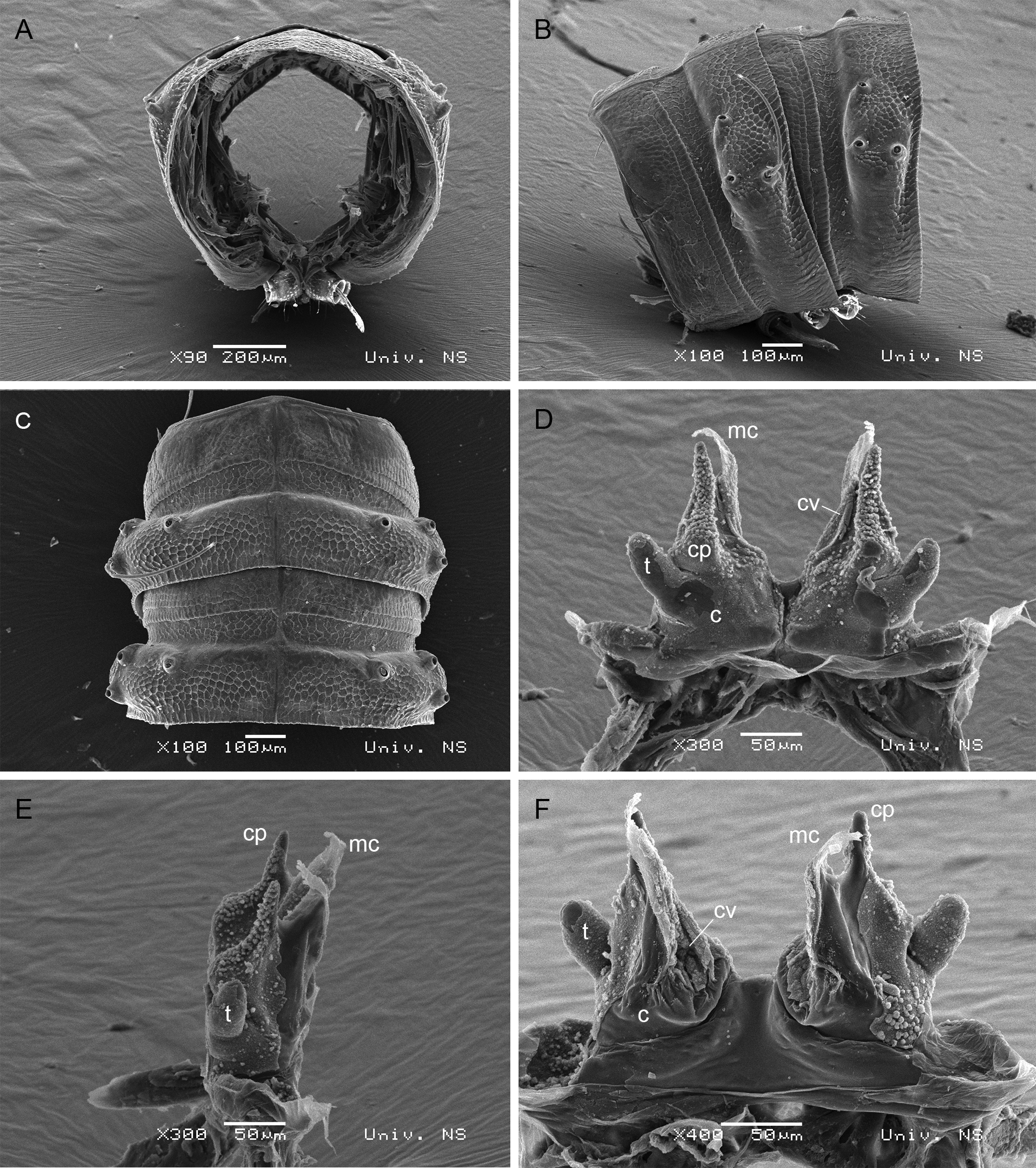
BODY SEGMENTS ( Figs 134 View FIGURE 134 , 135 View FIGURE 135 A–C). Lateral keels like lateral swellings. Macrochaetae long, apically with small spikes. CIX (pleurotergite 15) ~ 0.65; MIX (pleurotergite 15) ~ 2; PIX (pleurotergite 15) ~ 0.4; MA (pleurotergite 15) ~ 90˚.
TELSON. Epiproct with a pair of spinnerets and 3+3 setae (1+1 paramedian, 2+2 marginal). Hypoproct with 1+1 apical setae. Paraprocts with 3+3 marginal setae.
WALKING LEGS. In both sexes, leg-pairs 1 and 2 with tarsal combs; prefemora with several long and robust setae; femora and postfemora with a group of several long and robust setae.
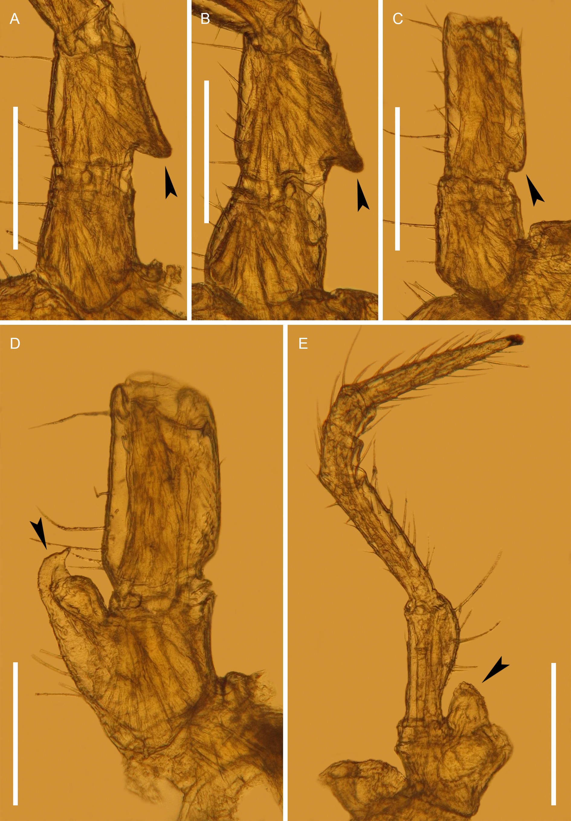
MALE SEXUAL CHARACTERS ( Fig. 136 View FIGURE 136 ). Leg-pairs 3–7 enlarged, especially so leg-pairs 3, 4 and 7. Leg-pairs 3 and 4 each with a basal exterior protrusion on prefemur. Leg-pair 5 with a well-developed, basal, subtriangular, oral protrusion on prefemur. Leg-pair 6 with a small, basal, exterior protrusion on prefemur. Leg-pair 7 with a highly characteristic, bird-shaped, coxal process. Leg-pair 10 with coxal glands and short, wide, coxal processes with one apical seta and small nipples, curved posteriorly; prefemora with a widening. Leg-pair 11 with coxal glands, no other peculiarities.
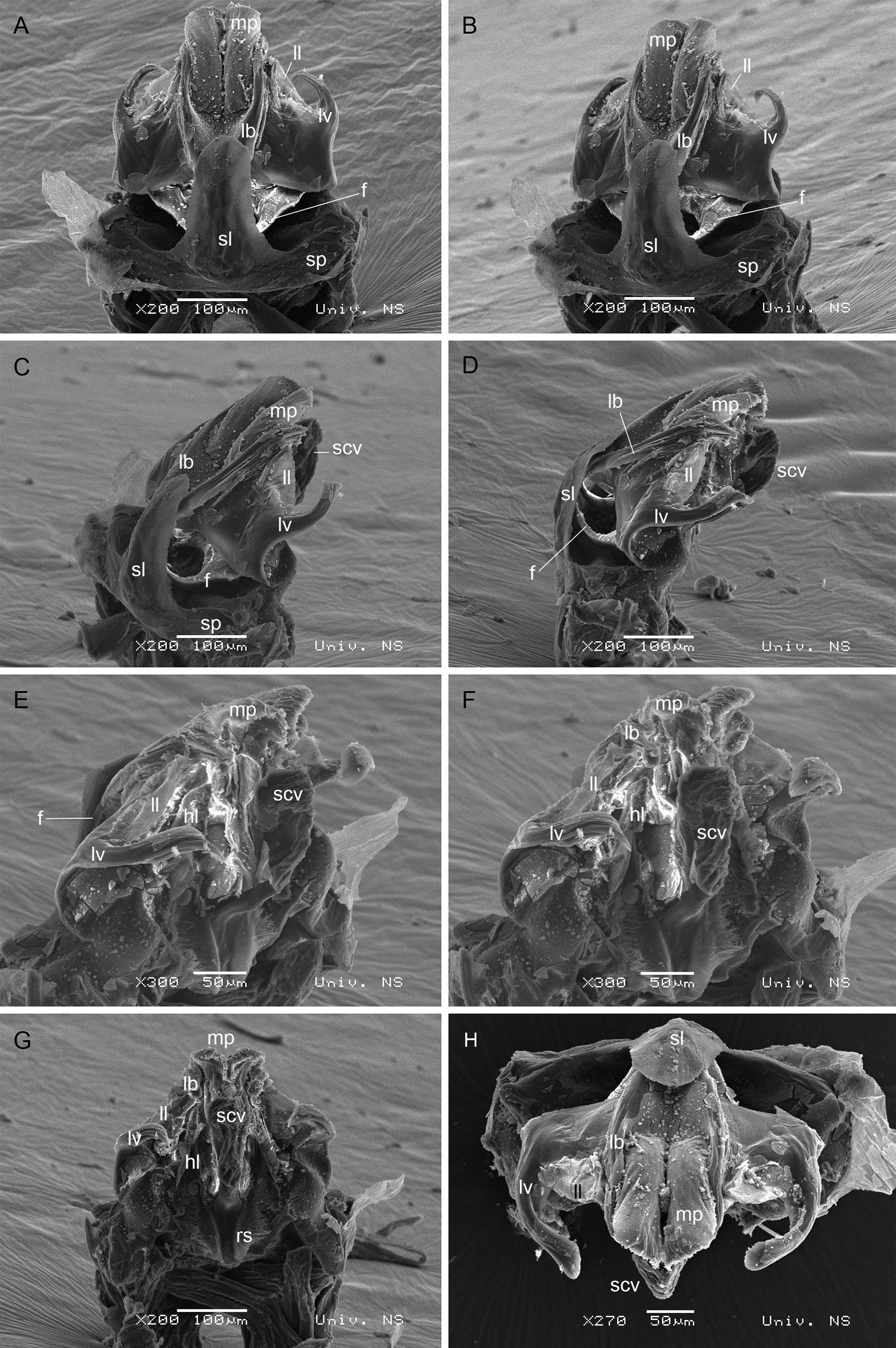
ANTERIOR GONOPODS ( Figs 137 View FIGURE 137 , 138 View FIGURE 138 ). Sternal plate ( sp) with a strongly developed, medial, hairless, spatulated, anterior lamella ( sl) (broken off in holotype). Gonopods divided distally. The most characteristic structure of the anterior gonopods is a pair of flagella ( f) arising from the base with free basal halves, while upper halves inserted in longitudinal bulges ( lb) lying between medial part ( mp) of the gonopods and lateral lamella ( ll), and are characterized by a posteriorly curved apex. Free basal halves of flagella protected by an anterior spatulated lamella ( sl). The lateralmost structure is a lateral lever ( lv) orientated posteromesally. Mesal edges of medial parts folded inside and forming projections ( ppr), the latter connected to a syncoxal vesicle ( scv). A pair of small hairy levers ( hl), a pair of small additional, mesally setose processes ( aps), as well as rows of setae ( rs) can be seen in posterior view.
POSTERIOR GONOPODS ( Fig. 135 View FIGURE 135 D–F). Coxites ( c) divided. Telopodites ( t) present on posterolateral sides. Coxal processes ( cp) with numerous nipples. Anterior side carrying a membranous crest ( mc). Coxal vesicles ( cv) present anteromesally.

| ZMUM |
Zoological Museum, University of Amoy |
No known copyright restrictions apply. See Agosti, D., Egloff, W., 2009. Taxonomic information exchange and copyright: the Plazi approach. BMC Research Notes 2009, 2:53 for further explanation.
|
Kingdom |
|
|
Phylum |
|
|
Class |
|
|
Order |
|
|
Family |
|
|
Genus |