
Trachygamasus gerdi, Witaliński, 2017
|
publication ID |
https://doi.org/10.11646/zootaxa.4303.3.6 |
|
publication LSID |
lsid:zoobank.org:pub:E36DA2B0-A0E3-427F-B101-B3833CBB077E |
|
DOI |
https://doi.org/10.5281/zenodo.6053230 |
|
persistent identifier |
https://treatment.plazi.org/id/534895DF-4177-4761-87F0-4825DBDD4E32 |
|
taxon LSID |
lsid:zoobank.org:act:534895DF-4177-4761-87F0-4825DBDD4E32 |
|
treatment provided by |
Plazi |
|
scientific name |
Trachygamasus gerdi |
| status |
sp. nov. |
Trachygamasus gerdi sp. nov.
( Figs 1–19 View FIGURE 1 View FIGURE 2 View FIGURES 3 – 13 View FIGURES 14 – 19 )
urn:lsid:zoobank.org:act:534895DF-4177-4761-87F0-4825DBDD4E32
Material examined. Holotype female (slide no. 2409), 1 September 2012, campus of Jagiellonian University , Kraków-Ruczaj, Poland, 50 1' N, 19 54' E, alt. ca GoogleMaps . 208 m a.s.l., wet rotten hay 30–50 cm thick, on grassland; 1 female (slide no. 2406), 2 males (slides no. 2407, 2409) paratypes, ibid., 1 male (slide no. 2698) paratype, 7.09.2016, Tylawa (Beskid Niski), south-eastern Poland, 49 27' N, 21 42' E, alt. ca GoogleMaps . 422 m a.s.l., rotten hay ca. 80 cm thick, on clearing in deciduous forest. Types are deposited in the Zoological Museum of the Jagiellonian University, Cracow, Poland .
Diagnosis. Both sexes moderately to well sclerotised, cuticle with many regularly distributed pits; gnathotectum trispinate with central prong broader and more solid than lateral ones, pointed in female and terminally rounded in male; gv1 gland pores not discernible, gland pores gv2 with three openings; setae Z V1 very short, located close to gv2 pores.
In the female, podonotum with three pairs of thickened and terminally pilose setae (j4, r3 and z5), opisthonotum with two such pairs of setae (Z1 and Z4). Sternal setae st2 shorter and thicker than others. Sternal shield and paragynia separated with poorly visible grooves oriented slightly obliquely rather than transversely. Epigynium triangular with convex lateral margins and concave posterior margin. Endogynium hyaline, delicate and poorly visible, elongated axially. Opisthogaster JV4 setae thicker and terminally pilose.
In the male, dorsal setae are simple, larger on opisthonotal shield; setae j4, r3 and z5 are thickened and terminally pilose as in the female. Only opisthonotal Z4 setae thickened and terminally pilose, Z1 simple. All sternal setae are similar and simple. Opisthogaster setation as in the female. Leg II with minute spurs: two on femur and one on genu and tibia. Sternal setae st1 far from anterior sternal margin. Genital opening well sclerotised, halfmoon shaped and located just behind anterior sternal margin. Genital lamina poorly sclerotised with hyaline, long protrusion difficult to discern.
Description. Female. Dorsal idiosoma. Podonotum and opisthonotum separated with poorly visible short suture located behind setae j6 and z6. Holotype (slide no. 2409) moderately sclerotised, brownish and pyriform in outline, 592 x 403 µm (length x width). Cuticle with many regularly distributed pits. Length of podonotal setae: 27–29 µm (j1), 9 µm (j2), 13 µm (j3), 34–35 µm (j4), 20 µm (j5), 20–21 µm (j6), 31–33 µm (r3), 34–35 µm (z5). Podonotal setae simple except j4, r3 and z5 which are thickened and terminally pilose; opisthonotal setae simple and moderately long, 21–26 µm except Z1 and Z4 which are longer (31 and 27 µm, respectively), thicker and terminally pilose; peritrematal groove expands to beyond coxa I; length of peritreme 290 µm.
Paratype (slide no. 2406) well sclerotised, brown and less pyriform than holotype, 600 x 390 µm (length x width). Cuticle with pits as in holotype. Length of podonotal setae: 21–24 µm (j1), 13 µm (j2), 12 µm (j3), 31–33 µm (j4), 17 µm (j5), 17–18 µm (j6), 29–30 µm (r3), 30–33 µm (z5). Podonotal setae ( Fig. 1 View FIGURE 1 ) simple except j4, r3 and z5 which are thickened and terminally pilose; opisthonotal setae simple and moderately long, 20–27 µm except Z1 and Z4 which are longer (29 and 27 µm, respectively), thicker and terminally pilose; length of peritrematal groove 270 µm.
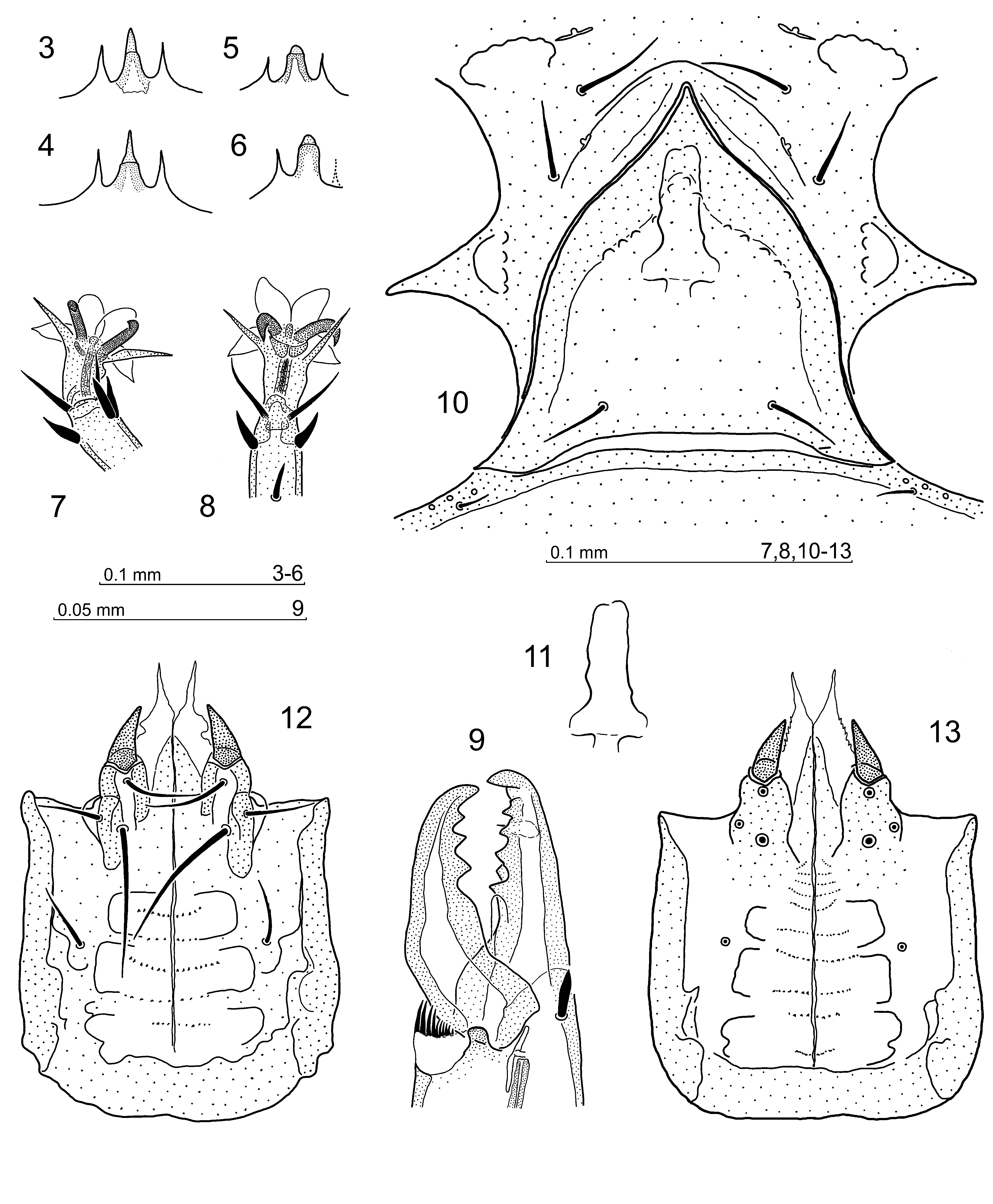
Gnathosoma . Gnathotectum trispinate ( Figs 3, 4 View FIGURES 3 – 13 ), central prong acuminate and more solid than the lateral ones, especially in the basal part. Corniculi conical, short. Hypostome similar to that in the male: hypognathal groove bears ca. 10 rows of denticles, hypostomatic setae simple, palpcoxal setae finely pectinate at external margin, posterior adaxial hypostomatic setae longest. Chelicera ( Fig. 9 View FIGURES 3 – 13 ): fixed digit with antiaxial row of four teeth, one tooth before and three behind pilus dentilis. One tooth located more adaxially is present between third and fourth tooth. Movable digit bears three teeth and its tip can be inserted into a depression in fixed digit. Pedipalp trochanter with seta v1 and v2 simple. Setae al on palp femur and genu spatulate.
Ventral idiosoma ( Fig. 2 View FIGURE 2 ). Presternal plates distinct, in the shape of a right triangle. Tritosternum present, biramous. Sternal shield with moderately visible reticulation in central part, well pronounced arcuate lines parallel to leg II openings, as well as two posteriorly open depressions situated laterally to sternal pores iv2 and two depressions between openings for leg III and IV ( Figs 2 View FIGURE 2 and 10 View FIGURES 3 – 13 ).
Genital region. Paragynia coalesce with sternal shield and groove between them is only visible in axial area, between setae st3. The groove runs slightly obliquely rather than transversely. Epigynial shield ( Figs 2 View FIGURE 2 , 10 View FIGURES 3 – 13 ) triangular, its lateral margins convex whereas posterior margin is concave. Endogynium ( Figs 10, 11 View FIGURES 3 – 13 ) hyaline and weakly visible, probably saccular with openings in posterior part.
Ventral chaetotaxy. Setae of sternogenital region—holotype: 20 µm (st1), 16 µm (st2), 26 µm (st3), 27 µm (st4), 26 µm (st5); paratype: 22 µm (st1), 17 µm (st2), 26 µm (st3), 25 µm (st4), 24 µm (st5). Opisthogaster setaeholotype: 25 µm (JV1), 34 µm (JV2), 38 µm (JV3), 38 µm (JV4), 16 µm (Ad), 14 µm (ZV1), 29 µm (ZV2), 38 µm (ZV3), 29 µm (ZV4); paratype: 22 µm (JV1), 31 µm (JV2), 35 µm (JV3), 39 µm (JV4), 14 µm (Ad), 13 µm (ZV1), 34 µm (ZV2), 37 µm (ZV3), 29 µm (ZV4). Sternal setae simple, but st2 shorter and thicker than others. Opisthogaster setae simple except for ZV4 which are thickened and terminally pilose. Setae ZV1 very short and located close to pores gv2.
Legs. Ambulacra of legs II–IV characteristic for genus: anterior pulvilli terminally rounded, whereas lateral pulvilli acuminate ( Figs 7, 8 View FIGURES 3 – 13 ). Other aspects of leg structure and setation unremarkable.
Male. Dorsal idiosoma. Paratypes (slides no. 2407, 2409, 2698) with holodorsal shield showing poorly discernible short suture behind podonotal setae j6. Idiosoma oval, well sclerotised and brown, 558–560 x 295–315 µm (N=2, slides no. 2409, 2698; third paratype on slide no. 2407 is broken, thus not available for measurements). Length of podonotal setae: 21–26 µm (j1), 9–10 µm (j2), 10–13 µm (j3), 27–34 µm (j4), 18–21 µm (j5), 17–20 µm (j6), 30–36 µm (r3), 30–34 µm (z5). Podonotal setae simple except j4, r3 and z5 which are thickened and terminally pilose; opisthonotal setae simple and moderately long, 20–27 µm, except Z4 which are 27 µm long, but thickened and terminally pilose; peritrematal groove 265–275 µm long and expands to beyond coxa I. Cuticle shows many regularly distributed pits.
Gnathosoma . Gnathotectum trispinate ( Figs 5, 6 View FIGURES 3 – 13 ), central prong with solid basal part, and a short, rounded apex. The lateral prongs acuminate as in female. Corniculi ( Figs 12, 13 View FIGURES 3 – 13 ) short and conical; hypognathal groove bears several ( Fig. 12 View FIGURES 3 – 13 ) up to 10 rows of denticles ( Fig. 13 View FIGURES 3 – 13 ). Hypostomatic setae simple, palpcoxal setae finely pectinate at external margin. Posterior adaxial hypostomatics longest. Chelicera ( Figs 14, 15 View FIGURES 14 – 19 ): fixed digit with one low tooth laterally to pilus dentilis and dentate lamellar ridge behind pilus dentilis. Movable digit with only one prominent tooth. Pedipalp trochanter with seta v1 and v2 simple. Setae al on palp femur and genu spatulate.
Ventral idiosoma. Tritosternum absent, presternal plates not discernible. Sternogenital shield anterior margin ( Fig. 16 View FIGURES 14 – 19 ) bears half-moon shaped genital opening flanked by thickened sternal cuticle ( Fig. 17 View FIGURES 14 – 19 ). Within orifice, a posteriorly-directed tongue-shaped cuticular thickening is present. Genital lamina poorly visible, anteriorly extending into a hyaline, wider lamellar basis with dentate margins and a narrow, long apically-barbed protrusion ( Fig. 17 View FIGURES 14 – 19 ). The protrusion is difficult to discern. Coxae of first legs inserted in deep concavities of anterior sternal margin ( Figs 16, 17 View FIGURES 14 – 19 ). Sternal setae length: 24–27 µm (st1), 22–25 µm (st2), 22–24 µm (st3), 21–22 µm (st4), 21–22 µm (st5). Opisthogaster setae length: 22–23 µm (JV1), 30–31 µm (JV2), 35–39 µm (JV3), 13–14 µm (JV4), 13 µm (Ad), 14–17 µm (ZV1), 38–42 µm (ZV2), 39–42 µm (ZV3), 35–37 µm (ZV4). Sternal and opisthogaster setae simple except for ZV4 which are thicker and terminally pilose. Idiosoma margin behind anus posteriorly convexed.
Legs. Leg I inserted in a deep concavity of anterior sternal margin; its coxa with thickened ridge protruding antero-ventrally ( Fig. 18 View FIGURES 14 – 19 ). Leg II ( Fig. 19 View FIGURES 14 – 19 ) spurred as follows: femur bears two minute spurs postero-ventrally, proximal spur is conical and higher than distal one, which is rather obtuse. Genual and tibial minute spurs conical and located more ventrally. Characteristic ambulacra of legs II–IV as in female. Other aspects of legs II, III and IV unremarkable.
Etymology. The species is dedicated to Prof. Dr. Dr. h.c. Gerd Alberti, outstanding acarologist and friend, who passed away on November 9, 2016.
Remarks. The newly described species may be closely related to T. borealis Ma & Wang , T. pusillus (Berlese) and T. medianus Tichomirov , mainly due to a similar type of gnathotectum, highly sclerotised cuticle with similarly distributed numerous pits, pyriform shape of idiosoma in female, forward protruding genital lamina projection in the male, as well as the presence of minute spurs on leg II. In T. pusillus the last two features are absent. The main difference concerns the shape of the epigynium and endogynium in the considered species, as well as the presence of distinct presternal plates on the sides of the male genital orifice in T. medianus ( Tichomirov, 1977) and in T. pusillus ( Holzmann, 1969) , and different structure of male chelicera in T. borealis ( Ma & Wang, 1996; Ma, 2010).


No known copyright restrictions apply. See Agosti, D., Egloff, W., 2009. Taxonomic information exchange and copyright: the Plazi approach. BMC Research Notes 2009, 2:53 for further explanation.
|
Kingdom |
|
|
Phylum |
|
|
Class |
|
|
SuperOrder |
Parasitiformes |
|
Order |
|
|
Family |
|
|
Genus |