
Akko rossi, Van, James L., Tassell & Baldwin, Carole C., 2004
|
publication ID |
https://doi.org/ 10.5281/zenodo.157403 |
|
DOI |
https://doi.org/10.5281/zenodo.5612855 |
|
persistent identifier |
https://treatment.plazi.org/id/03B687D4-743F-FF8B-FEF2-FB66FD84C62C |
|
treatment provided by |
Plazi |
|
scientific name |
Akko rossi |
| status |
sp. nov. |
Akko rossi View in CoL sp. nov.
Fig. 1 View FIGURE 1
Holotype: USNM 371780, 90 mm SL, female, Gulf of Fonseca, El Salvador, trawl, 13°12.89’N, 87°51.29’W to 13°13.74’N, 87°49.81’W, 7.0– 9.5 m, mud.
Description: Morphometric data for the holotype (the only known specimen) are given in Table 1 View TABLE 1 . Characters given in the Diagnosis are not repeated here.
Median fins: The second dorsal and anal fins both have 16 elements, the first element a spine in both. The caudal fin is lanceolate with 17 segmented rays, 4 upper and 5 lower procurrent rays. Median fin rays are serially branched with 4 to 5 branches.
Caudal skeleton: Hypurals 1–2 are fused to one another but not to other elements in the caudal skeleton. Hypurals 3–4 are fused to one another and to the terminal half centrum. A small parhypural supports the lowermost segmented ray, hypural 5 is free, and the single epural supports the uppermost segmented ray.
Paired fins: The pectoral fin is short, ending posteriorly before the posterior extension of the pelvic fin, well in front of the anus; the pectoral fin has 19 dichotomously branched rays. Pelvic fins are united forming a disk with a welldeveloped anterior frenum. The pelvic base is formed into a muscular pedicel with strong muscle bundles extending from the vicinity of the pelvic process to the vicinity of the pelvic spine, with some of the fibers inserting on the spines. Rays 1, 2, and 3 are serially branched; rays 4 and 5 dichotomously branched.
A. rossi A. brevis males (n=12) A. brevis females (n=9) Head: The mouth is large, the opening at an angle of 50° to the horizontal. No rostral frenum is present, and the upper jaw is only slightly protrusible. The oral velum has dorsal and ventral projections at the midline. The anterior nostril is a short thickened tube. The posterior nostril is a slit. There is no pseudobranch. The eye is tiny with a large lens occupying most of the eye. The gill opening extends from the level of the sixth pectoral fin ray ventrally to the pelvicfin base, terminating in a membrane supported by the second branchiostegal ray. The cephalic canal system has a small pore medial and just posterior to the anterior nostril (B’) and a second small pore posterior to the eye (G’).
Sensory papillae: The sensory papillae are slightly abraded, and no drawing is included, but the pattern is the same as that of Akko brevis ( Fig 2 View FIGURE 2 ). Horizontal row b has an elongate anterior extension ending under the eye; row d is complete, not divided into two sections, and row ot extends ventrally onto the branchiostegals, ending just past the second branchiostegal ray. The first vertical papillae row is complete, extending from the eye to near row d. Additional vertical rows are divided by row b, with two rows above and four rows below. Only the last vertical row below row b extends below the level of row d. Dorsal rows x 1 and x 2 are continuous or nearly so as is row n.
Jaws: The upper jaw has a single row of large depressible teeth, 16 on each premaxilla in the holotype. The lower jaw has two rows of teeth, the outer row consisting of 11 large evenly spaced teeth, the inner row with smaller widely spaced teeth.
Vertebral column: There are 11 precaudal and 17 caudal vertebrae. Two analfin pterygiophores precede the anteriormost haemal spine. The dorsal pterygiophore pattern is 3 221110.
Scales: Scales are small, cycloid, embedded, and nonoverlapping anteriorly, becoming larger and overlapping posteriorly. The body is scaled from the predorsal area over the operculum to the base of the caudal fin, with about 115 scales in the lateral series.
Genitalia: The female genital papilla is short and broad, opening through a slit.
Pigment: The head and dorsal portion of the trunk are brown in the preserved specimen. The ventral section, below the midline, is pale tan anteriorly, becoming tan to light brown posteriorly. The fins are dark brown, becoming light brown or speckled brown at the base only. The pigment is restricted to the interradial membranes in all fins except the first dorsal, in which the membrane covering the spines is also brown. The genital papilla is brown, the tip and remainder of the surrounding ventral area white.
Etymology: The specific epithet is in honor of our friend and colleague, D. Ross Robertson, of the Smithsonian’s Tropical Research Institute in Panama, who has made substantial contributions to our understanding of diversity of tropical eastern Pacific shorefishes.
Distribution: The only known specimen of Akko rossi was collected from the Gulf of Fonseca, El Salvador.
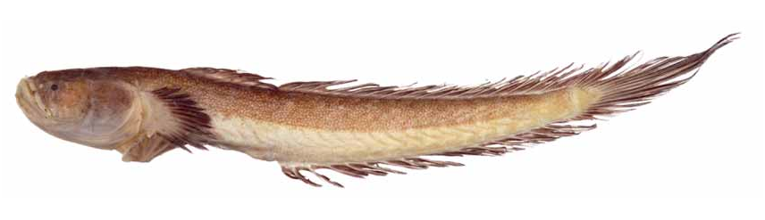
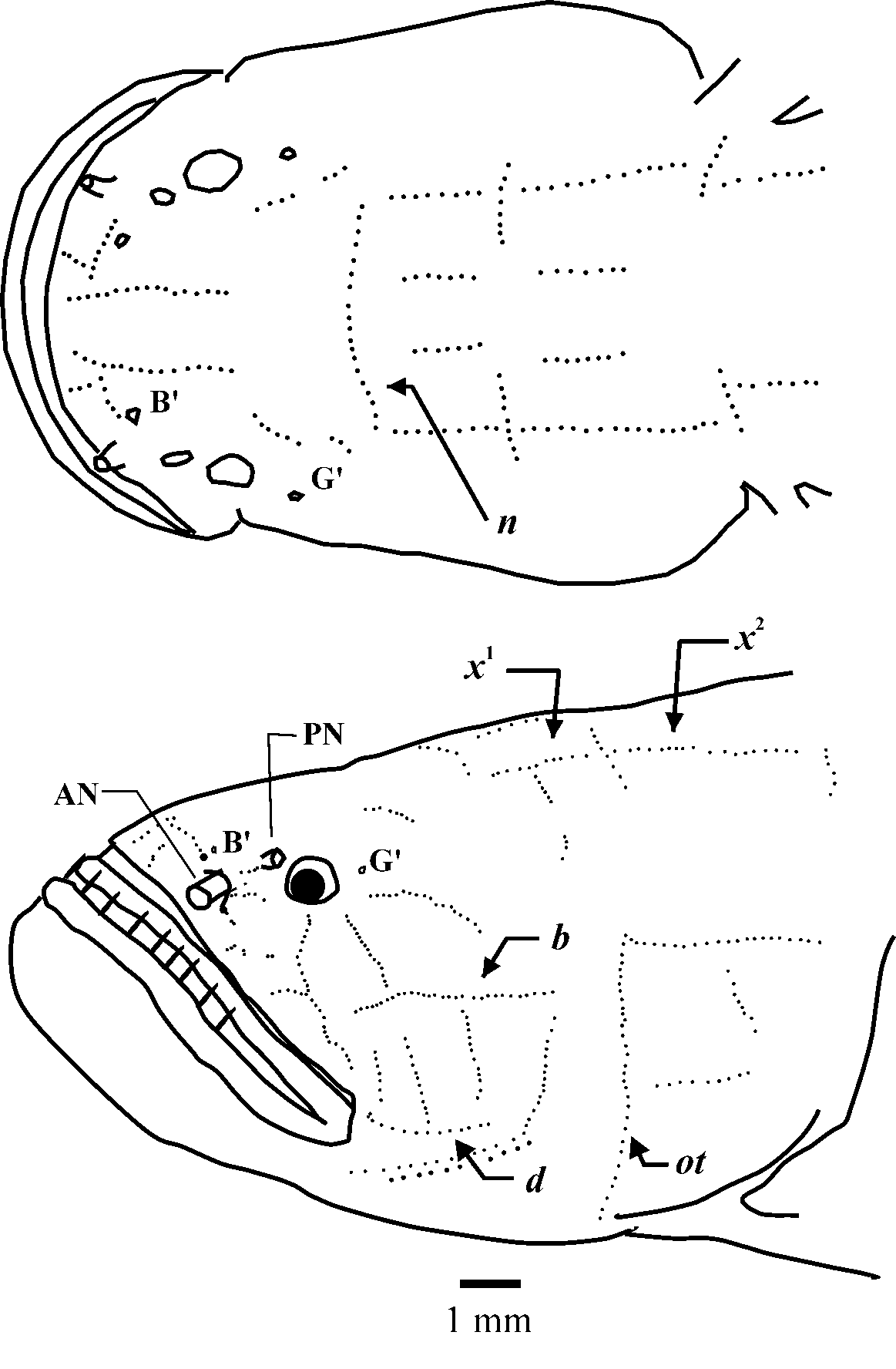
TABLE 1. Morphometric Data for Akko rossi and Akko brevis. Standard length is in mm; interorbital distance and eye diameter are in percent head length; other measurements are in percent standard length
| Holotype | range | mean | range | mean | |
|---|---|---|---|---|---|
| Standard length | 90.8 | 27.7 – 58.9 | 46.6 | 29.3 – 52.7 | 39.0 |
| Preanus length | 43.6 | 40.6 – 49.0 | 43.6 | 42.3 – 48.6 | 44.7 |
| Head length | 23.7 | 22.9 – 28.8 | 25.0 | 23.2 – 27.8 | 25.5 |
| Depth at anus | 12.2 | 13.2 – 15.3 | 14.4 | 13.6 – 16.5 | 14.5 |
| Caudalfin length | 32.3 | 18.2 – 35.2 | 31.6 | 23.0–39.7 | 34.7 |
| Pectoralfin length | 13.1 | 13.4 – 16.9 | 14.9 | 12.2 – 18.4 | 15.4 |
| Pelvicfin length | 17.7 | 14.5 – 19.8 | 17.5 | 16.3 – 20.3 | 18.1 |
| Interorbital distance | 35.1 | 24.8 – 35.4 | 30.0 | 25.6 – 32.4 | 29.2 |
| Eye diameter | 4.8 | 5.4 – 6.8 | 6.1 | 5.7 – 7.6 | 6.5 |
| USNM |
Smithsonian Institution, National Museum of Natural History |
No known copyright restrictions apply. See Agosti, D., Egloff, W., 2009. Taxonomic information exchange and copyright: the Plazi approach. BMC Research Notes 2009, 2:53 for further explanation.
|
Kingdom |
|
|
Phylum |
|
|
Class |
|
|
Order |
|
|
Family |
|
|
Genus |