
Scapholeberis kingi Sars, 1888
|
publication ID |
https://doi.org/10.5281/zenodo.214313 |
|
DOI |
https://doi.org/10.5281/zenodo.5680404 |
|
persistent identifier |
https://treatment.plazi.org/id/03B687AA-FFBF-5A4B-25DC-980FFC35FA42 |
|
treatment provided by |
Plazi (2016-04-12 21:22:09, last updated 2024-11-26 05:54:19) |
|
scientific name |
Scapholeberis kingi Sars, 1888 |
| status |
|
3. Scapholeberis kingi Sars, 1888
Fig. 5 View FIGURE 5
Synonymy. Scapholeberis kingi Sars, 1888 , p. 68; Chiang & Du, 1979, p. 145–146, Fig. 97; Dumont & Pensaert 1983, p. 24–25, Fig. 2 View FIGURE 2 : 3; Fig. 4 View FIGURE 4 : 4; Fig. VI: 1–2; Pl. 1: 8; Pl. 2: 4; Pl. 3: 5, 7, 9; Pl. 4: 1–7; Pl. 5: 1–2, 4; Fig. 10 View FIGURE 10 : 3; Pl. 6: 6–8; Fig. 12 View FIGURE 12 Fig. 21 View FIGURE 21 : 4; Kotov et al. 2011a, p. 405.
Scapholeberis kingi n.sp. in Sars 1903, p. 8–10, Pl. 1: figs 2a–c.
Scapholeberis rammneri Dumont & Pensaert in Yoon 2010, p. 64–66, Fig. 34.
Type locality. “South Creek and Paramatta, New South Wales, Australia ” ( Dumont & Pensaert 1983).
Localities in Korea. 3, 5, 6a–b, 7a–b, 8, 9, 10, 13, 14 (see Fig. 1 and Table 1 View TABLE 1 ).
Parthenogenetic female. Brownish in colour. Body with dorsal margin interrupted by a cervical incision, postero-dorsal angle well-expressed, posterion margin slightly convex, postero-ventral angle with a strong spine - mucro, which is of 0.2 of valve length ( Fig. 5 View FIGURE 5 A). Head rather large, lacking a horn, rostrum trilobate in ventral view ( Fig. 5 View FIGURE 5 B), middle lobe with "a hyaline membrane in front" in terminology of Dumont & Pensaert (1983), compound eye very large, occupies distalmost portion of head ( Fig. 5 View FIGURE 5 A). A ridge departs from the insertion of the second antenna and extends to the side of the head—seen frontally, it appear as a pair of shallow depressions, "auricles" in terminology of Dumont & Pensaert (1983). An elongate frontal head pore on the rostrum ( Fig. 5 View FIGURE 5 B, arrow). Valves with reticulations as vertical lines near the posterior margin. A projection on ventral valve margin before the system of setae located on a flat portion, "sucker-plate" in terminology of Dumont & Pensaert (1983). A broad hyaline membrane extends beyond the posterior valve rim ( Fig. 5 View FIGURE 5 –D). Postabdomen slightly widened distally, preanal margin long, preanal angle obtuse, anal margin straight, postanal angle not expressed, postanal margin very short ( Fig. 5 View FIGURE 5 E). About 3–5 single postanal teeth, followed by clusters of spinules proximally (on anal margin), numerous series of minute setules laterally ( Fig. 5 View FIGURE 5 F). In distal dorsal external pecten 2–4 proximalmost denticles specially strong and sparsely located. First antenna short, with antennular sensory seta and 9 terminal aesthetascs ( Fig. 5 View FIGURE 5 G). Antenna II long, antennal formula: setae 0-1-3/1-1-3 ( Fig. 5 View FIGURE 5 H). Limb I as shown in Fig. 5 View FIGURE 5 I. Other limbs not studied. Size in our material 0.5–1.0 mm.
Notes. According to Dumont & Pensaert (1983), this taxon is distributed in Australia, SE Asia, India, Middle East and Africa. It is known from the Far East of Russia ( Kotov et al. 2011b), China ( Chiang & Du 1979) and Japan ( Mizuno & Takahashi 1991), so its presence in Korea was expected. Yoon (2010) described S. mucronata ( O. F. Müller, 1776) and S. rammneri Dumont & Pensaert, 1983 from Korea, but we did not see these species in our samples. Probably this author misidentified S. kingi as S. rammneri . Unfortunately, descriptions and illustrations by Kim (1988), Kim & Yoon (1987) and Yoon (2010) do not allow us to assign their " S. mucronata " to any species, because most taxonomically important characters were not mentioned.
Chiang, S. & Du, N. (1979) Fauna Sinica. Crustacea. Freshwater Cladocera. Science Press, Academia Sinica, Peking, China, 297 pp.
Dumont, H. J. & Pensaert, J. (1983) A revision of the Scapholeberinae (Crustacea: Cladocera). Hydrobiologia, 100, 3 - 45.
Yoon, S. M. & Kim, H. S. (1987) A systematic study on the freshwater Cladocera from Korea. The Korean Journal of Systematic Zoology, 3, 175 - 207. [in Korean]
Kim, I. H. (1988) Key to the Korean freshwater Cladocera. Korean Journal of Systematic Zoology, Special Issue, 2, 43 - 65. [In Korean]
Kotov, A. A., Korovchinsky, N. M., Sinev, A. Y. & Smirnov, N. N. (2011 a). Cladocera (Crustacea, Branchiopoda) of the Zeya basin (Amurskaya Area, Russian Federation). 3. Systematic-faunistic and zoogeographic analysis. Zoologichesky Zhurnal, 90, 402 - 411.
Kotov, A. A., Sinev, A. Y., Korovchinsky, N. M., Smirnov, N. N., Bekker, E. I. & Sheveleva, N. G. (2011 b) Cladocera (Crustacea, Branchiopoda) of the Zeya basin (Amurskaya Area, Russian Federation). 1. New taxa for fauna of Russia. Zoologichesky Zhurnal, 90, 131 - 142.
Mizuno, T. & Takahashi, E., eds., 1991. An illustrated guide to freshwater zooplankton in Japan. Tokai University Press, Tokyo, 534 pp. [in Japanese]
Sars, G. O. (1888) Additional notes on Australian Cladocera, raised from dried mud. Forhandlinger i Videnskabs - Selskabet i Christiania, 1888, 7, 1 - 74.
Sars, G. O. (1903) Fresh-water Entomostraca from China and Sumatra. Archiv for Mathematik og Naturvidenskab, 25, 1 - 44.
Yoon, S. M. (2010) Arthropoda: Branchiopoda: Anostraca, Notostraca, Spinicaudata, Laevicaudata, Ctenopoda, Anomopoda, Haplopoda Branchiopods. Invertebrate fauna of Korea, 21 (2), 1 - 156.

FIGURE 2. Sida ortiva Korovchinsky, 1979, parthenogenetic female from Seon You Dong, locality 11: A, lateral view; B, head; C, postero-ventral portion of valves; D, postabdomen; E, its distal portion; F, antenna I; G, antenna II. Scale bars: 0.1 mm.
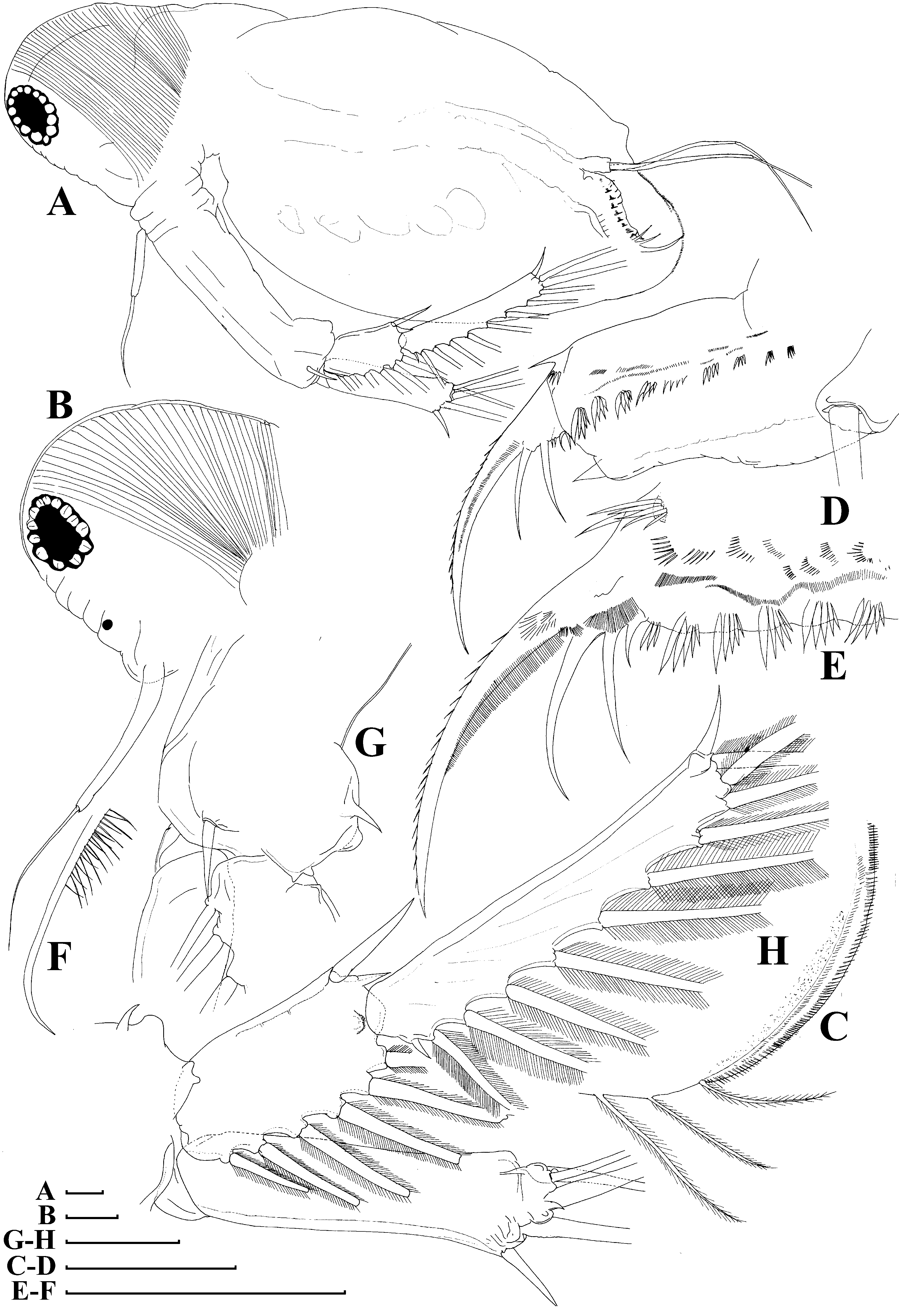
FIGURE 4. Pseudosida cf. szalayi (Daday, 1898), parthenogenetic female from Bak Sil Ji 3, locality 8. A, lateral view; B, head; C, postero-ventral portion of valve, inner view; D, postabdomen; E, its distal portion; F, distal sensory seta of antenna I; G, distal portion of basal segment of antenna II; H, branches of antenna II. Scale bars: 0.1 mm.

FIGURE 5. Scapholeberis kingi Sars, 1888, parthenogenetic female from Bak Sil Ji 1, locality 6 a: A, lateral view; B, head, ventral view; C, postero-ventral valve portion, external view; D, posterior margin, inner view; E, postabdomen and abdomen; F, postabdominal claw; G, antenna I; H, antenna II, posterior view; I, limb I. Scale bars: 0.1 mm.
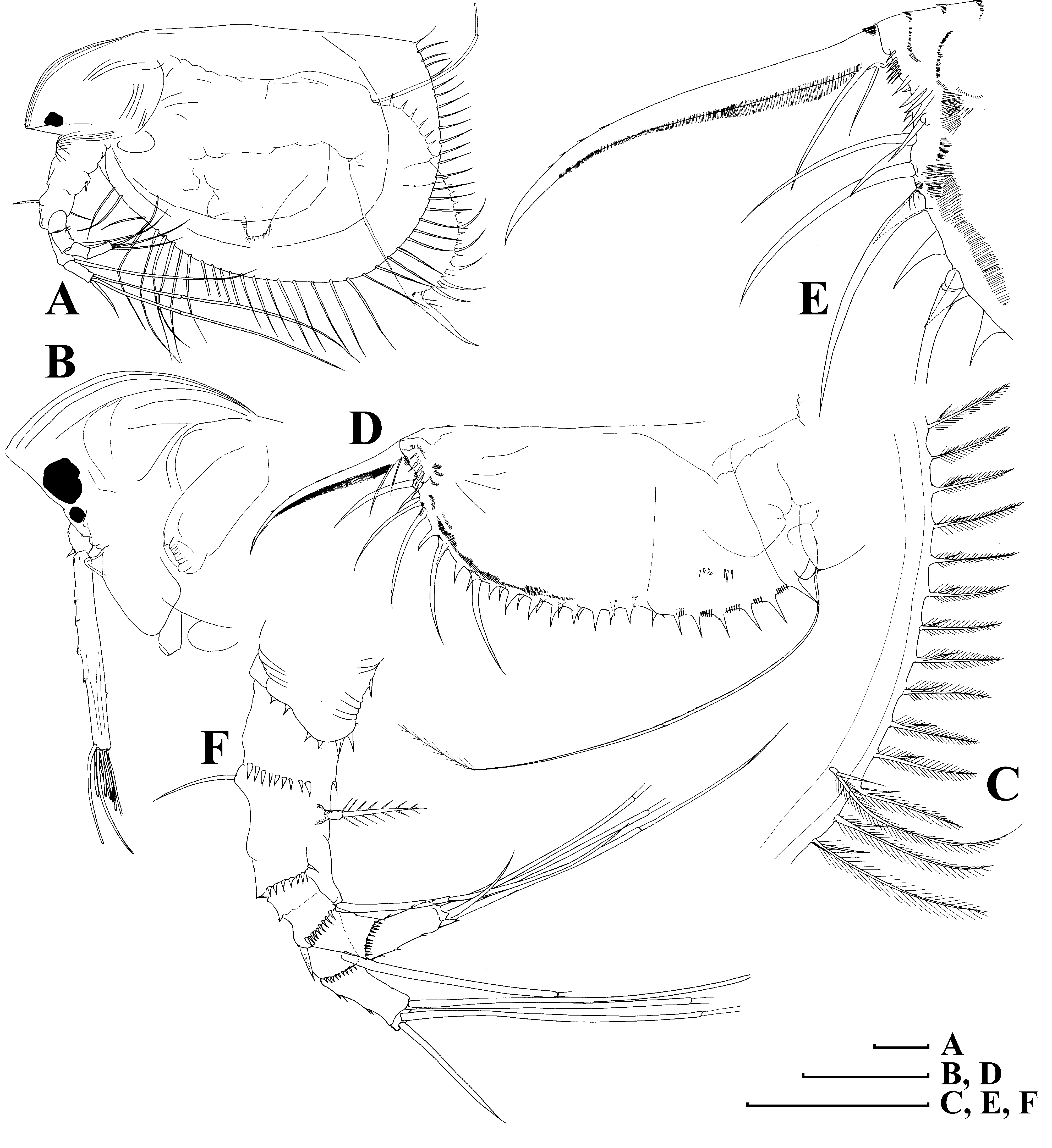
FIGURE 10. Ilyocryptus spinifer Herrick, 1882, parthenogenetic female from Bak Sil Ji 3. locality 8: A, lateral view; B, head; C, posterior valve margin; D, postabdomen; E, its distal portion; F, antenna II. Scale bars: 0.1 mm.
FIGURE 12. Macrothrix triserialis Brady, 1886, appendages of parthenogenetic female from Bak Sil Ji 1, locality 6 a: A, antenna I; B, antenna II, anterior view; C, setae of endopod proximal segment; D, distal portion of limb I. Scale bars: 0.1 mm.

FIGURE 21. Camptocercus uncinatus Smirnov, 1971, parthenogenetic female from Bak Sil Ji 1, locality 6 a: A, lateral view; B, head; C, labrum; D, armature of postero-ventral valve margin, inner view; E, armature of posterior valve margin, inner view; F, postabdomen; G, postabdominal claw; H, exopod of antenna II; I, distal portion of limb I. Scale bars: 0.1 mm.
No known copyright restrictions apply. See Agosti, D., Egloff, W., 2009. Taxonomic information exchange and copyright: the Plazi approach. BMC Research Notes 2009, 2:53 for further explanation.
|
Kingdom |
|
|
Phylum |
|
|
Class |
|
|
Order |
|
|
Genus |
1 (by plazi, 2016-04-12 21:22:09)
2 (by ImsDioSync, 2016-12-21 02:34:39)
3 (by ImsDioSync, 2016-12-21 02:36:42)
4 (by ImsDioSync, 2018-06-29 22:01:46)
5 (by ImsDioSync, 2019-03-29 23:17:36)
6 (by ExternalLinkService, 2019-09-26 19:23:57)
7 (by ExternalLinkService, 2021-11-09 17:29:56)
8 (by ExternalLinkService, 2021-11-10 06:23:56)
9 (by ExternalLinkService, 2021-11-12 11:34:29)
10 (by ExternalLinkService, 2021-11-12 11:34:29)
11 (by plazi, 2023-10-26 09:02:15)