
Moinodaphnia macleayi ( King, 1853 )
|
publication ID |
https://doi.org/10.5281/zenodo.214313 |
|
DOI |
https://doi.org/10.5281/zenodo.5680408 |
|
persistent identifier |
https://treatment.plazi.org/id/03B687AA-FFBD-5A48-25DC-98F3FC00FC1C |
|
treatment provided by |
Plazi (2016-04-12 21:22:09, last updated 2024-11-26 05:54:19) |
|
scientific name |
Moinodaphnia macleayi ( King, 1853 ) |
| status |
|
5. Moinodaphnia macleayi ( King, 1853) View in CoL
Fig. 7 View FIGURE 7
Synonymy. Moina macleayi King, 1853 , p. 251–252, Pl. 5.
Moinodaphnia macleayi (King) View in CoL in Goulden 1968, p. 84–87, Figs 45–46; Smirnov 1976, p. 187–189, Figs 165–166; Chiang & Du 1979, p. 161–162, Fig. 108.
Moina submucronata Brady, 1886 View in CoL , p. 294, Pl. 37: figs 4–5.
Type locality. "Pond near Elizabeth Bay, Sydney" ( King 1853), New South Wales, Australia.
Locality in Korea. 6a (see Fig. 1 and Table 1 View TABLE 1 ).
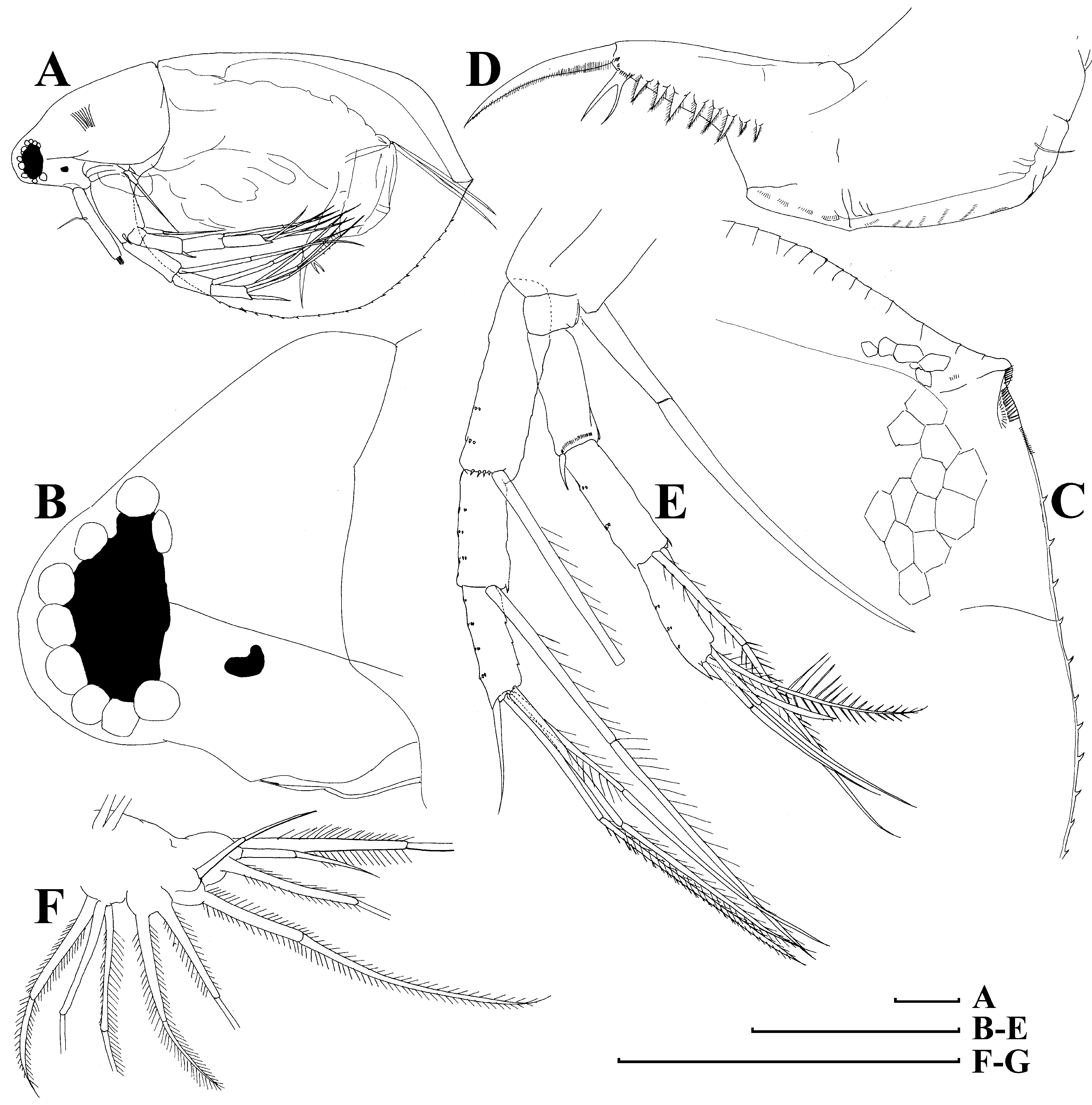
Parthenogenetic female. Body subovoid, dorsal margin with a wear cervical incision, postero-dorsal angle as a rounded triangle ( Fig. 7 View FIGURE 7 A–B), posterior margin fluently turned to ventral margin without any angle, ventral margin convex; no hairs anywhere else on the head or valves. Body compressed laterally, with a well-expressed median keel on dorsal portion of valves. Head small, sub-triangular, with a shallow supra-ocular depression above large compound eye which fills the tip of the head; ocellus small ( Fig. 7 View FIGURE 7 B). Valves reticulated, ventral margin with denticles, a pair of submarginal hooks at the point where the valves come together ( Fig. 7 View FIGURE 7 C). Postabdomen with wide preanal and narrow postanal portions, preanal margin large and lacking long setules, preanal angle distinct, anal margin straight, postanal angle expressed, postanal margin straight ( Fig. 7 View FIGURE 7 D). On postanal portion, eight-nine feathered lateral teeth plus a long, bident tooth near base of postabdominal claw. Postabdominal claw long, with two pectens of thin setules. Antenna I long, cylindrical, antennular sensory seta originated somewhat proximally to its middle; nine very short aesthetascs distally ( Fig. 7 View FIGURE 7 A). Antenna II thin and long, basal segment with a long seta on posterior side ( Fig. 7 View FIGURE 7 E). Antennal formula: setae 0-0-1-3/1-1-3, spines 0-1-0-1/0-0-1. Apical spine on exopod specially long, so, it is sometimes counted as a seta ( Smirnov, 1976), but we think that antennal formula as suggested here is more correct for homologization of setae and spines on antenna II on Moinodaphnia and other anomopods. Limb I as illustrated in Fig. 7 View FIGURE 7 F. Size in our material 0.7–1.2 mm.
Notes. According to Smirnov (1976), Moinodaphnia macleayi is a circumtropical species. Most probably Korea is the northernmost area of its distribution. The species is also known from China ( Chiang & Du 1979) and Japan ( Mizuno & Takahashi 1991). This is the first record for Korea.
Brady, G. S. (1886) Notes on Entomostraca collected by Mr. A. Haly in Ceylon. Journal of the Linnean Society of London, Zoology, 19, 293 - 317.
Chiang, S. & Du, N. (1979) Fauna Sinica. Crustacea. Freshwater Cladocera. Science Press, Academia Sinica, Peking, China, 297 pp.
Goulden, C. E. (1968) The systematics and evolution of the Moinidae. Transactions of the American Philosophical Society Held at Philadelphia, New Series, 58, 1 - 101.
King, R. L. (1853) On some of the species of Daphnidae found in New South Wales. Papers and Proceedings of the Royal Society of Tasmania, 2, 243 - 253.
Mizuno, T. & Takahashi, E., eds., 1991. An illustrated guide to freshwater zooplankton in Japan. Tokai University Press, Tokyo, 534 pp. [in Japanese]
Smirnov, N. N. (1976) Macrothricidae and Moinidae of the World fauna. Fauna SSSR, novaya seriya, Rakoobraznye, 1 (3), 1 - 237. [In Russian]
No known copyright restrictions apply. See Agosti, D., Egloff, W., 2009. Taxonomic information exchange and copyright: the Plazi approach. BMC Research Notes 2009, 2:53 for further explanation.
|
Kingdom |
|
|
Phylum |
|
|
Class |
|
|
Order |
|
|
Family |
|
|
Genus |
Moinodaphnia macleayi ( King, 1853 )
| Kotov, Alexey A., Jeong, Hyun Gi & Lee, Wonchoel 2012 |
Moina submucronata
| Brady 1886 |
1 (by plazi, 2016-04-12 21:22:09)
2 (by ImsDioSync, 2016-12-21 02:34:39)
3 (by ImsDioSync, 2016-12-21 02:36:42)
4 (by ImsDioSync, 2018-06-29 22:01:46)
5 (by ImsDioSync, 2019-03-29 23:17:36)
6 (by ExternalLinkService, 2019-09-26 19:23:57)
7 (by ExternalLinkService, 2021-11-09 17:29:56)
8 (by ExternalLinkService, 2021-11-10 06:23:56)
9 (by ExternalLinkService, 2021-11-12 11:34:29)
10 (by ExternalLinkService, 2021-11-12 11:34:29)
11 (by plazi, 2023-10-26 09:02:15)