
Ilyocryptus cuneatus Štifter, 1988
|
publication ID |
https://doi.org/10.5281/zenodo.214313 |
|
DOI |
https://doi.org/10.5281/zenodo.5680411 |
|
persistent identifier |
https://treatment.plazi.org/id/03B687AA-FFBC-5A4F-25DC-9D83FC64FDF1 |
|
treatment provided by |
Plazi (2016-04-12 21:22:09, last updated 2024-11-26 05:54:19) |
|
scientific name |
Ilyocryptus cuneatus Štifter, 1988 |
| status |
|
6. Ilyocryptus cuneatus Štifter, 1988 View in CoL
Figs 8–9 View FIGURE 8 View FIGURE 9
Synonymy. Ilyocryptus cuneatus Štifter, 1988 , p. 292–296, Figs 7–19 View FIGURE 7 View FIGURE 8 View FIGURE 9 View FIGURE 10 View FIGURE 11 View FIGURE 12 View FIGURE 13 View FIGURE 14 View FIGURE 15 View FIGURE 16 View FIGURE 17 View FIGURE 18 View FIGURE 19 ; Tanaka 2001, p. 221, Fig. 3 View FIGURE 3 A–E; Kotov & Štifter 2006, p. 76–81, Figs 2 View FIGURE 2 G, 35–38; Kotov et al. 2011a, p. 405.
Type locality. “West Bohemia, Březová reservoir on the river Teplá, 5 km south of Karlovy Vary” ( Štifter 1988), Czech Republic.
Localities in Korea. 6a, 7b, 8, 14 (see Fig. 1 and Table 1 View TABLE 1 ).
Parthenogenetic female. Body triangular-ovoid, postero-dorsal angle expressed ( Fig. 8 View FIGURE 8 A). In anterior view, body thick, with thick and low dorsal keel, without lateral horns. Moulting incomplete. Ocellus small ( Fig. 8 View FIGURE 8 B). Head shield with mandibular articulation as a small projection ( Fig. 8 View FIGURE 8 C). Setae at ventral margin plumose ( Fig. 8 View FIGURE 8 D). Each seta at posterior margin with a long, stout basal part, than with a series of 2–4 stout spine-like setules along one side in its basal portion, and with fine hairs in distal portion ( Fig. 8 View FIGURE 8 E). Anus opens in the middle between base and distal extremity of postabdomen; preanal teeth 10–12, subequal in size, both doubled and single in the same specimen, few denticles on postabdomen base laterally ( Fig. 8 View FIGURE 8 F). Paired spines shorter than large lateral setae. Postabdominal claw with a single denticle in distal part, no denticles in its middle part, distal and proximal spines on claw base subequal in size; setules situated on base of claw ventrally long ( Fig. 8 View FIGURE 8 G). Antenna I relatively long, with transverse ridges ( Fig. 8 View FIGURE 8 B). Proximal segment with a finger-like projection and few low hillocks ( Fig. 8 View FIGURE 8 H). The longest aesthetasc about half the distal segment. Antenna II short, distal burrowing spine relatively short, with tip almost reaching or reaching distal border of basal segment ( Fig. 9 View FIGURE 9 A). Both antennal branches short ( Fig. 9 View FIGURE 9 B–C). Apical swimming setae relatively short, armed with short setules distally ( Fig. 9 View FIGURE 9 D). Proximal lateral swimming seta shorter than distal one, both without hooks on tips and armed along one side with setules analogous to these of apical setae, and more stout and long setules along other side ( Fig. 9 View FIGURE 9 E–F). Spine on second segment of exopod equal to or somewhat shorter than half of third segment ( Fig. 9 View FIGURE 9 B–C). A large seta near ejector hooks of limb I. Gnathobase I as a setulated hillock. Limb VI with row of long setules along inner margin subdivided into six bundles by small incisions on the margin. Size in our material 0.61–0.83 mm.
Notes. The species is widely distributed in the Northern Palaearctic and Nearctic ( Kotov & Štifter 2006) and found close to Korea: Japan ( Tanaka 2001) and the Amur basin in Far East of Russia, where it is quite common ( Kotov et al. 2011a). We found it also to be common in the Korean Peninsula, which could be the southernmost area of its distribution in Asia. Unfortunately, there is no information on its presence in China, and previous descriptions of ilyocryptids ( Chiang & Du 1979) are not detailed enough to separate sordidus -like species sensu Kotov & Elías-Gutiérrez (2009). Korean specimens are somewhat different from European ones in: (1) only few denticles on postabdomen base laterally; (2) ridges, although ill-expressed, on distal segment of antenna I. It is possible that populations from the Far East form a separate taxon, but this idea could be confirmed only in the course of a global revision of the cuneatus -like populations in the Palaearctic.
Chiang, S. & Du, N. (1979) Fauna Sinica. Crustacea. Freshwater Cladocera. Science Press, Academia Sinica, Peking, China, 297 pp.
Kotov, A. A. & Stifter, P. (2006) Cladocera: family Ilyocryptidae (Branchiopoda: Cladocera: Anomopoda). Guides to the identification of the microivertebrates of the Continental Waters of the world, Vol. 22, Kenobi Productions, Ghent & Backhuys Publishers, Leiden, 172 pp.
Kotov, A. A. & Elias-Gutierrez, M. (2009) A phylogenetic analysis of Ilyocryptus Sars, 1862 (Cladocera: Ilyocryptidae). International Review of Hydrobiology, 94, 208 - 225.
Kotov, A. A., Korovchinsky, N. M., Sinev, A. Y. & Smirnov, N. N. (2011 a). Cladocera (Crustacea, Branchiopoda) of the Zeya basin (Amurskaya Area, Russian Federation). 3. Systematic-faunistic and zoogeographic analysis. Zoologichesky Zhurnal, 90, 402 - 411.
Stifter, P. (1988) Two new species of the genus Ilyocryptus (Cladocera, Crustacea) confused with I. sordidus Lievin. V e stnik c eskoslovenske spole c nosti zoologicke, 52: 290 - 301.
Tanaka, S. (2001) Three species of the genus Ilyocryptus (Anomopoda, Branchiopoda) occurring in Japan. Limnology, 2, 219 - 222.

FIGURE 2. Sida ortiva Korovchinsky, 1979, parthenogenetic female from Seon You Dong, locality 11: A, lateral view; B, head; C, postero-ventral portion of valves; D, postabdomen; E, its distal portion; F, antenna I; G, antenna II. Scale bars: 0.1 mm.

FIGURE 3. Sida crystallina (O. F. Müller, 1776), parthenogenetic female from Cheok Ji Ri, locality 12: A, lateral view; B, head; C, postero-ventral portion of valves; D, postabdomen; E, its distal portion; F, antenna I; G, antenna II. Scale bars: 0.1 mm.
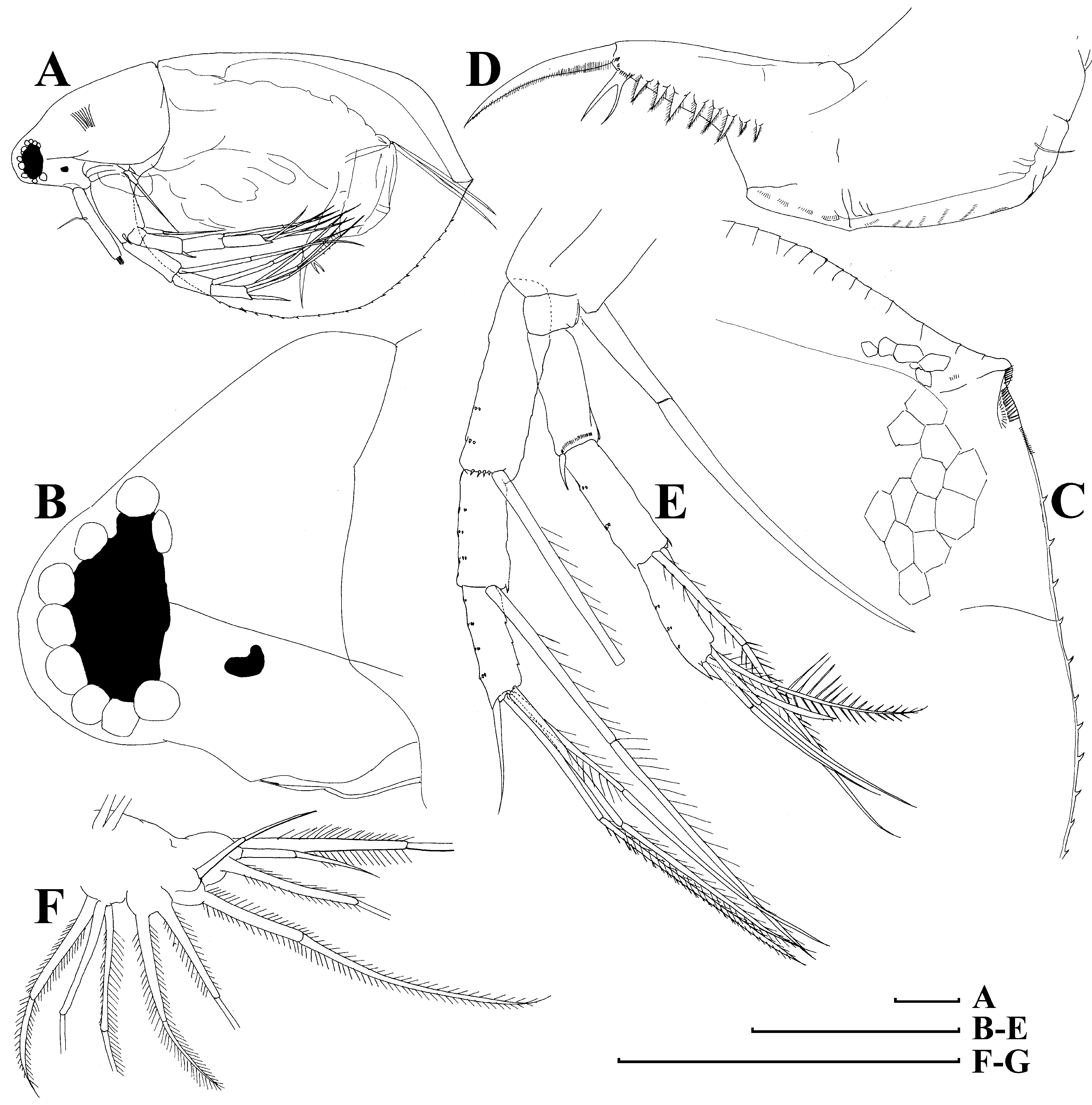
FIGURE 7. Moinodaphnia macleayi (King, 1853), parthenogenetic female from Bak Sil Ji 1, locality 6 a: A, lateral view; B, head; C, postero-dorsal portion; D, postabdomen; E, antenna II; F, limb I. Scale bars: 0.1 mm.
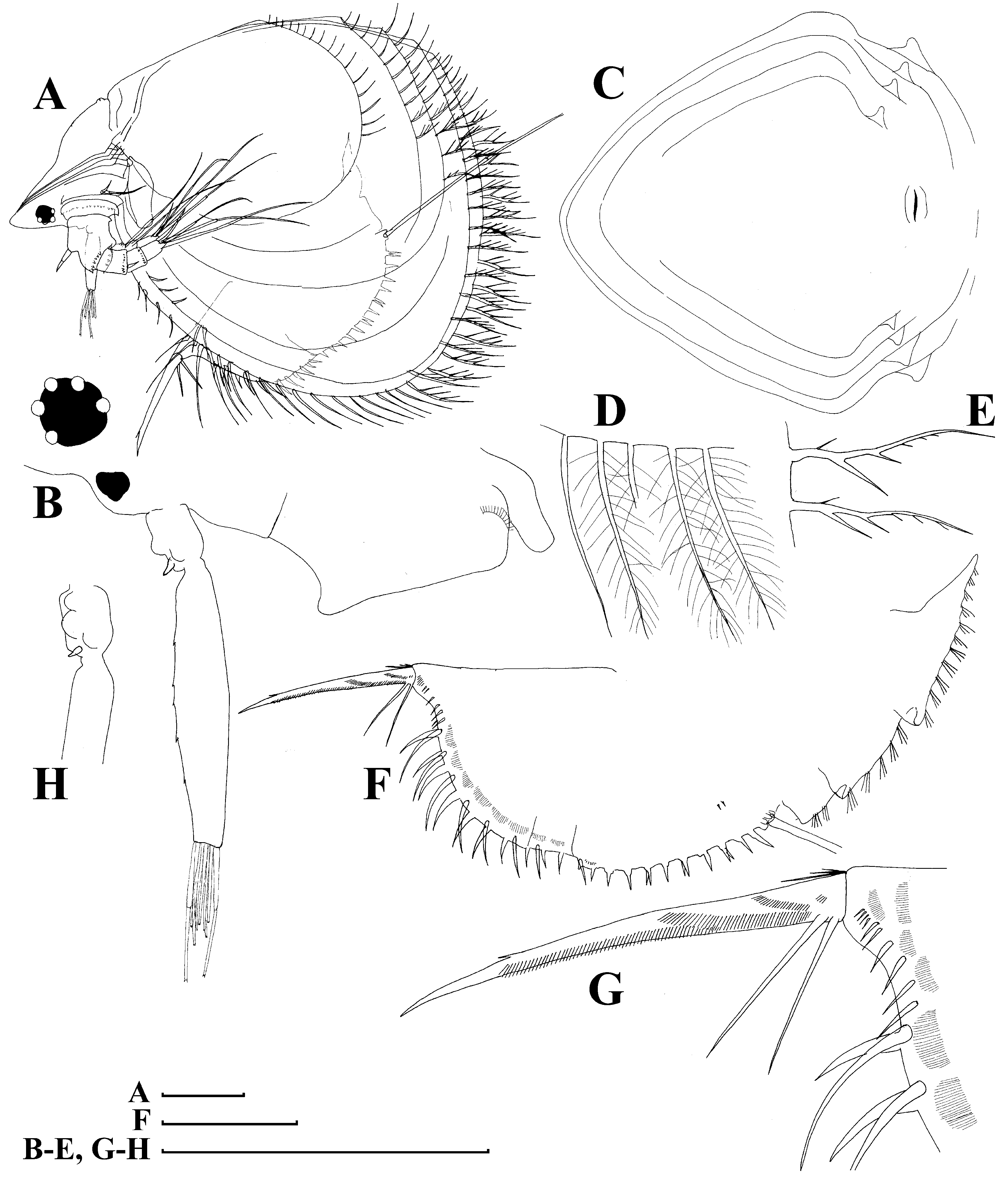
FIGURE 8. Ilyocryptus cuneatus Štifter, 1988, parthenogenetic female from Bak Sil Ji 1, locality 6 a: A, lateral view; B, head, lateral view; C, head shield; D, setae at middle of ventral margin; E, setae at posterior valve margin; F, postabdomen; G, its distal portion and postabdominal claw; H, proximal portion of antenna I. Scale bars: 0.1 mm.
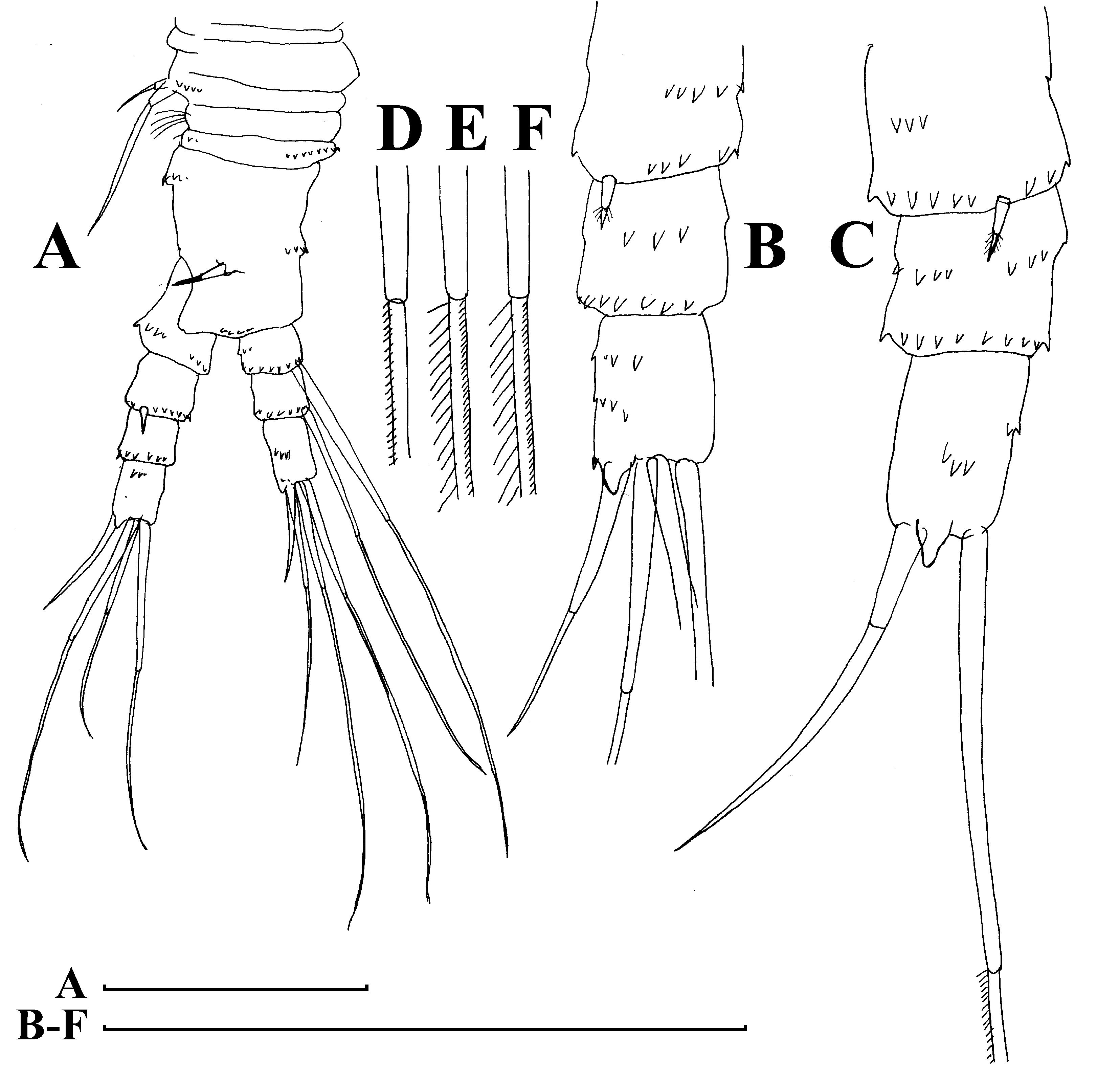
FIGURE 9. Ilyocryptus cuneatus Štifter, 1988, antenna II of parthenogenetic female from Bak Sil Ji 1, locality 6 a: A, anterior view; B – C, exopods of two different specimens; D, apical swimming seta; E – F, distal and proximal lateral swimming seta. Scale bars: 0.1 mm.
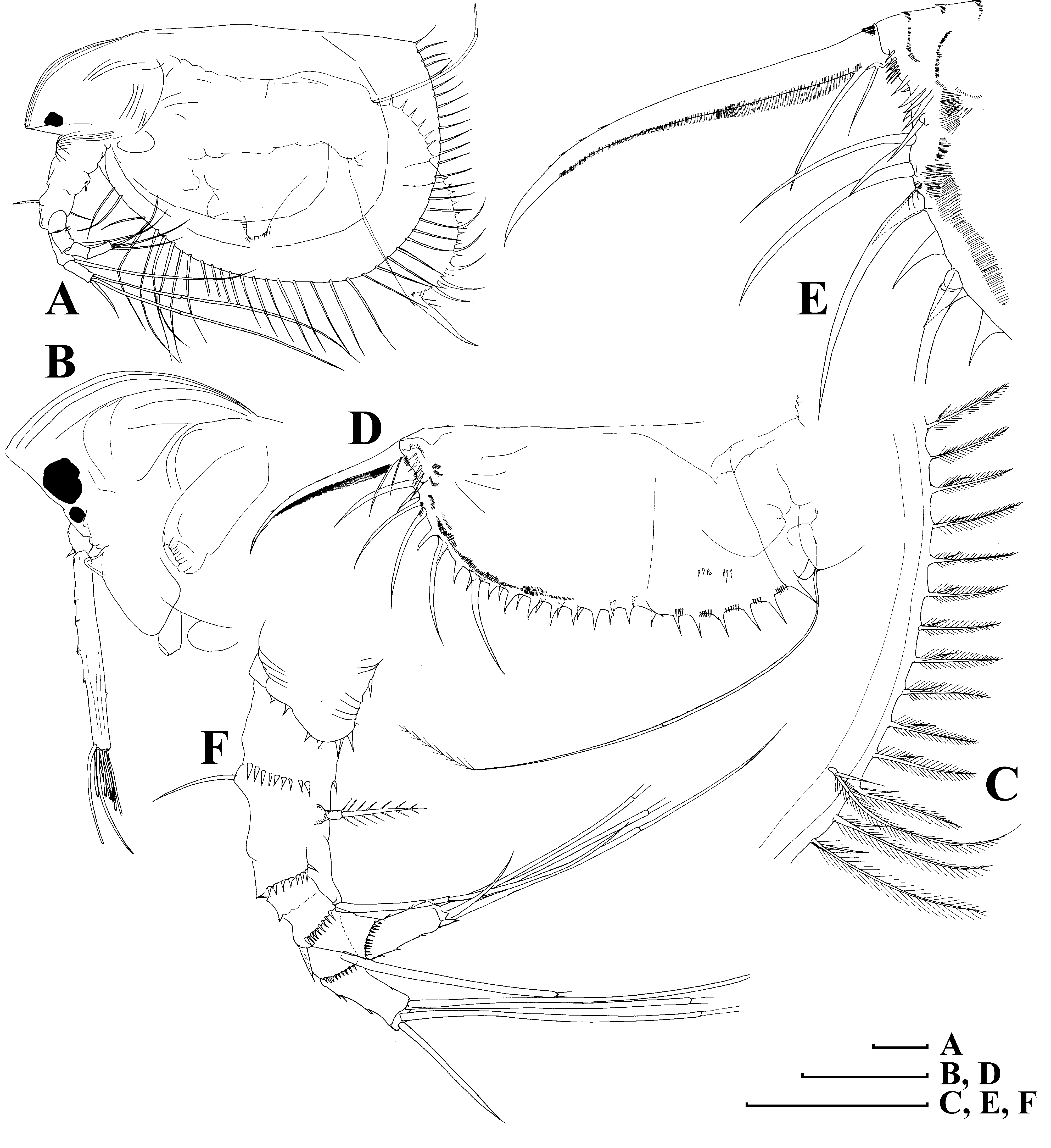
FIGURE 10. Ilyocryptus spinifer Herrick, 1882, parthenogenetic female from Bak Sil Ji 3. locality 8: A, lateral view; B, head; C, posterior valve margin; D, postabdomen; E, its distal portion; F, antenna II. Scale bars: 0.1 mm.
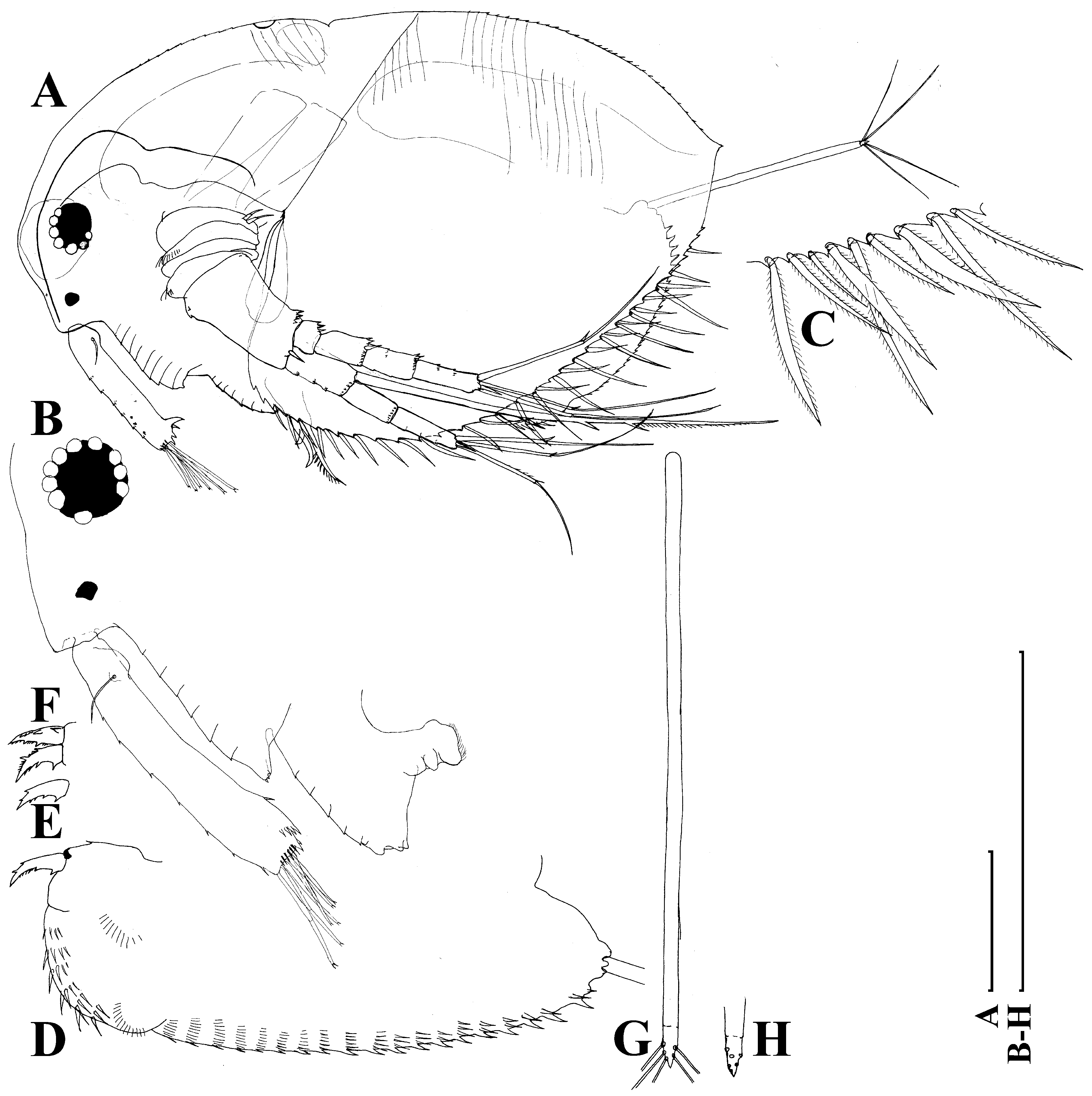
FIGURE 11. Macrothrix triserialis Brady, 1886, parthenogenetic female from Bak Sil Ji 1, locality 6 a: A, lateral view; B, head, lateral view; C, setae at postero-vetral valve margin; D, postabdomen; E – F, postabdominal claws; G – H, postabdominal seta and its distal portion. Scale bars: 0.1 mm.
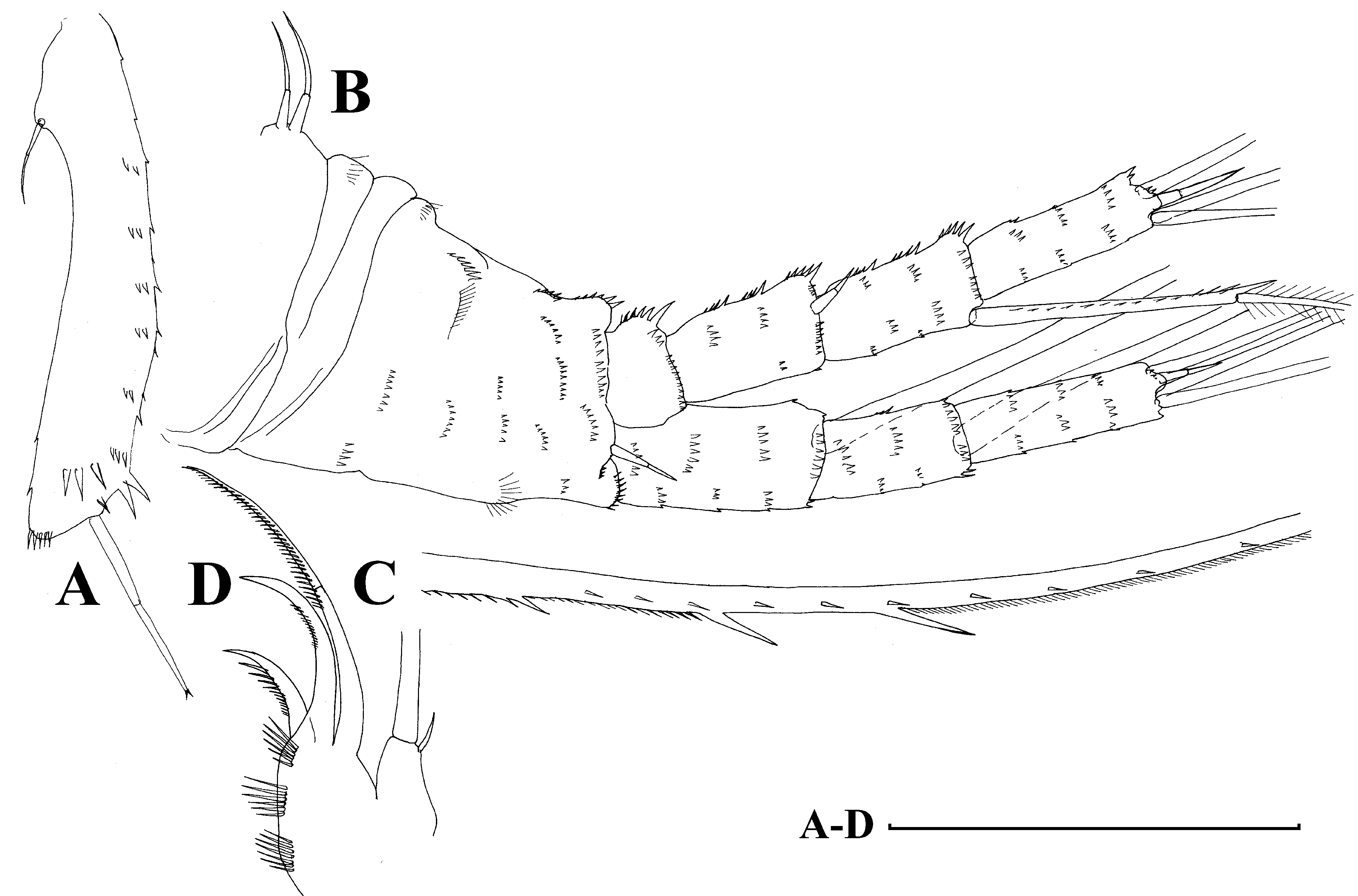
FIGURE 12. Macrothrix triserialis Brady, 1886, appendages of parthenogenetic female from Bak Sil Ji 1, locality 6 a: A, antenna I; B, antenna II, anterior view; C, setae of endopod proximal segment; D, distal portion of limb I. Scale bars: 0.1 mm.
FIGURE 13. Macrothrix rosea (Jurine, 1820), parthenogenetic female from Ho Tan wetland: A, lateral view; B, head, lateral view; C – D, setae at postero-vetral valve margin; E, postabdomen; F, postabdominal claw; G – H, postabdominal seta and its distal portion. Scale bars: 0.1 mm.

FIGURE 14. Macrothrix rosea (Jurine, 1820), appendages of parthenogenetic female from Ho Tan wetland: A, antenna I; B, antenna II, anterior view; C – D, setae of endopod proximal segment; E, distal portion of limb I. Scale bars: 0.1 mm.

FIGURE 15. Bosmina fatalis Burckhardt, 1924, parthenogenetic female from Yong Yeon Ji, locality 4: A, lateral view; B, head, anterior view; C – D, lateral head pore; E, postero-ventral angle of valve, inner view; F, postabdomen. Scale bars: 0.1 mm.
FIGURE 16. Bosmina longirostris (O. F. Müller, 1776), parthenogenetic female from Nu Gyo Ri, locality 19: A, lateral view; B, lateral head pore; C, postabdomen. Scale bars: 0.1 mm.
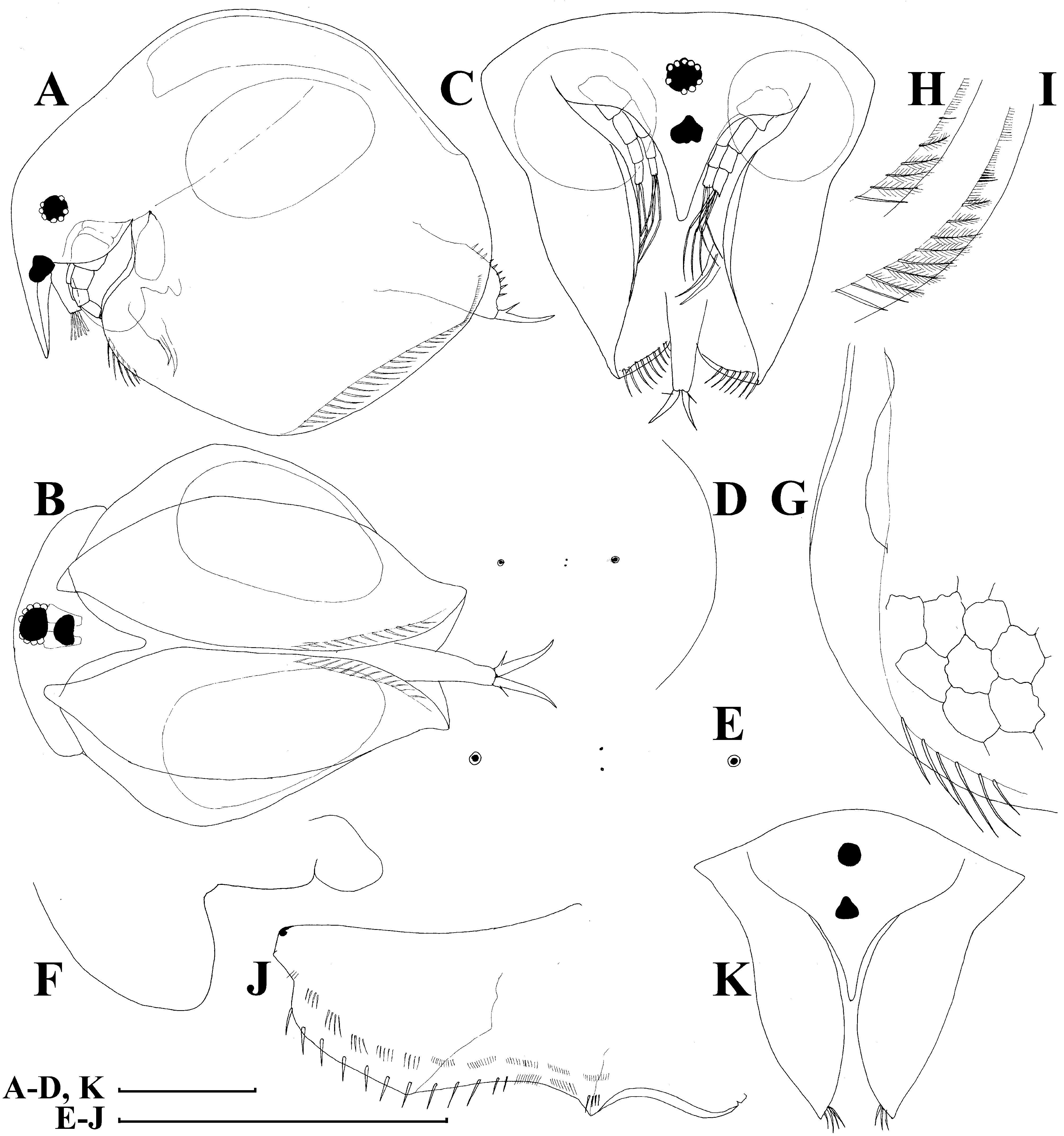
FIGURE 17. Chydorus irinae Smirnov & Sheveleva, 2010, parthenogenetic female from Bak Sil Ji 1, locality 6 a: A – C, adult, lateral, ventral and anterior view; D – E, dorsal head pores; F, labrum; G, anterior portion of valve; H – I, postero-ventral portion of valve, inner view; J, postabdomen; K, juvenile, anterior view. Scale bars: 0.1 mm.
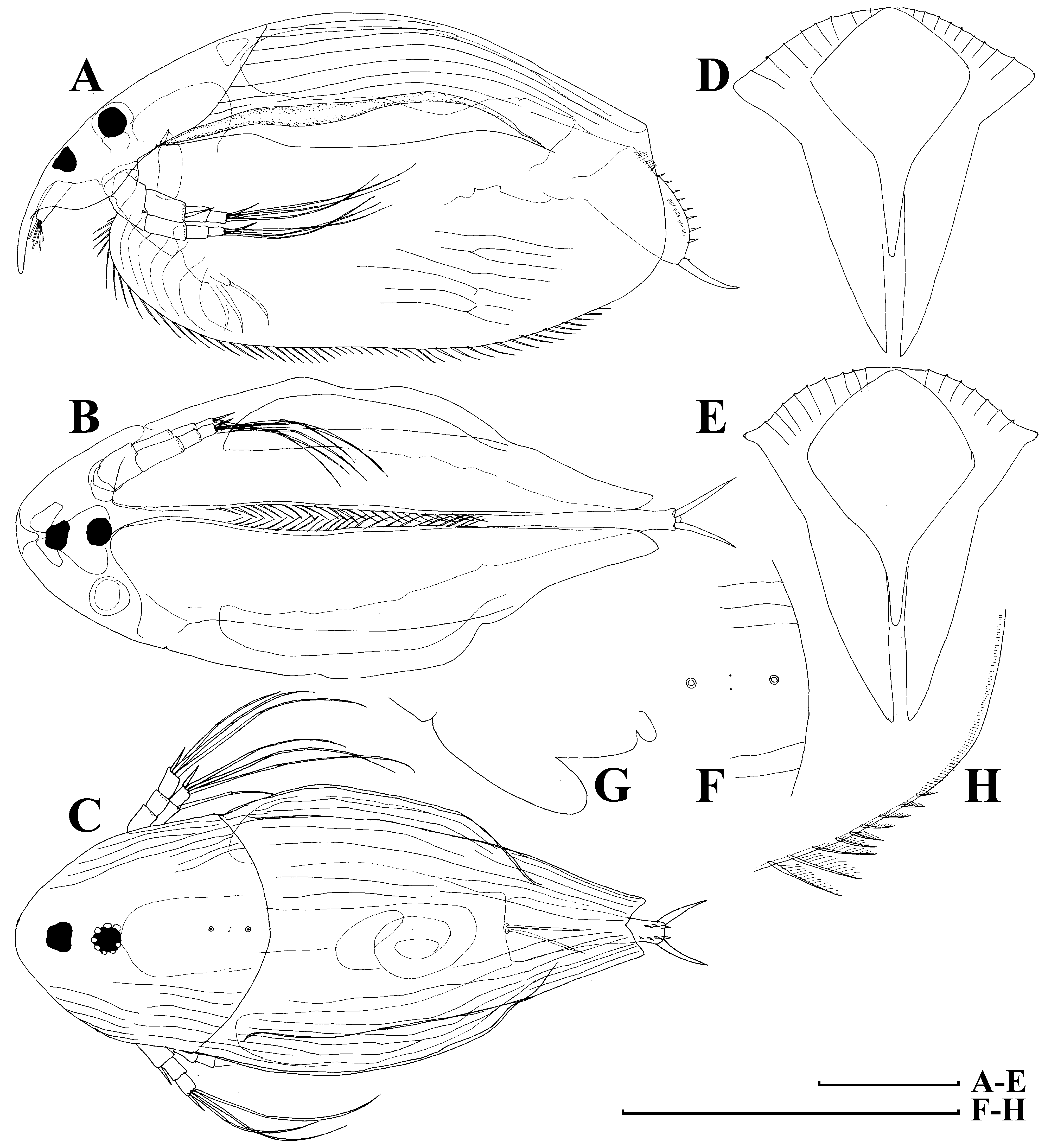
FIGURE 18. Disparalona ikarus Kotov & Sinev, 2011, adult parthenogenetic female from Bak Sil Ji 1, locality 6 a: A – C, lateral, ventral and dorsal view; D – E, anterior view of two different individuals; F, dorsal head pores; G, labrum; H, postero-ventral valve portion, inner view. Scale bars: 0.1 mm.

No known copyright restrictions apply. See Agosti, D., Egloff, W., 2009. Taxonomic information exchange and copyright: the Plazi approach. BMC Research Notes 2009, 2:53 for further explanation.
|
Kingdom |
|
|
Phylum |
|
|
Class |
|
|
Order |
|
|
Family |
|
|
Genus |
1 (by plazi, 2016-04-12 21:22:09)
2 (by ImsDioSync, 2016-12-21 02:34:39)
3 (by ImsDioSync, 2016-12-21 02:36:42)
4 (by ImsDioSync, 2018-06-29 22:01:46)
5 (by ImsDioSync, 2019-03-29 23:17:36)
6 (by ExternalLinkService, 2019-09-26 19:23:57)
7 (by ExternalLinkService, 2021-11-09 17:29:56)
8 (by ExternalLinkService, 2021-11-10 06:23:56)
9 (by ExternalLinkService, 2021-11-12 11:34:29)
10 (by ExternalLinkService, 2021-11-12 11:34:29)
11 (by plazi, 2023-10-26 09:02:15)