
Macrothrix triserialis Brady, 1886
|
publication ID |
https://doi.org/10.5281/zenodo.214313 |
|
DOI |
https://doi.org/10.5281/zenodo.5680419 |
|
persistent identifier |
https://treatment.plazi.org/id/03B687AA-FFB8-5A52-25DC-9FB7FE8FFC21 |
|
treatment provided by |
Plazi (2016-04-12 21:22:09, last updated 2024-11-26 05:54:19) |
|
scientific name |
Macrothrix triserialis Brady, 1886 |
| status |
|
10. Macrothrix triserialis Brady, 1886 View in CoL
Fig. 11–12 View FIGURE 11 View FIGURE 12
Synonymy. Macrothrix triserialis Brady, 1886 , p. 295, Pl. 37: figs 16–20; Smirnov 1992, p. 51–55, Figs 191–205, 219, 225; Dumont et al. 2002, p. 6–7, Figs 18–20 View FIGURE 18 View FIGURE 19 View FIGURE 20 .
Not M. triserialis Brady in Chiang & Du 1979, Fig. 127.
See intensive synonymy in Smirnov (1992).
Type locality. "Colombo" ( Brady 1886), Sri Lanka.
Locality in Korea. 6a (see Fig. 1 and Table 1 View TABLE 1 ).
Parthenogenetic female. Body subovoid, dorsal margin in general regularly curved, with well-expressed ocular dome, shallow depression behind dorsal head pore, postero-dorsal as a pointed angle, lying somewhat above longitudinal body axis ( Fig. 11 View FIGURE 11 A). Whole surface of head and valves covered with striae, with rare or numerous anastomousings. Body significantly compressed laterally, with sharp dorsal keel. Ventral head margin somewhat convex, with a projection anterior to labrum, which is sub-rectangular, with a series of tubercles at apex; ocellus small ( Fig. 11 View FIGURE 11 B). Setae at ventral margin plumose, organized in short series of three setae of different size located at different angles, the first seta thick ( Fig. 11 View FIGURE 11 C). Postabdomen subovoid, with rounded distal extremity, distinct heel basally, preanal margin with short transversal series of short to medium-sized setules; in basal portion these setules always longer and more robust ( Fig. 11 View FIGURE 11 D). Postabdominal claw small, outer dorsal row of two large denticles: a ‘basal spine’ and second denticle, plus a small denticles distally; inner dorsal row with numerous denticles, two of them remarkably more robust than the rest; ventral row of two denticles ( Fig. 11 View FIGURE 11 D–F). Postabdominal seta with a very short distal segment, no setules on basal segment ( Fig. 11 View FIGURE 11 G–H). Antenna I rod-like, sensory seta externally at a distance more than antennular diameter from antennule joint, about 7 transverse rows of denticles on anterior surface of antennule; nine short aesthetascs, two of them significantly larger than the rest ( Fig. 11 View FIGURE 11 B, 12A). Antenna II with two small basal sensory setae of subequal size. Basal segment with distal burrowing spine equal in length to basal segment of exopod, naked; swimming setae 0-0-1-3/1-1-3, spines 0-1-0-1/0-0-1 ( Fig. 12 View FIGURE 12 B). Largest seta (on basal endopod segment) with three strong spinules in middle portion, and a row of short setules in basal and distal portions ( Fig. 12 View FIGURE 12 C). True spine on second segment of exopod with third length of this segment. Additional spines on exopod segments small, decreasing gradually in dorsal direction ( Fig. 12 View FIGURE 12 B). On limb I, ODL with long apical seta, and small lateral seta; IDL with tree series of strong setules, and three bisegmented setae of different size, unilaterally setulated distally, two smaller two smaller ones hook-shaped ( Fig. 12 View FIGURE 12 D). Other limbs as in females from type locality as described by Dumont et al. (2002). Size in our material 0.55–0.73 mm.
Notes. The Macrothrix rosea-triserialis group was revised during the last decade, and it was found that M. triserialis lives only in the tropics and subtropics of the Old World ( Dumont et al. 2002; Kotov et al. 2004). Kim & Yoon (1987) and Yoon (2010) reported for Korea only a single species from this group, the palaearctic M. rosea (Jurine, 1820) (as Echinisca ). We confirm its presence in Korea (see Figs 13–14 View FIGURE 13 View FIGURE 14 of M. rosea from Ho Tan wetland). M. triserialis differs from the latter in: (1) more expressed projection anterior to labrum; (2) thinner first seta in each triplet on valve; (3) very short distal segment of postabdominal seta; (4) three strong spinules in the middle of largest antennal seta. The finding of M. triserialis in Korea‘s non-subtropical climate (i.e. winters with negative temperatures) was quite surprising. Taking into consideration that " M. triserialis " in Fig. 192 by Chiang & Du 1979 is apparently M. rosea , the presence of M. triserialis in China needs to be confirmed. The latter we now consider as a tropicopolitan species (=species widely distributed in the tropics, but capable of penetrating more northern territories).
Dumont, H. J., Silva-Briano, M. & Babu, K. K. S. (2002) A re-evaluation of the Macrothrix rosea-triserialis group, with the description of two new species (Crustacea Anomopoda: Macrothricidae). Hydrobiologia, 467, 1 - 44.
Brady, G. S. (1886) Notes on Entomostraca collected by Mr. A. Haly in Ceylon. Journal of the Linnean Society of London, Zoology, 19, 293 - 317.
Chiang, S. & Du, N. (1979) Fauna Sinica. Crustacea. Freshwater Cladocera. Science Press, Academia Sinica, Peking, China, 297 pp.
Yoon, S. M. & Kim, H. S. (1987) A systematic study on the freshwater Cladocera from Korea. The Korean Journal of Systematic Zoology, 3, 175 - 207. [in Korean]
Kotov, A. A., Garfias-Espejo T. & Elias-Gutierrez, M. (2004) Separation of two Neotropical species: Macrothrix superaculeata (Smirnov, 1982) versus M. elegans Sars, 1901 (Macrothricidae, Anomopoda, Cladocera). Hydrobiologia, 517, 61 - 88.
Smirnov, N. N. (1992) The Macrothricidae of the world. Guides to the identification of the microivertebrates of the Continental Waters of the world, Vol. 1, SPB Academic Publishing, The Hague, 143 pp.
Yoon, S. M. (2010) Arthropoda: Branchiopoda: Anostraca, Notostraca, Spinicaudata, Laevicaudata, Ctenopoda, Anomopoda, Haplopoda Branchiopods. Invertebrate fauna of Korea, 21 (2), 1 - 156.
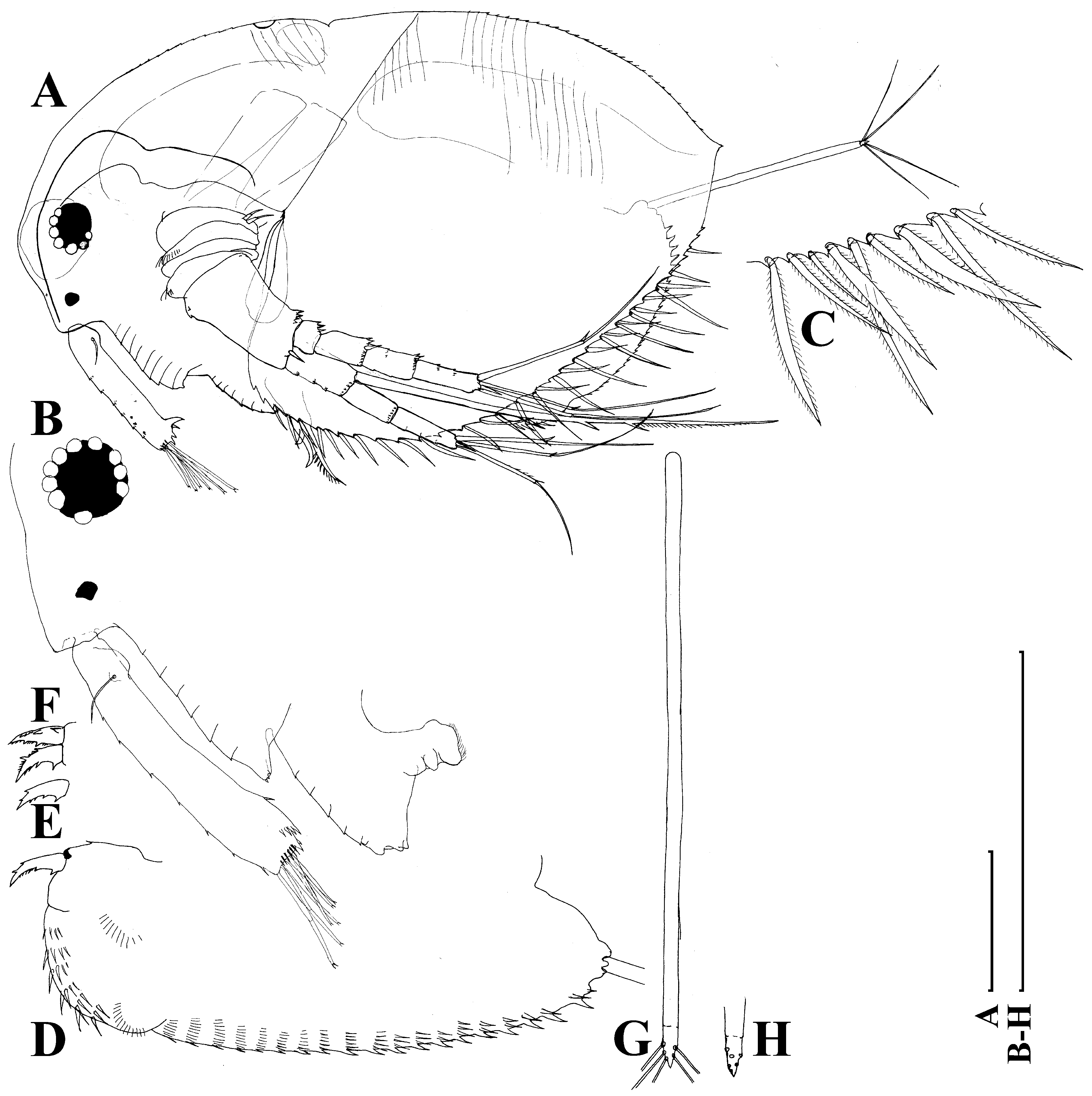
FIGURE 11. Macrothrix triserialis Brady, 1886, parthenogenetic female from Bak Sil Ji 1, locality 6 a: A, lateral view; B, head, lateral view; C, setae at postero-vetral valve margin; D, postabdomen; E – F, postabdominal claws; G – H, postabdominal seta and its distal portion. Scale bars: 0.1 mm.
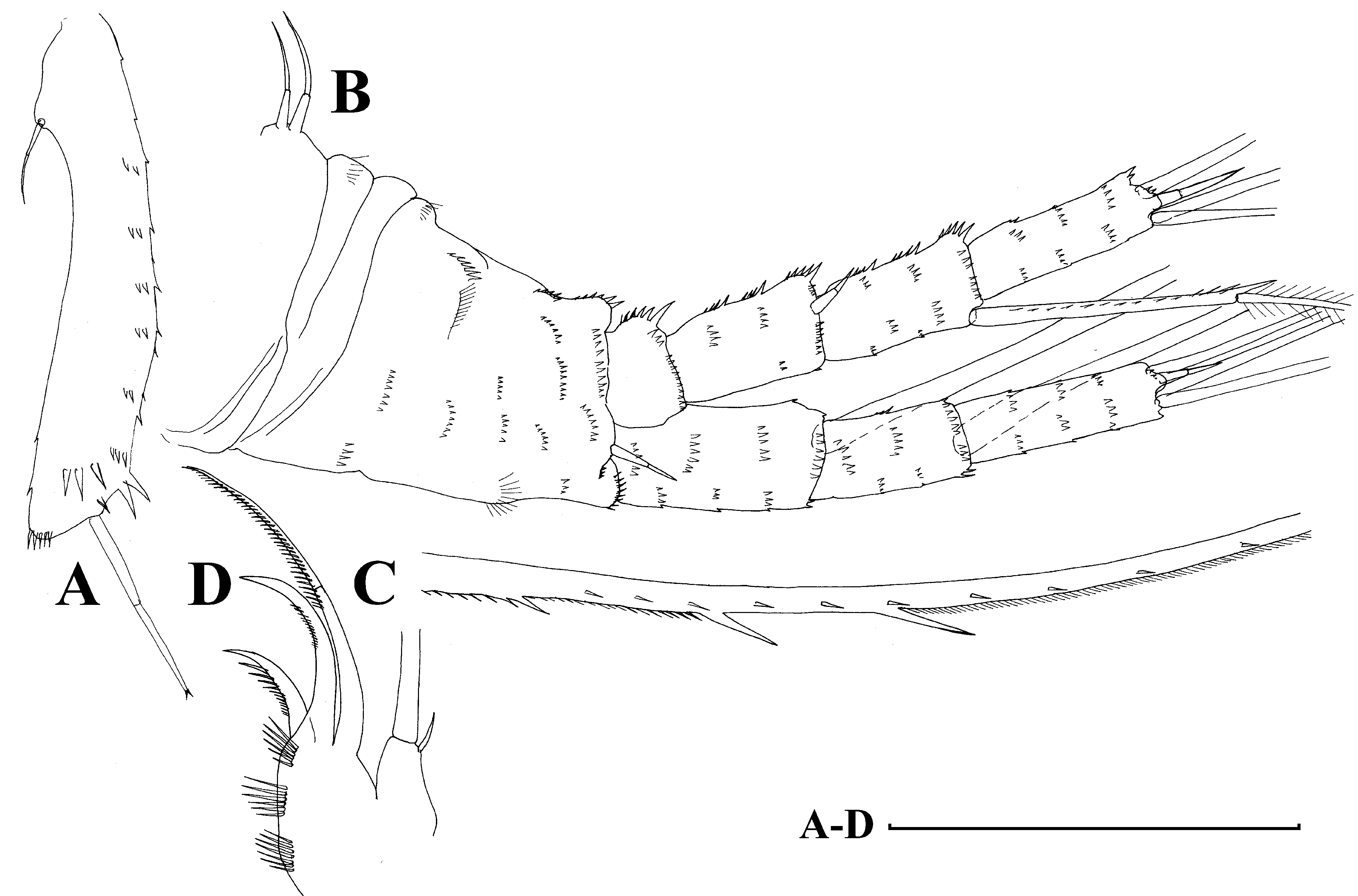
FIGURE 12. Macrothrix triserialis Brady, 1886, appendages of parthenogenetic female from Bak Sil Ji 1, locality 6 a: A, antenna I; B, antenna II, anterior view; C, setae of endopod proximal segment; D, distal portion of limb I. Scale bars: 0.1 mm.
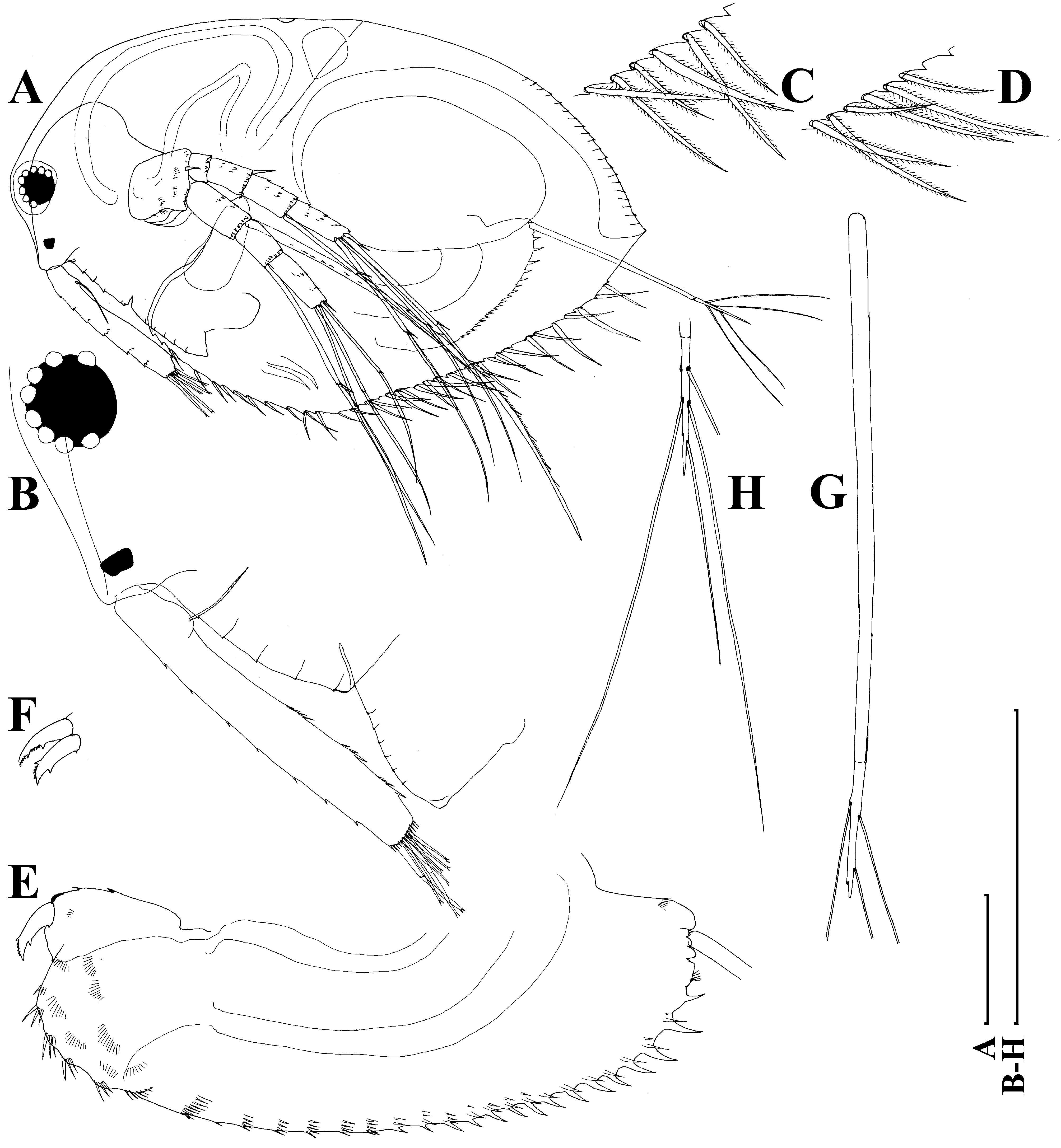
FIGURE 13. Macrothrix rosea (Jurine, 1820), parthenogenetic female from Ho Tan wetland: A, lateral view; B, head, lateral view; C – D, setae at postero-vetral valve margin; E, postabdomen; F, postabdominal claw; G – H, postabdominal seta and its distal portion. Scale bars: 0.1 mm.

FIGURE 14. Macrothrix rosea (Jurine, 1820), appendages of parthenogenetic female from Ho Tan wetland: A, antenna I; B, antenna II, anterior view; C – D, setae of endopod proximal segment; E, distal portion of limb I. Scale bars: 0.1 mm.
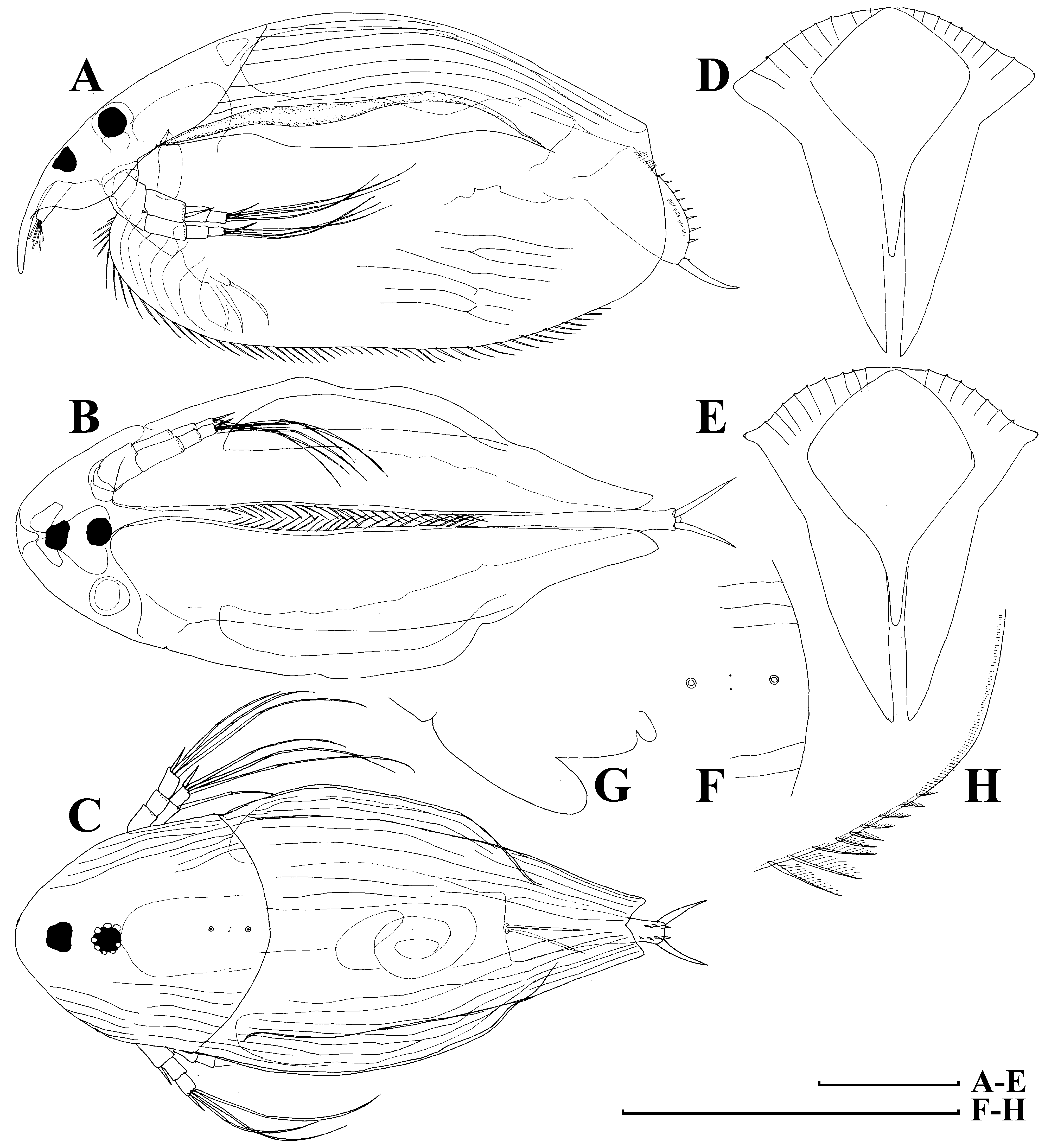
FIGURE 18. Disparalona ikarus Kotov & Sinev, 2011, adult parthenogenetic female from Bak Sil Ji 1, locality 6 a: A – C, lateral, ventral and dorsal view; D – E, anterior view of two different individuals; F, dorsal head pores; G, labrum; H, postero-ventral valve portion, inner view. Scale bars: 0.1 mm.

FIGURE 19. Disparalona ikarus Kotov & Sinev, 2011, adult (A – C) and juvenile (D – E) parthenogenetic female from Bak Sil Ji 1, locality 6 a: A, postabdomen; B, antenna I; C, distal portion of limb I; D, juvenile, lateral view; E, its postabdomen. Scale bars: 0.1 mm.

No known copyright restrictions apply. See Agosti, D., Egloff, W., 2009. Taxonomic information exchange and copyright: the Plazi approach. BMC Research Notes 2009, 2:53 for further explanation.
|
Kingdom |
|
|
Phylum |
|
|
Class |
|
|
Order |
|
|
Family |
|
|
Genus |
1 (by plazi, 2016-04-12 21:22:09)
2 (by ImsDioSync, 2016-12-21 02:34:39)
3 (by ImsDioSync, 2016-12-21 02:36:42)
4 (by ImsDioSync, 2018-06-29 22:01:46)
5 (by ImsDioSync, 2019-03-29 23:17:36)
6 (by ExternalLinkService, 2019-09-26 19:23:57)
7 (by ExternalLinkService, 2021-11-09 17:29:56)
8 (by ExternalLinkService, 2021-11-10 06:23:56)
9 (by ExternalLinkService, 2021-11-12 11:34:29)
10 (by ExternalLinkService, 2021-11-12 11:34:29)
11 (by plazi, 2023-10-26 09:02:15)