
Bunocephalus hartti, Carvalho & Cardoso & Friel & Reis, 2015
|
publication ID |
https://doi.org/ 10.1590/1982-0224-20140152 |
|
DOI |
https://doi.org/10.5281/zenodo.5138059 |
|
persistent identifier |
https://treatment.plazi.org/id/03B68783-1F0B-FFA7-C9DB-FF63FB49C96E |
|
treatment provided by |
Carolina |
|
scientific name |
Bunocephalus hartti |
| status |
sp. nov. |
Bunocephalus hartti View in CoL , new species
urn:lsid:zoobank.org:act:510B1ECB-BF95-4174-B7BC-856AB3BE3FCA
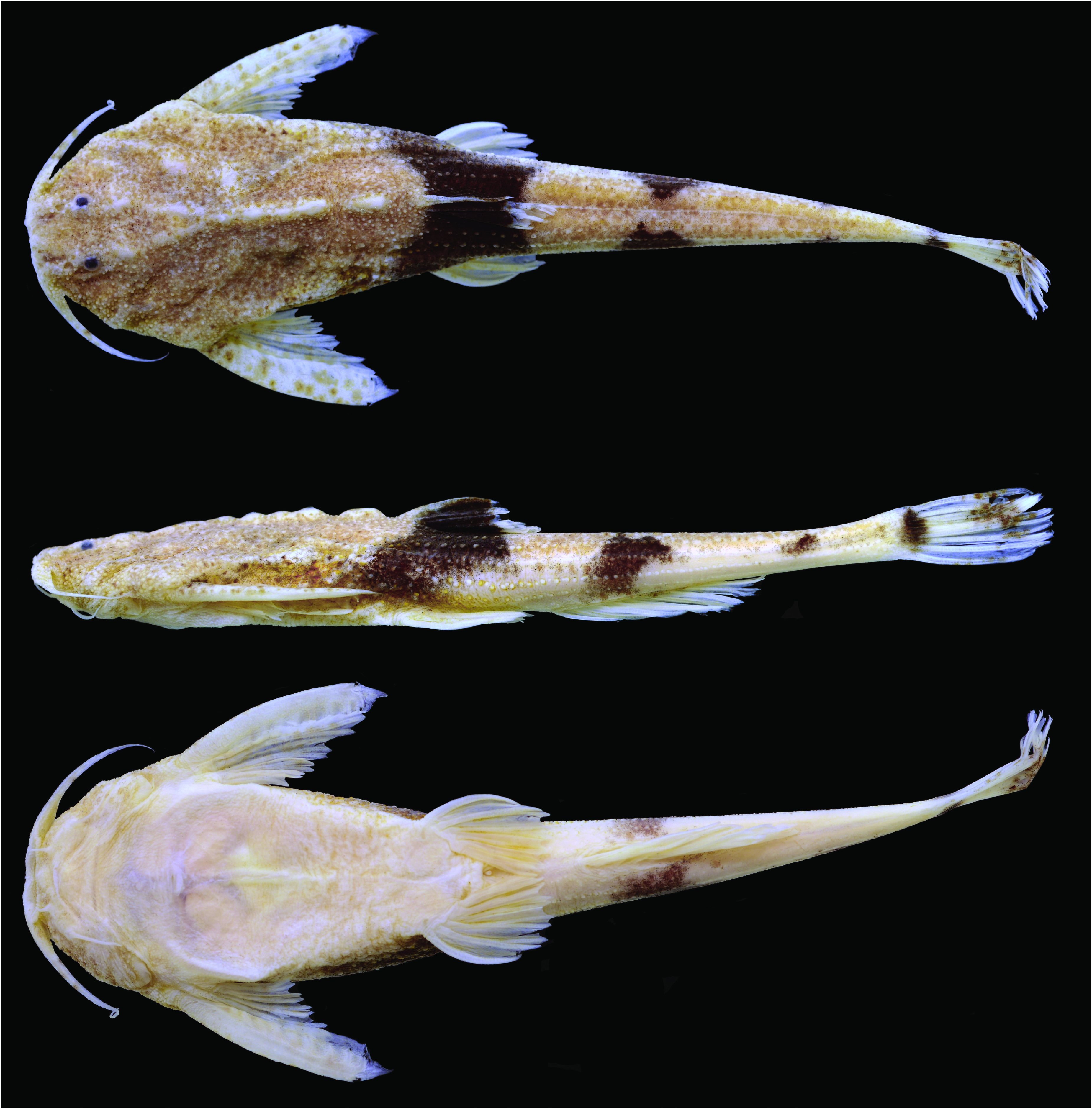
Fig. 1 View Fig , Table 1 View Table 1
Bunocephalus larai View in CoL non Ihering, 1930.- Mees, 1989:238 [doubtfully referred]. - Friel, 2003: 263 [referred as undescribed].
Bunocephalus View in CoL sp. n. -Alves & Pompeu, 2001: 185 [listed].
Bunocephalus sp. N. 1. -Alves & Pompeu, 2005: 597 [listed as undescribed].
Bunocephalus View in CoL sp. A. - Barbosa & Soares, 2009: 162 [listed].
Holotype. MZUSP 62745 View Materials , 54.8 mm SL, Brazil, Minas Gerais, Presidente Juscelino, rio Cipó at Fazenda Duas Barras , 18°41’04”S 43°59’18”W, 1 Jul 2000, C. B. M. Alves & P. S. Pompeu. GoogleMaps
Paratypes. All from Minas Gerais, Brazil, rio São Francisco basin: CAS 53522, 1 About CAS , 57.7 mm SL, creek tributary to rio das Velhas ca. 35 miles north of Belo Horizonte , 2 Feb 1941, T. D. White and others. MCP 45236, 1 View Materials , 39.9 mm SL, Augusto de Lima municipality, rio Curimataí , tributary to rio das Velhas, 17°59’32”S 44°10’47”W, 9 Jul 2009, C. G. Leal & D. C. de Carvalho. MCP 48280, 1 View Materials c&s, 21.5 mm SL, Iguatama municipality, ribeirão São Miguel, tributary to rio São Francisco , 20°12’04”S 45°39’12”W, 26 Sep 2003, B. P. Nogueira and others. MNRJ 31385 View Materials , 4 View Materials (1c&s), 37.2- 42.6 mm SL, Piumhi municipality, mouth of rio Piumhi , 20°20’31”S 45°59’03”W, 28 Feb 2007, P. L. Gallo, L. H. Silva, D. L. Z. Kantek & W. A. M. Perez. MZUSP 39443 View Materials , 1 View Materials , 45.5 mm SL, rio Formoso , tributary to rio São Francisco , 8 Feb 1988, Y. Sato. MZUSP 39480 View Materials , 1 View Materials , 40.0 mm SL, rio São Francisco at mouth of rio Formoso , approx. 17°26’S 44°57’W, 8-10 Feb 1988, Y. Sato. MZUSP 64227 View Materials , 3 View Materials , 36.0- 44.7 mm SL, Juataba municipality, rio Paraopeba between Juataba and Betim, approx. 19°56’S 44°18’W, 6 Nov 2000, C. B. M. Alves GoogleMaps .
Diagnosis. Bunocephalus hartti is distinguished from all congeners by the absence of serrations along the anterior margin of the pectoral-fin spine in adults ( Fig. 2a View Fig ; vs. presence of serrations along the anterior margin in adults, Fig. 2c View Fig ). Bunocephalus hartti can be further distinguished from most congeners, except for B. verrucosus , by having the last dorsal-fin ray completely or almost completely adnate to the dorsum (vs. dorsal-fin ray completely free or with less than half extension connected to the dorsum).
Description. Morphometric data summarized in Table 1 View Table 1 . Maximum body size moderate to small compared to congeners (maximum observed size 57.7 mm SL). Dorsal, left lateral and ventral views of body in Fig. 1 View Fig . Head and body depressed, lateral profile ascending from tip of snout to dorsal-fin origin, with bony skull ornamentations in between. Posterodorsal profile of body straight and descending from dorsal-fin origin to near base of caudal fin, becoming slightly convex anterior to caudal-fin base. Ventral body profile convex from mouth to insertion of pelvic fin; concave from this point to anal-fin origin, straight and ascending from anal-fin origin to base of caudal fin, slightly concave at caudal-fin base. Caudal peduncle slender, somewhat rounded in cross section, but flattened dorsally and ventrally, shallowest at midpoint between end of anal fin and caudal-fin origin.
Skull ornamentation weakly developed. Eye small and positioned dorsolaterally. Skin covering eye dense and pale. Anterior nostril located terminally at tip of snout, associated with fleshy tube projecting beyond upper lip. Posterior nostril without flap, opening anteromedially near eye. Mouth subterminal, upper lip more prominent relative to lower lip. All barbels simple, unbranched; maxillary barbel reaching or slightly surpassing insertion of pectoralfin spine, posterolateral mental barbel twice as long as anteromedial one. Opercular opening reduced to small valvular slit located just anterior and medially to insertion of pectoral-fin spine. Axial slit pore present, dorsoventrally inclined underneath posterior cleithral process. Adult males with digitiform testes. Integument covered with large unculiferous tubercles, forming series of aligned longitudinal rows on posterior portion of body. Large and well-defined rows of tubercles on caudal peduncle, one on middorsum, and three on lateral of body. Other rows poorly defined.
Osteological description based on two cleared and stained specimens (21.5-42.2 mm SL, see paratype list). Anterior margin of mesethmoid slightly concave, anterolateral projection slightly pronounced ( Fig. 3a View Fig ). Ethmoid cartilage separate from articular facet of palatine. Frontal with lateral projections forming dorsal margin of eye. Frontal posteriorly projected laterally to posterior cranial fontanel and contacting supraoccipital, epiphyseal bar present. Supratemporal fossa present at middle portion of contact between pterotic and supraoccipital bones. Pterotic with laterally expanded and pointed bony shelf. Premaxilla with somewhat rectangular shape, bearing few teeth on its posteromedial margin. Dentary slender, abutting counterpart at medial portion, symphyseal portion slightly expanded, teeth present along anterior half of dorsal margin. Ascending process of Meckel’s cartilage present. Coronomeckelian bone present. Hyomandibula associated with preopercle and posterior portion of mandibular laterosensory canal, supraopercle absent. Cartilaginous contact of hyomandibula with neurocranium restricted to sphenotic bone. Anterodorsal process of hyomandibula developed, contacting ventral surface of sphenotic. Opercular condyle of hyomadibula well developed. Metapterygoid present, contacting quadrate and hyomandibula. Endopterygoid present, somewhat triangular in shape, located underneath contact of palatine and lateral ethmoid. Posterior margin of palatine cartilaginous and rounded. Opercle “L” shaped, posterior arm larger than ventral arm. Interopercle present, triangular in shape and firmly attached to ventral arm of opercle. Dorsal hypohyal absent. Anterior ceratohyal with expanded lamina on anteroventral margin, contacting posterior ceratohyal by means of cartilage and interdigitated suture. Posterior ceratohyal with foramen on midventral portion. Interhyal present. Four branchiostegal rays. Urohyal present, pointed anteriorly, with foramen and well developed lateral wings. First and second pharyngobranchials absent; third and fourth present and ossified. First hypobranchial ossified, second and third cartilaginous. Second and third basibranchial ossified, fourth cartilaginous. Third epibranchial bearing uncinated process. Gill rakers present in all branchial arches, but limited to few on first and second arches. Pharyngeal teeth well developed on upper tooth plate; about two rows of teeth on fifth ceratobranchial limited to its medial margin.
Dorsal lamina of Weberian complex reaching dorsal surface of body, lateral profile of lamina ascending posteriorly with anterior concavity and bony knob at middle portion and elevated crest posteriorly. Parapophysis of fourth vertebra forming broad lamina over swim bladder, contacting parapophysis of fifth vertebrae extensively. Parapophysis of fifth vertebra long, extending to lateral body surface transverse to main body axis. Distal portion of fifth parapophysis expanded. Total vertebrae 35. Vertebra bearing horizontal transverse processes from centrum nine to 31. Hemal spine contacting anal-fin pterygiophores bifid. Four to five pairs of ribs (modally five), on vertebrae six to nine or ten. Abdominal vertebrae foramina (hemal arches) for hemal canal on 6 th or 7 th and posteriorly on 10 th vertebrae.
Dorsal fin with five or six rays (modally five), without spinelet. First ray unbranched followed by four or five branched rays. Membrane of last dorsal-fin ray adnate to dorsum. Anterior nuchal plate absent, middle nuchal plate contacting posterior nuchal plate laterally. Posterior nuchal plate not developed laterally, lateral limit not extending beyond contact with middle nuchal plate. Pectoral fin with one rigid spine and five branched soft rays. Pectoral spine curved, feeble serrations present in anterior portion in juveniles, but absent in adults ( Figs. 2a,b View Fig ). Serrations along posterior margin of spine increasing in number with larger body sizes, maximum of 10 serrations on posterior margin. Two ossified plus one cartilaginous pectoral-fin radial. Postcoracoid process of pectoral girdle extending slightly posterior to postcleithral process in lateral view. Pelvic fin with six soft rays, second and third rays longest, not reaching anal-fin origin, first ray unbranched. Posterior margin of basipterygium jagged. Lateral cartilage of basipterygium extending from its anteriormost portion to contact with last pelvic-fin ray. Anal fin with seven to nine rays (modally eight), first two or three unbranched, third of length of last anal-fin ray extension adnate by membrane to body. Caudal fin with ten principal rays, five associated with upper lobe and five with ventral lobe, posterior margin of caudal fin convex. Lowermost and uppermost caudal-fin rays unbranched, with proximal expansion and slightly shorter than branched middle rays. Caudal fin with two procurrent rays on upper and lower lobes, anterior procurrent ray small, triangular in shape, posterior procurrent ray longer and spine like. Posterior margin of upper hypural plate extending posteriorly further than lower hypural plate (hypurals one and two fused with parhypural). Second ural half-centrum well developed. Adipose fin absent.
Nasal canal ossified and positioned laterally to mesethmoid, one or two separate tubular ossifications around canal. Antorbital present, with anterior limb pointed, extending anterior to anterior margin of premaxilla. Antorbital mesial limb rounded and associated with laterosensory canal. Infraorbital canal present with three tubular ossifications, canal exiting antorbital, passing below eye margin and entering neurocranium through sphenotic. Mandibular canal interrupted, with two tubular ossifications lateral to posterior portion of dentary, and two tubular ossifications near to contact with preopercle. Extrascapular present. Lateral line not associated with fourth parapophyses, anterior portion running just aside margin of parapophyses. Lateral line complete, extending variably to caudal peduncle, formed by simple tubes, median portion presenting small inconspicuous hooks.
Color in alcohol. Head and body light brown dorsally, ventral portions lighter brown to yellowish pale. Four saddles of dark coloration on dorsal surface of body. First dark saddle at level of dorsal fin, second at anal fin vertical, third at middle caudal peduncle and fourth at origin of caudal fin. Second, third and fourth saddles sometimes not connected at middorsal line. Dorsal fin mostly dark brown with light distal margin; pectoral fin whitish cream to hyaline with small light brown spots, pelvic and anal fins mostly hyaline, without conspicuous dark spots. Caudal fin hyaline with dark blotch at proximal portion and scattered black spots on distal third sometimes forming band.
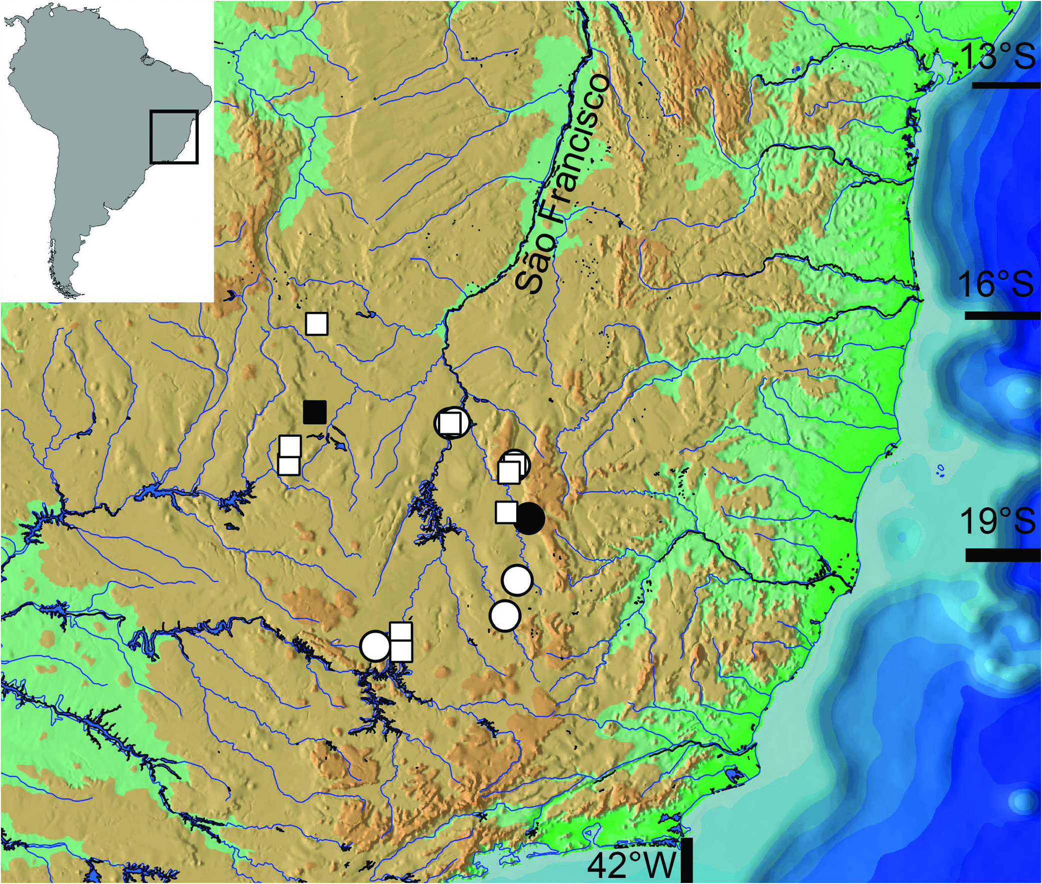
Distribution. Known from several tributaries of the upper and middle rio São Francisco basins including the das Velhas, Paraopeba and Formoso rivers in Minas Gerais State, Brazil ( Fig. 4 View Fig ).
Etymology. The epithet hartti is a patronym honoring Charles Frederick Hartt, a Canadian-American geologist, and first professor of Geology at Cornell University. Hartt worked extensively in Brazil, and a few of his notable accomplishments include the publication of “Geology and physical geography of Brazil ” ( Hartt, 1870), and serving as the founder and director of the section of geology at the Museu Nacional of Brazil from 1866 to 1867.
Conservation status. Bunocephalus hartti is known from an Extent of Occurrence (EOO) of approximately 34,000 km 2, and despite some areas within its range suffer continuing decline in habitat quality because of contamination from the city of Belo Horizonte and also mining and agriculture, there is no evidence of its habitat being severely fragmented or occurring extreme fluctuations in range or number of individuals. Considering that no specific threats to the species were detected, B. hartti is categorized as Least Concern (LC) according to the International Union for Conservation of Nature (IUCN) categories and criteria ( IUCN Standards and Petitions subcommittee, 2014).


No known copyright restrictions apply. See Agosti, D., Egloff, W., 2009. Taxonomic information exchange and copyright: the Plazi approach. BMC Research Notes 2009, 2:53 for further explanation.
|
Kingdom |
|
|
Phylum |
|
|
Class |
|
|
Order |
|
|
Family |
|
|
Genus |
Bunocephalus hartti
| Carvalho, Tiago P., Cardoso, Alexandre R., Friel, John P. & Reis, Roberto E. 2015 |
Bunocephalus
| Barbosa J & Soares & Perfil 2009: 162 |
Bunocephalus larai
| Friel 2003: 263 |